

Search Count: 864
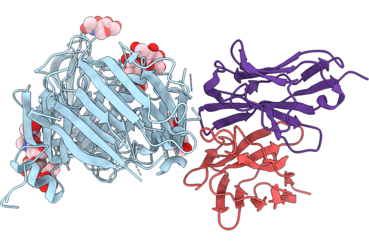 |
Cryo-Em Structure Of N19 Fab : D0D-S1A
Organism: Porcine epidemic diarrhea virus, Sus scrofa
Method: ELECTRON MICROSCOPY Resolution:2.30 Å Release Date: 2026-08-05 Classification: VIRAL PROTEIN/IMMUNE SYSTEM Ligands: NAG |
Organism: Porcine epidemic diarrhea virus, Sus scrofa
Method: ELECTRON MICROSCOPY
Release Date: 2026-08-05
Ligands: NAG
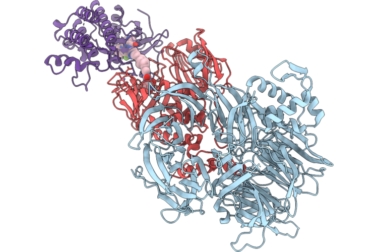 |
Cryoem Structure Of Irak4:Kt-474:Crbn-Ddb1 Ternary Complex
Organism: Homo sapiens
Method: ELECTRON MICROSCOPY Release Date: 2026-07-29 Classification: Transferase/DNA-Binding Protein Ligands: ZN, A1CDC |
Organism: Homo sapiens
Method: ELECTRON MICROSCOPY
Release Date: 2026-07-29
Ligands: ZN, A1CDC
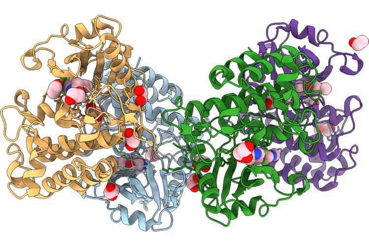 |
Crystal Structure Of Irak4 With Compound 1, An Analog Of Kt-474 Tbm
Organism: Homo sapiens
Method: X-RAY DIFFRACTION Resolution:2.05 Å Release Date: 2026-07-29 Classification: TRANSFERASE Ligands: A1CDD, EDO, GOL |
Organism: Homo sapiens
Method: X-RAY DIFFRACTION
Release Date: 2026-07-29
Ligands: A1CDD, EDO, GOL
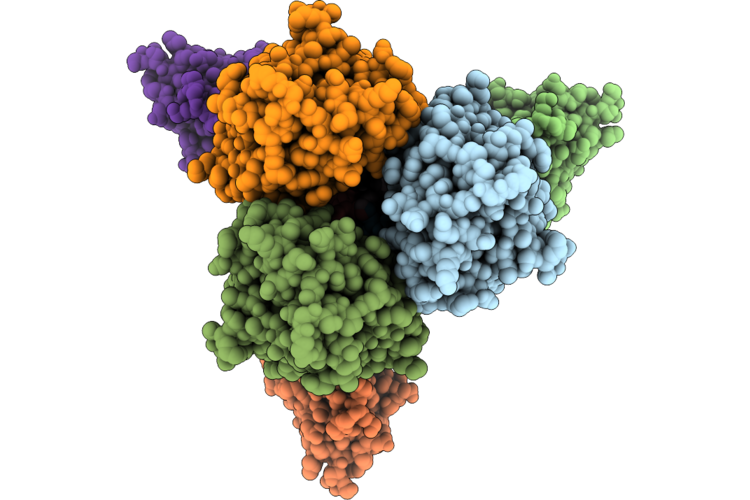 |
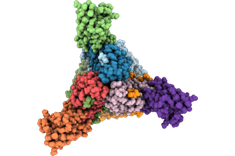
Cryo Em Structure Of Hemagglutinin From Puerto Rico/8/1934 (H1N1) In Complex With A Single Domain Antibody
Organism: Influenza a virus (strain a/puerto rico/8/1934 h1n1), Vicugna pacos
Method: ELECTRON MICROSCOPY Release Date: 2026-06-24 Classification: VIRAL PROTEIN |
Organism: Influenza a virus (strain a/puerto rico/8/1934 h1n1), Vicugna pacos
Method: ELECTRON MICROSCOPY
Release Date: 2026-06-24
 |
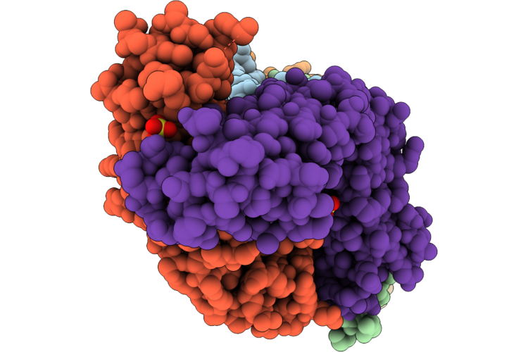
Cryo Em Structure Of Hemagglutinin From Puerto Rico/8/1934-New York/55/2004 (H3N2) In Complex With A Single Domain Antibody
Organism: Influenza a virus (a/new york/55/2004(h3n2)), Vicugna pacos
Method: ELECTRON MICROSCOPY Resolution:2.70 Å Release Date: 2026-06-24 Classification: VIRAL PROTEIN |
Organism: Influenza a virus (a/new york/55/2004(h3n2)), Vicugna pacos
Method: ELECTRON MICROSCOPY
Release Date: 2026-06-24
 |
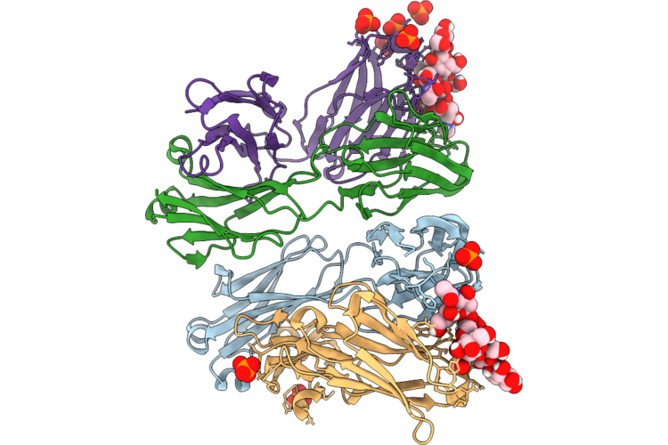
Tgfb1 In Complex With Nis793 Fab
Organism: Homo sapiens
Method: X-RAY DIFFRACTION Resolution:2.75 Å Release Date: 2026-06-17 Classification: CYTOKINE Ligands: SO4 |
Organism: Homo sapiens
Method: X-RAY DIFFRACTION
Release Date: 2026-06-17
Ligands: SO4
 |
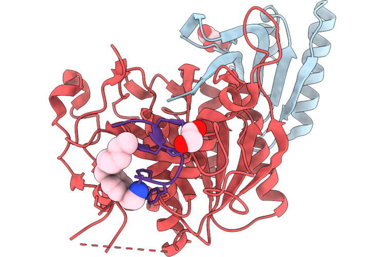
Hca450-21.1 Fab And Gd2 Complex
Organism: Mus musculus
Method: X-RAY DIFFRACTION Resolution:1.69 Å Release Date: 2026-06-17 Classification: RECOMBINATION Ligands: PO4 |
Organism: Mus musculus
Method: X-RAY DIFFRACTION
Release Date: 2026-06-17
Ligands: PO4
 |
Pcsk9 In Complex With Cyclic Peptide 21
Organism: Homo sapiens, Synthetic construct
Method: X-RAY DIFFRACTION Resolution:1.83 Å Release Date: 2026-06-03 Classification: HYDROLASE Ligands: GOL, A1C8P, GOA |
Organism: Homo sapiens, Synthetic construct
Method: X-RAY DIFFRACTION
Release Date: 2026-06-03
Ligands: GOL, A1C8P, GOA
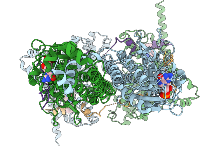 |
Cryo-Em Structure Of Bovine Phosphodiesterase 6 Bound To Cb-5083
Organism: Bos taurus
Method: ELECTRON MICROSCOPY Release Date: 2026-05-27 Classification: SIGNALING PROTEIN Ligands: PCG, ZN, MG, JDP |
Organism: Bos taurus
Method: ELECTRON MICROSCOPY
Release Date: 2026-05-27
Ligands: PCG, ZN, MG, JDP
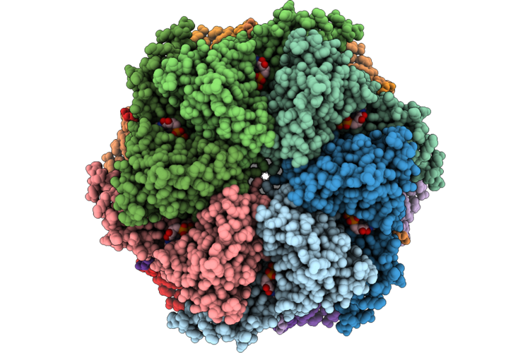 |
Cryo-Em Structure Of Human P97/Vcp Bound To Inhibitor Gnd-135
Organism: Homo sapiens
Method: ELECTRON MICROSCOPY Release Date: 2026-05-27 Classification: HYDROLASE/HYDROLASE INHIBITOR Ligands: ADP, A1CBF |
Organism: Homo sapiens
Method: ELECTRON MICROSCOPY
Release Date: 2026-05-27
Ligands: ADP, A1CBF
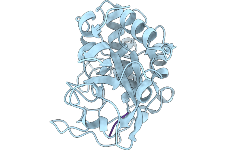 |
Crystal Structure Of Human Cathepsin B With A L-Peptide Substrate
Organism: Homo sapiens, Synthetic construct
Method: X-RAY DIFFRACTION Resolution:1.50 Å Release Date: 2026-05-06 Classification: PEPTIDE BINDING PROTEIN |
Organism: Homo sapiens, Synthetic construct
Method: X-RAY DIFFRACTION
Release Date: 2026-05-06
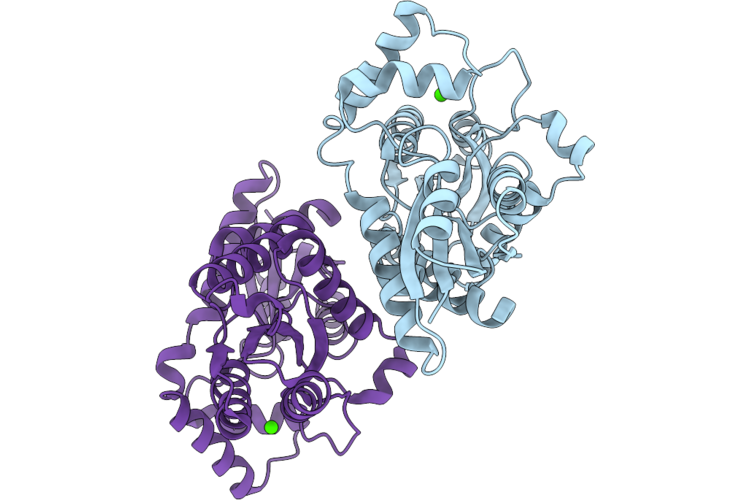 |
The Structure Of Coproporphyrin Ferrochelatase(Cpfc)In Staphylococcus Aureus.
Organism: Staphylococcus aureus (strain nctc 8325 / ps 47)
Method: X-RAY DIFFRACTION Resolution:2.19 Å Release Date: 2026-05-06 Classification: METAL BINDING PROTEIN Ligands: CA |
Organism: Staphylococcus aureus (strain nctc 8325 / ps 47)
Method: X-RAY DIFFRACTION
Release Date: 2026-05-06
Ligands: CA
 |
Crystal Structure Of Human Cathepsin B With A D-Peptide Substrate
Organism: Homo sapiens, Synthetic construct
Method: X-RAY DIFFRACTION Resolution:1.50 Å Release Date: 2026-05-06 Classification: PEPTIDE BINDING PROTEIN |
Organism: Homo sapiens, Synthetic construct
Method: X-RAY DIFFRACTION
Release Date: 2026-05-06
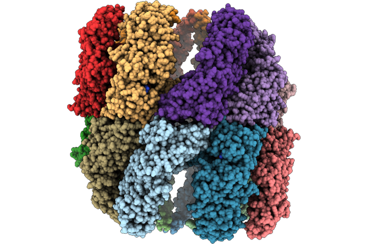 |
Bovine Flagellar Tric
Organism: Bos taurus
Method: ELECTRON MICROSCOPY Release Date: 2026-04-29 Classification: CHAPERONE Ligands: MG, ADP |
Organism: Bos taurus
Method: ELECTRON MICROSCOPY
Release Date: 2026-04-29
Ligands: MG, ADP
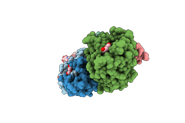 |
Crystal Structure Of The A/Puerto Rico/8/1934 (H1N1) Influenza Virus Hemagglutinin In Complex With Fusion Inhibitor Clips Peptide Cp121132 (Cp-S1)
Organism: Influenza a virus (a/puerto rico/8/1934(h1n1)), Synthetic construct
Method: X-RAY DIFFRACTION Resolution:2.35 Å Release Date: 2026-03-18 Classification: VIRAL PROTEIN Ligands: NAG, PEG, FUC, SEZ |
Organism: Influenza a virus (a/puerto rico/8/1934(h1n1)), Synthetic construct
Method: X-RAY DIFFRACTION
Release Date: 2026-03-18
Ligands: NAG, PEG, FUC, SEZ
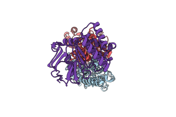 |
Lolcde In Complex With Smt-738_3
Organism: Escherichia coli k-12
Method: ELECTRON MICROSCOPY Resolution:2.88 Å Release Date: 2026-03-18 Classification: TRANSPORT PROTEIN Ligands: A1ER4 |
Organism: Escherichia coli k-12
Method: ELECTRON MICROSCOPY
Release Date: 2026-03-18
Ligands: A1ER4
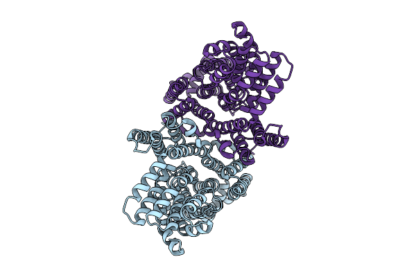 |
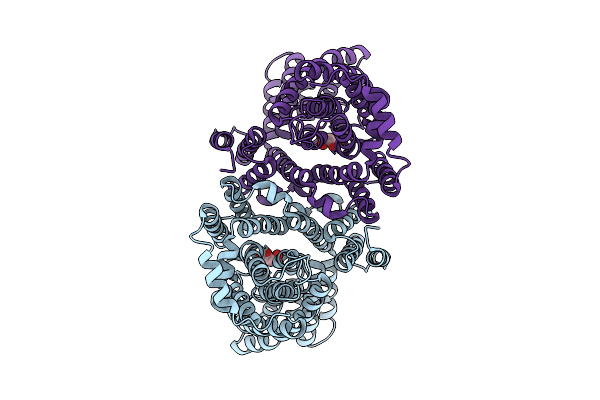
Plant Chloroplast Dicarboxylate Transporter Atdit1
Organism: Arabidopsis thaliana
Method: ELECTRON MICROSCOPY Release Date: 2026-03-11 Classification: TRANSPORT PROTEIN |
Organism: Arabidopsis thaliana
Method: ELECTRON MICROSCOPY
Release Date: 2026-03-11
 |
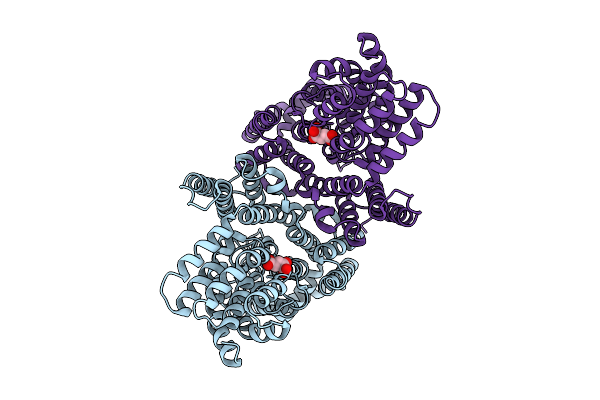
Plant Chloroplast Dicarboxylate Transporter Atdit1 Bound With Oaa
Organism: Arabidopsis thaliana
Method: ELECTRON MICROSCOPY Release Date: 2026-03-11 Classification: TRANSPORT PROTEIN Ligands: OAA |
Organism: Arabidopsis thaliana
Method: ELECTRON MICROSCOPY
Release Date: 2026-03-11
Ligands: OAA
 |
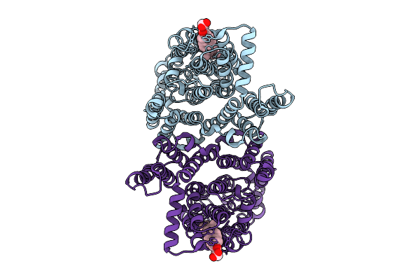
Plant Chloroplast Dicarboxylate Transporter Atdit1 Bound With 2-Og
Organism: Arabidopsis thaliana
Method: ELECTRON MICROSCOPY Release Date: 2026-03-11 Classification: TRANSPORT PROTEIN Ligands: AKG |
Organism: Arabidopsis thaliana
Method: ELECTRON MICROSCOPY
Release Date: 2026-03-11
Ligands: AKG
 |
Plant Chloroplast Dicarboxylate Transporter Atdit2.1
Organism: Arabidopsis thaliana
Method: ELECTRON MICROSCOPY Release Date: 2026-03-11 Classification: TRANSPORT PROTEIN Ligands: Y01 |
Organism: Arabidopsis thaliana
Method: ELECTRON MICROSCOPY
Release Date: 2026-03-11
Ligands: Y01