
Search Count: 1,45,905
All
Selected
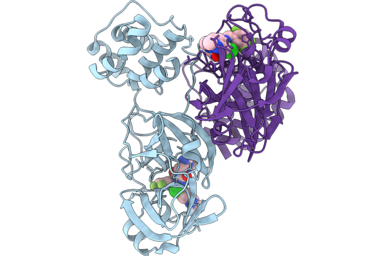 |
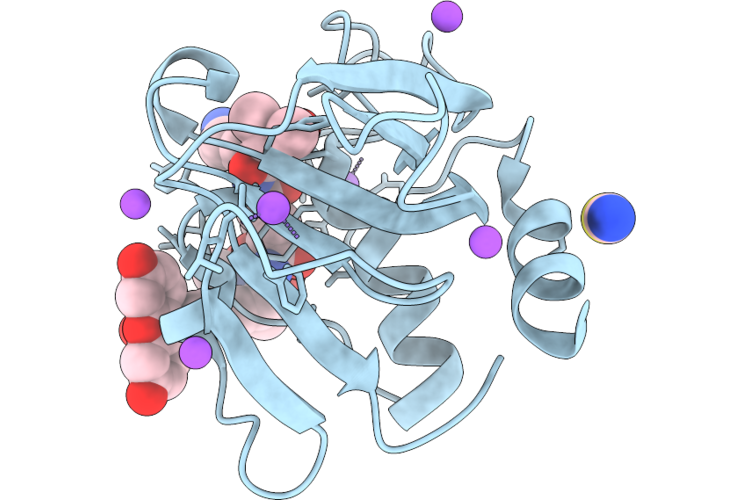
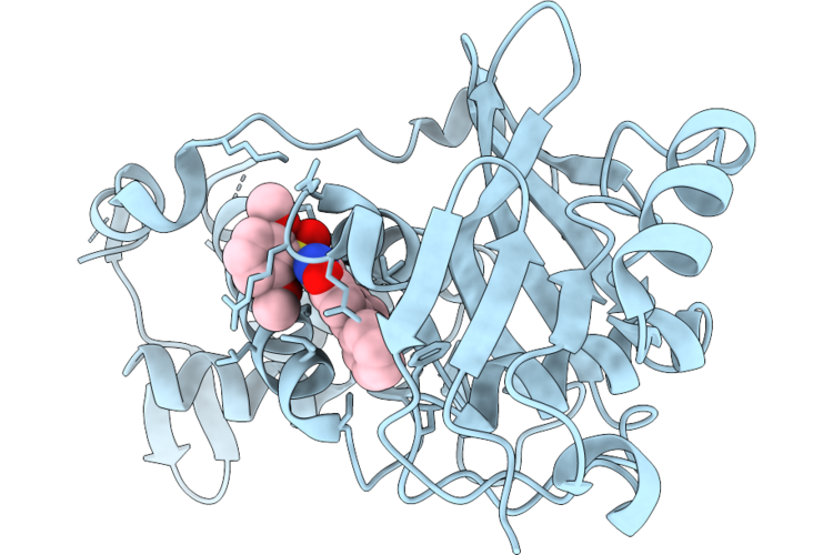
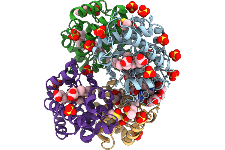
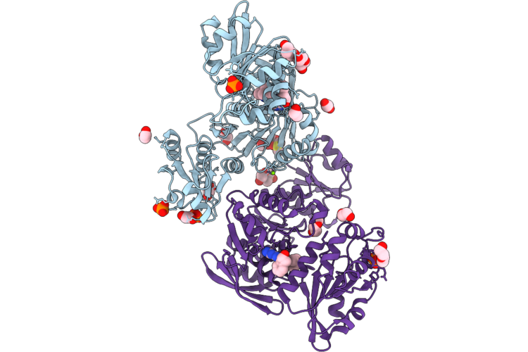
Room Temperature X-Ray Structure Of Sars Cov-2 Main Protease Intermediate Precursor With Ensitrelvir (Esv)
Organism: Homo sapiens
Method: X-RAY DIFFRACTION Resolution:2.05 Å Release Date: 2026-05-06 Classification: HYDROLASE/HYDROLASE INHIBITOR Ligands: 7YY |
 |
Organism: Bos taurus
Method: ELECTRON MICROSCOPY Resolution:3.06 Å Release Date: 2026-05-06 Classification: HYDROLASE Ligands: VIA, PCG, MG, ZN |
 |
Organism: Bacillus thuringiensis
Method: X-RAY DIFFRACTION Resolution:2.00 Å Release Date: 2026-05-06 Classification: TRANSFERASE Ligands: UDP, NOV |
 |
Organism: Staphylococcus aureus
Method: X-RAY DIFFRACTION Resolution:1.25 Å Release Date: 2026-05-06 Classification: HYDROLASE |
 |
Organism: Staphylococcus aureus
Method: X-RAY DIFFRACTION Resolution:1.15 Å Release Date: 2026-05-06 Classification: HYDROLASE |
 |
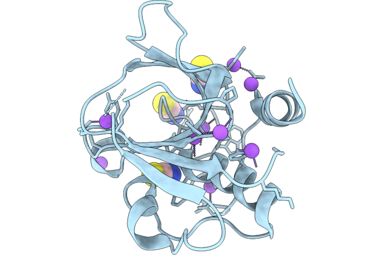
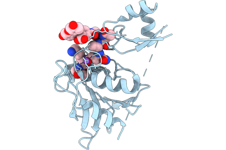
Chap Domain Of Staphylococcus Aureus-Specific Lysin L1 Covalently Complexed To Pep1A-Cmk Substrate Mimic
Organism: Staphylococcus aureus
Method: X-RAY DIFFRACTION Resolution:1.09 Å Release Date: 2026-05-06 Classification: HYDROLASE Ligands: A1DEZ, NA, SCN |
 |
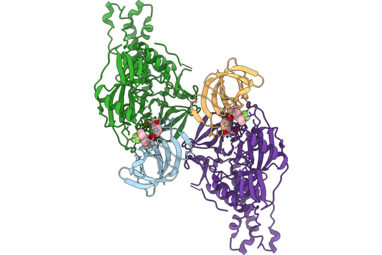
Staphylococcus Aureus-Specific Lysin L1-3 (Lysm-Chap) Covalently Complexed To Pep1A-Cmk Substrate Mimic
Organism: Staphylococcus aureus
Method: X-RAY DIFFRACTION Resolution:2.83 Å Release Date: 2026-05-06 Classification: HYDROLASE Ligands: A1DEZ, NA |
 |
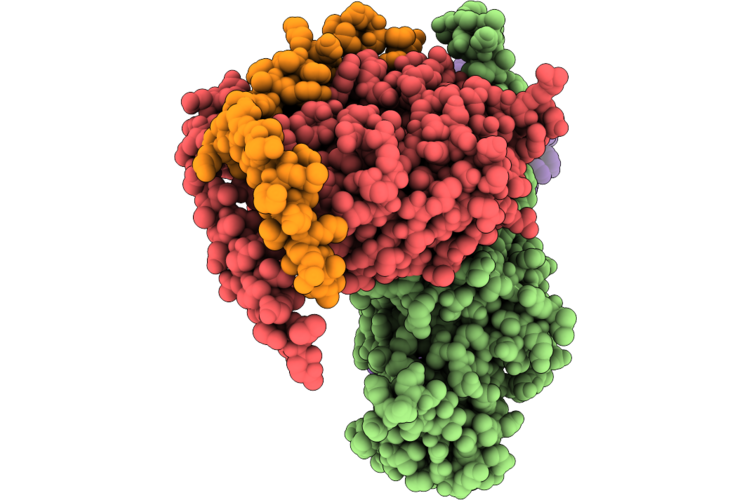
Cryo-Em Structure Of Crbn In Complex With Hbs1L And Tng-4857 (Focused Refinement)
Organism: Homo sapiens
Method: ELECTRON MICROSCOPY Release Date: 2026-05-06 Classification: CYTOSOLIC PROTEIN Ligands: ZN, A1C9W |
 |
Organism: Homo sapiens
Method: ELECTRON MICROSCOPY Resolution:2.90 Å Release Date: 2026-05-06 Classification: HYDROLASE Ligands: CPL, NAG |
 |
Organism: Homo sapiens, Mus musculus, Bos taurus, Escherichia coli, Synthetic construct
Method: ELECTRON MICROSCOPY Release Date: 2026-05-06 Classification: SIGNALING PROTEIN |
 |
Organism: Rattus norvegicus, Bos taurus, Homo sapiens, Synthetic construct
Method: ELECTRON MICROSCOPY Release Date: 2026-05-06 Classification: SIGNALING PROTEIN/IMMUNE SYSTEM Ligands: CLR |
 |
Organism: Mus musculus
Method: ELECTRON MICROSCOPY Release Date: 2026-05-06 Classification: CELL CYCLE Ligands: ATP, GTP, MG, ZN |
 |
Crystal Structure Of Myst Histone Acetyltransferase Kat6A In Complex With Inhibitor Compound 9
Organism: Homo sapiens
Method: X-RAY DIFFRACTION Resolution:2.29 Å Release Date: 2026-05-06 Classification: TRANSCRIPTION Ligands: A1MGC |
 |
Organism: Homo sapiens
Method: X-RAY DIFFRACTION Resolution:1.73 Å Release Date: 2026-05-06 Classification: HYDROLASE Ligands: HEM, GOL, A1ITR, DMS, CMO, ACT, SO4, PGE |
 |
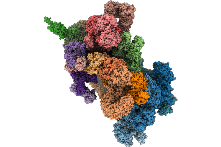
Cryo-Em Structure Of The Cul1-Rbx1-Skp1-Fbxo22 Scf Ubiquition Ligase In Complex With Nsd2 Via Unc10088
Organism: Homo sapiens
Method: ELECTRON MICROSCOPY Release Date: 2026-05-06 Classification: LIGASE Ligands: A1J20 |
 |
Cryo-Em Structure Of The Cul1-Rbx1-Skp1-Fbxo22 Scf Ubiquition Ligase In Complex With Nsd2, Unc10088 And Bach1
Organism: Homo sapiens
Method: ELECTRON MICROSCOPY Release Date: 2026-05-06 Classification: LIGASE Ligands: A1J20 |
 |
Cryo-Em Structure Of The Cul1-Rbx1-Skp1-Fbxo22 Scf Ubiquition Ligase In Complex With Nsd2 Via Unc10415667
Organism: Homo sapiens
Method: ELECTRON MICROSCOPY Release Date: 2026-05-06 Classification: LIGASE Ligands: A1J21 |
 |
Crystal Structure Of Udp-N-Acetylmuramate-L-Alanine Ligase (Murc) From Pseudomonas Aeruginosa In Complex With Compound Osa_001175 (Wyh77)
Organism: Pseudomonas aeruginosa
Method: X-RAY DIFFRACTION Resolution:2.40 Å Release Date: 2026-05-06 Classification: LIGASE Ligands: PO4, MG, A1J52, EDO, DMS, PEG |
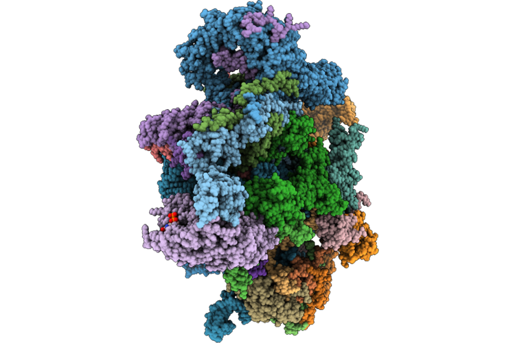 |
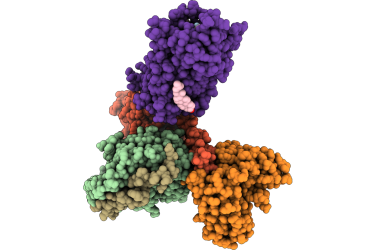
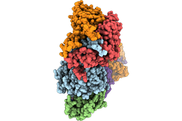
Assembly Intermediate Of Human Mitochondrial Ribosome Small Subunit In Complex With Noa1 And Partial Rbfa (State N2)
Organism: Homo sapiens
Method: ELECTRON MICROSCOPY Release Date: 2026-05-06 Classification: RIBOSOME Ligands: MG, K, ZN, FES, ATP, GDP |
 |
Organism: Leishmania braziliensis, Bos taurus
Method: X-RAY DIFFRACTION Resolution:1.90 Å Release Date: 2026-05-06 Classification: HYDROLASE Ligands: PEG, ACT, GOL, EDO, CL |