10OZ image
Deposition Date 2026-01-30
Release Date 2026-05-06
Last Version Date 2026-05-06
Entry Detail
PDB ID:
10OZ
Keywords:
Title:
Structure of PDE5 PDE6 chimera
Biological Source:
Source Organism(s):
Bos taurus (Taxon ID: 9913)
Method Details:
Experimental Method:
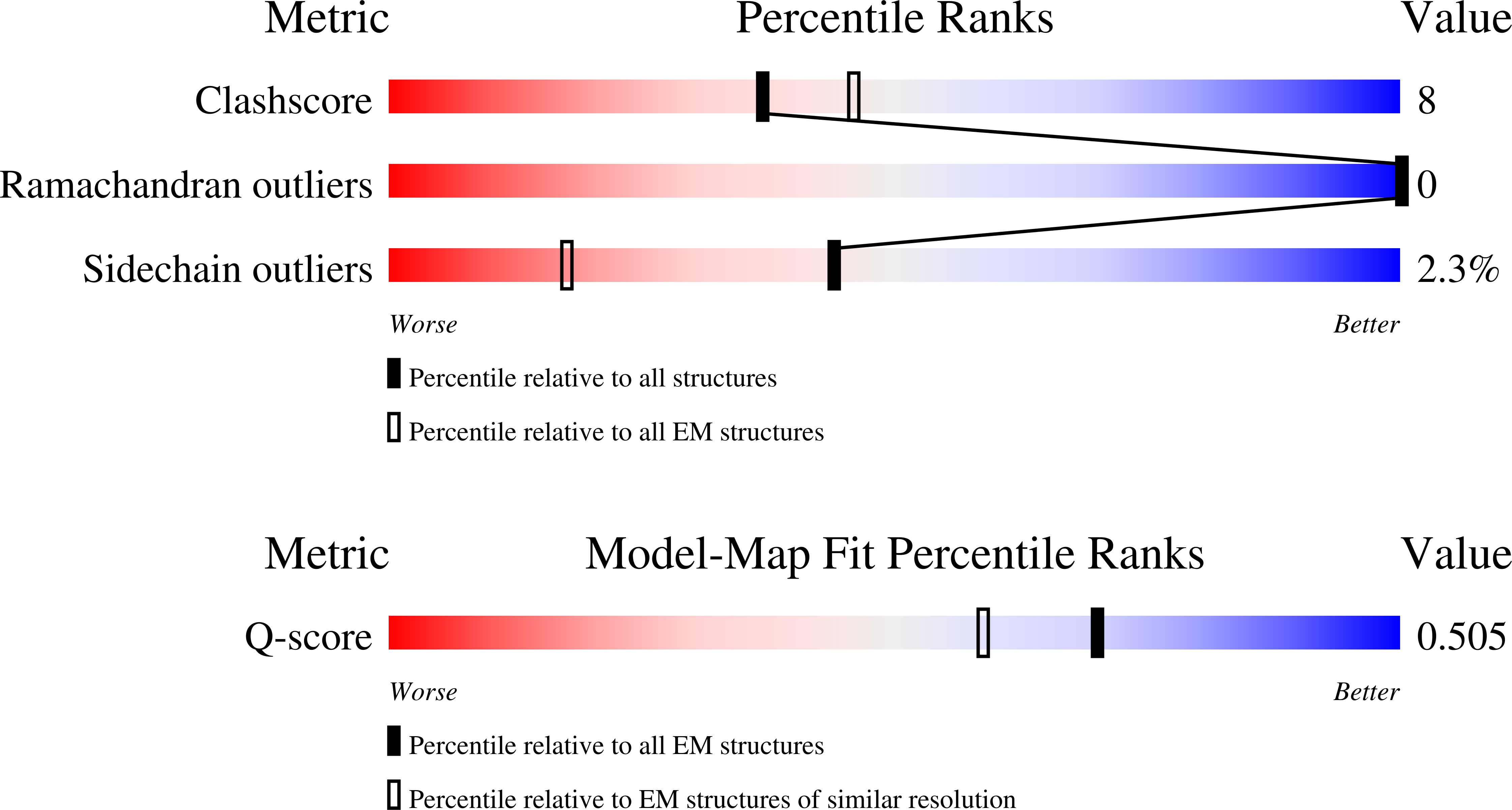
Resolution:
3.06 Å
Aggregation State:
PARTICLE
Reconstruction Method:
SINGLE PARTICLE
Macromolecular Entities
Protein Blast
Polymer Type:polypeptide(L)
Molecule:cGMP-specific 3',5'-cyclic ph
Gene (Uniprot):PDE6C, PDE5A
Chain IDs:A, B
Chain Length:800
Number of Molecules:2
Biological Source:Bos taurus
Structures with similar UniProt ID
Protein Blast
Polymer Type:polypeptide(L)
Molecule:Retinal rod rhodopsin-sensiti
Gene (Uniprot):PDE6G
Chain IDs:C, D
Chain Length:107
Number of Molecules:2
Biological Source:Bos taurus
Primary Citation
Structural basis of phosphodiesterase-5 conformational organization revealed by a PDE6/PDE5 Chimera.
J.Biol.Chem. ? 111467 111467 (2026)
PMID: 42001945 DOI: 10.1016/j.jbc.2026.111467

Abstact

Phosphodiesterase 5 (PDE5) plays critical role in the nitric oxide-cGMP signaling pathway. Consequently, PDE5 catalytic site inhibitors are widely used in the treatment of erectile dysfunction and pulmonary arterial hypertension. Despite a wealth of structural data on the individual PDE5 catalytic domain with bound drug molecules, understanding of the structural organization of the full-length enzyme and its allosteric activation by noncatalytic cGMP-binding is lacking. To begin to understand the structural organization of PDE5, we solved a cryo-EM structure of a chimeric PDE enzyme (PDE6C/5) comprised of the regulatory domains of cone PDE6C and the PDE5 catalytic domain. The PDE6C/5 structure revealed the protein in the open state conformation similar to that of PDE6, suggesting a comparable conformation for the cGMP-bound PDE5 molecule. The H- and M-loops outlying the catalytic pocket, which are conformationally variable in the structures of isolated PDE5 catalytic domain, are immobilized in the PDE6/5 chimera via the interaction of the H-loop with a linker helix LH2. Decreased dynamics of these loops may underlie the higher catalytic activities of the full-length PDE5 and PDE6C/5 compared to that of the isolated PDE5 catalytic domain. Furthermore, the PDE6C/5 structure defines the folding requirement of the PDE6 catalytic domain for chaperone-dependent maturation that is important for vision.

Legend

Protein

Chemical

Disease

Primary Citation of related structures
Feedback Form
Name
Email
Institute
Feedback