
Search Count: 423
 |
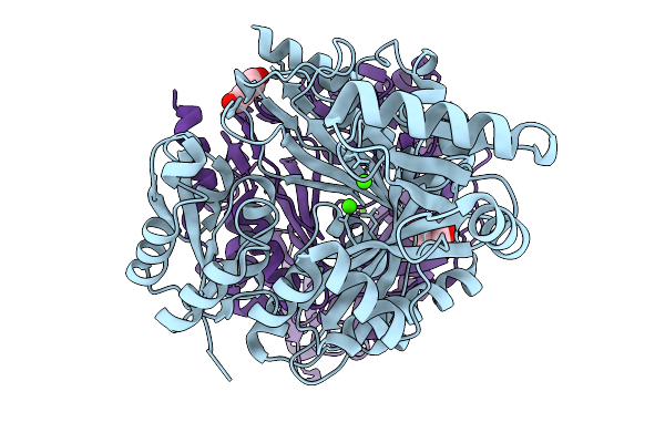
Organism: Lachesis muta
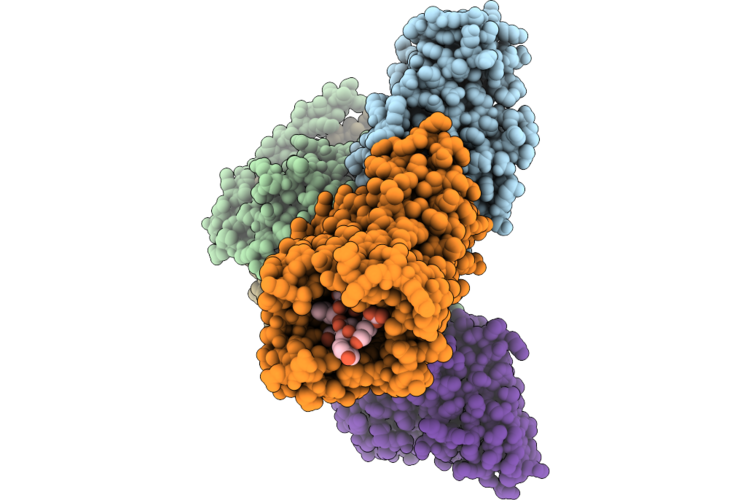
Method: X-RAY DIFFRACTION Resolution:2.36 Å Release Date: 2026-05-13 Classification: TOXIN,HYDROLASE Ligands: MES, CA |
 |
Organism: Homo sapiens, Synthetic construct, Escherichia coli
Method: ELECTRON MICROSCOPY Release Date: 2026-04-29 Classification: SIGNALING PROTEIN |
 |
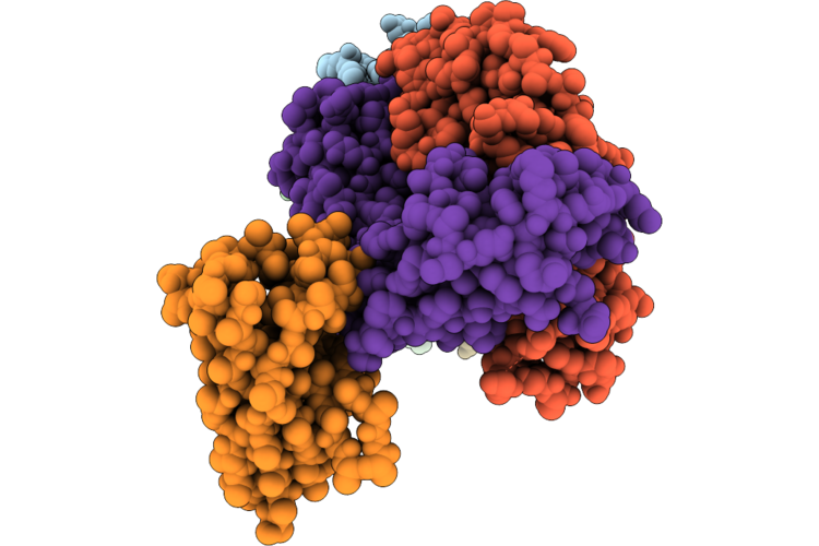
Cryoem Structure Of The Themis:Grb2 Complex With Bound Promacrobody 256, Local Refinement
Organism: Homo sapiens, Synthetic construct, Escherichia coli
Method: ELECTRON MICROSCOPY Resolution:3.20 Å Release Date: 2026-04-29 Classification: SIGNALING PROTEIN |
 |
Organism: Homo sapiens, Escherichia coli, Oplophorus gracilirostris
Method: ELECTRON MICROSCOPY Resolution:2.80 Å Release Date: 2026-04-29 Classification: SIGNALING PROTEIN/IMMUNE SYSTEM Ligands: CLR |
 |
Organism: Homo sapiens, Escherichia coli, Oplophorus gracilirostris
Method: ELECTRON MICROSCOPY Resolution:2.70 Å Release Date: 2026-04-29 Classification: SIGNALING PROTEIN Ligands: CLR |
 |
Organism: Homo sapiens, Lama glama
Method: ELECTRON MICROSCOPY Release Date: 2026-04-22 Classification: IMMUNE SYSTEM/Hydrolase |
 |
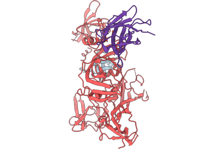
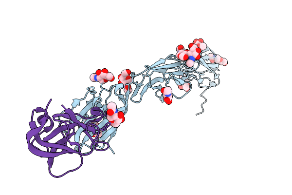
Crystal Structure Of A Zikv E Glycoprotein Di-Diii Vaccine Candidate In Complex With Human Neutralizing Antibody Mz4
Organism: Homo sapiens, Zika virus zikv/h. sapiens/frenchpolynesia/10087pf/2013
Method: X-RAY DIFFRACTION Resolution:2.85 Å Release Date: 2026-04-15 Classification: VIRAL PROTEIN Ligands: NAG |
 |
Cardioderma Bat Coronavirus Ky43 Receptor Binding Domain In Complex With Human Ceacam6
Organism: Homo sapiens, Cardioderma bat coronavirus
Method: X-RAY DIFFRACTION Resolution:3.01 Å Release Date: 2026-04-08 Classification: VIRAL PROTEIN Ligands: NAG |
 |
Cardioderma Bat Coronavirus 2B Receptor Binding Domain In Complex With Human Ceacam6
Organism: Homo sapiens, Cardioderma bat coronavirus
Method: X-RAY DIFFRACTION Resolution:2.99 Å Release Date: 2026-04-08 Classification: VIRAL PROTEIN Ligands: NAG |
 |
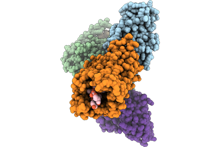
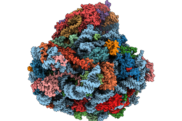
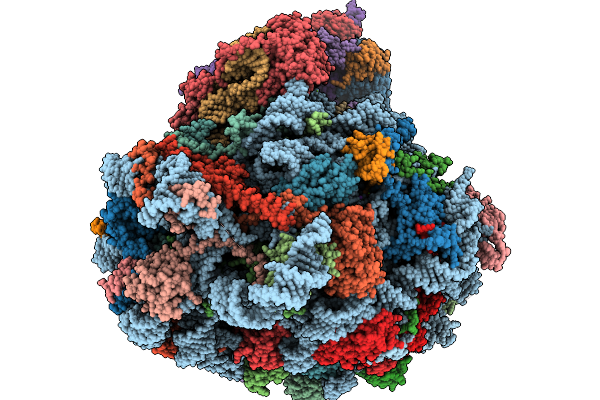
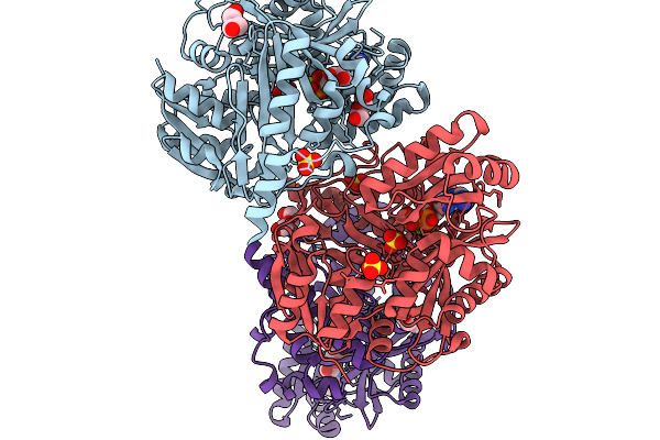
Structure Of The Arabidopsis Thaliana 80S Ribosome In Complex With P- And E-Site Trnas And Mrna
Organism: Arabidopsis thaliana
Method: ELECTRON MICROSCOPY Resolution:1.82 Å Release Date: 2026-02-04 Classification: RIBOSOME Ligands: MG, K, TER, SPD, EPE, ZN |
 |
Structure Of The Arabidopsis Thaliana 80S Ribosome In Complex With P- And E-Site Trnas, Mrna, And Thermospermine
Organism: Arabidopsis thaliana
Method: ELECTRON MICROSCOPY Resolution:2.20 Å Release Date: 2026-02-04 Classification: RIBOSOME Ligands: TER, MG, K, SPD, EPE, ZN |
 |
Structure Of The Arabidopsis Thaliana 80S Ribosome Ovac Mutant In Complex With P- And E-Site Trnas, Mrna, And Thermospermine
Organism: Arabidopsis thaliana
Method: ELECTRON MICROSCOPY Resolution:2.25 Å Release Date: 2026-02-04 Classification: RIBOSOME Ligands: TER, MG, K, SPD, EPE, ZN |
 |
Structure Of The Arabidopsis Thaliana 80S Ribosome Ovac Mutant In Complex With P- And E-Site Trnas And Mrna
Organism: Arabidopsis thaliana
Method: ELECTRON MICROSCOPY Resolution:2.25 Å Release Date: 2026-02-04 Classification: RIBOSOME Ligands: MG, K, TER, SPD, EPE, ZN |
 |
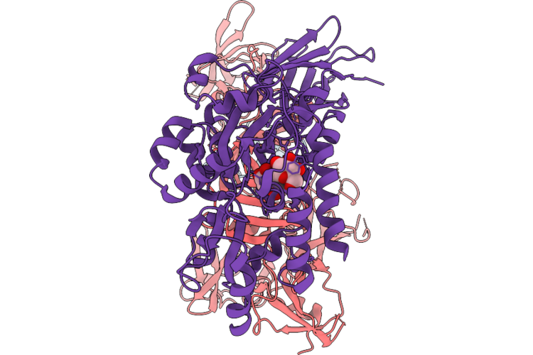
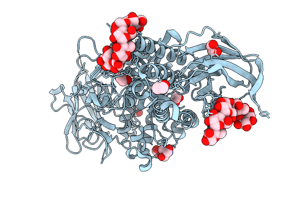
Organism: Faecalibacterium duncaniae
Method: X-RAY DIFFRACTION Resolution:1.70 Å Release Date: 2025-12-31 Classification: TRANSFERASE Ligands: TRS, GOL, EDO, PEG |
 |
Organism: Faecalibacterium duncaniae
Method: X-RAY DIFFRACTION Resolution:1.60 Å Release Date: 2025-12-31 Classification: TRANSFERASE Ligands: TRS, EDO |
 |
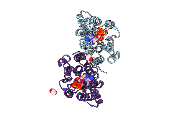
X-Ray Structure Analysis Of Human Complement Component 5 Te Domain In Complex With The Peptide Ra30303
Organism: Homo sapiens, Synthetic construct
Method: X-RAY DIFFRACTION Resolution:2.00 Å Release Date: 2025-12-24 Classification: IMMUNE SYSTEM Ligands: GOL, 8VH |
 |
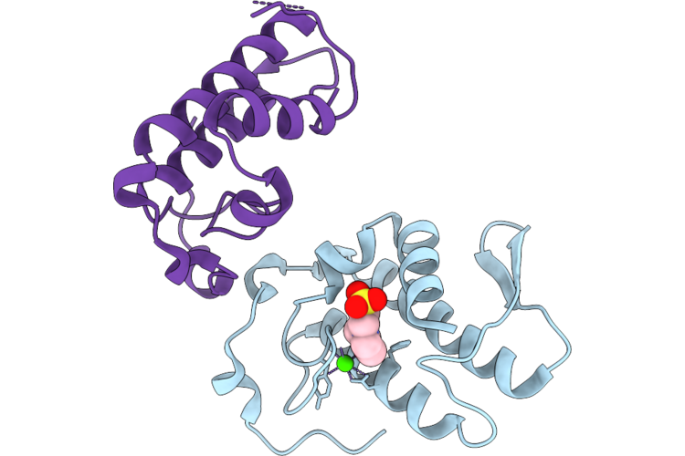
Organism: Homo sapiens
Method: X-RAY DIFFRACTION Resolution:1.90 Å Release Date: 2025-12-10 Classification: HYDROLASE Ligands: ZN, PPS, EDO, ACY |
 |
Organism: Ankistrodesmus falcatus
Method: X-RAY DIFFRACTION Resolution:1.75 Å Release Date: 2025-11-19 Classification: LIGASE Ligands: PEG, CA, MG |
 |
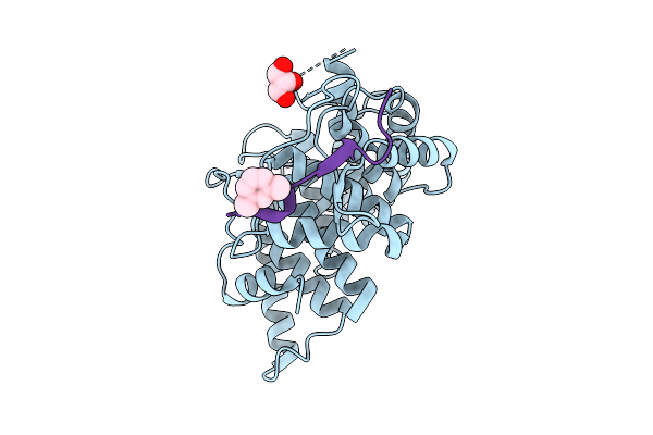
Crystal Structure Of Biotin Carboxylase From Ankistrodesmus In Complex With Adp
Organism: Ankistrodesmus falcatus
Method: X-RAY DIFFRACTION Resolution:1.90 Å Release Date: 2025-11-19 Classification: LIGASE Ligands: MG, ADP, PEG, SO4 |
 |
Organism: Vicugna pacos
Method: SOLUTION NMR Release Date: 2025-11-12 Classification: PROTEIN BINDING |