Deposition Date
2024-12-19
Release Date
2026-05-13
Last Version Date
2026-05-13
Entry Detail
PDB ID:
9MLE
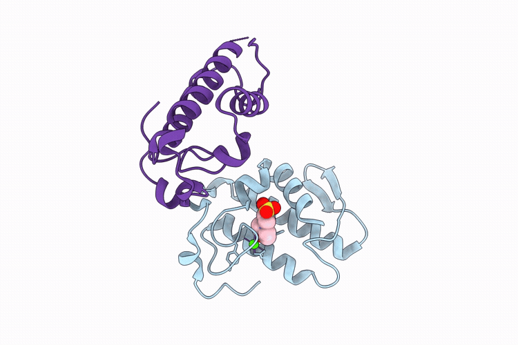
Title:
Crystal structure of Asp49 Phospholipase A2 isolated from Lachesis muta
Biological Source:
Source Organism(s):
Lachesis muta (Taxon ID: 8752)
Method Details:
Experimental Method:
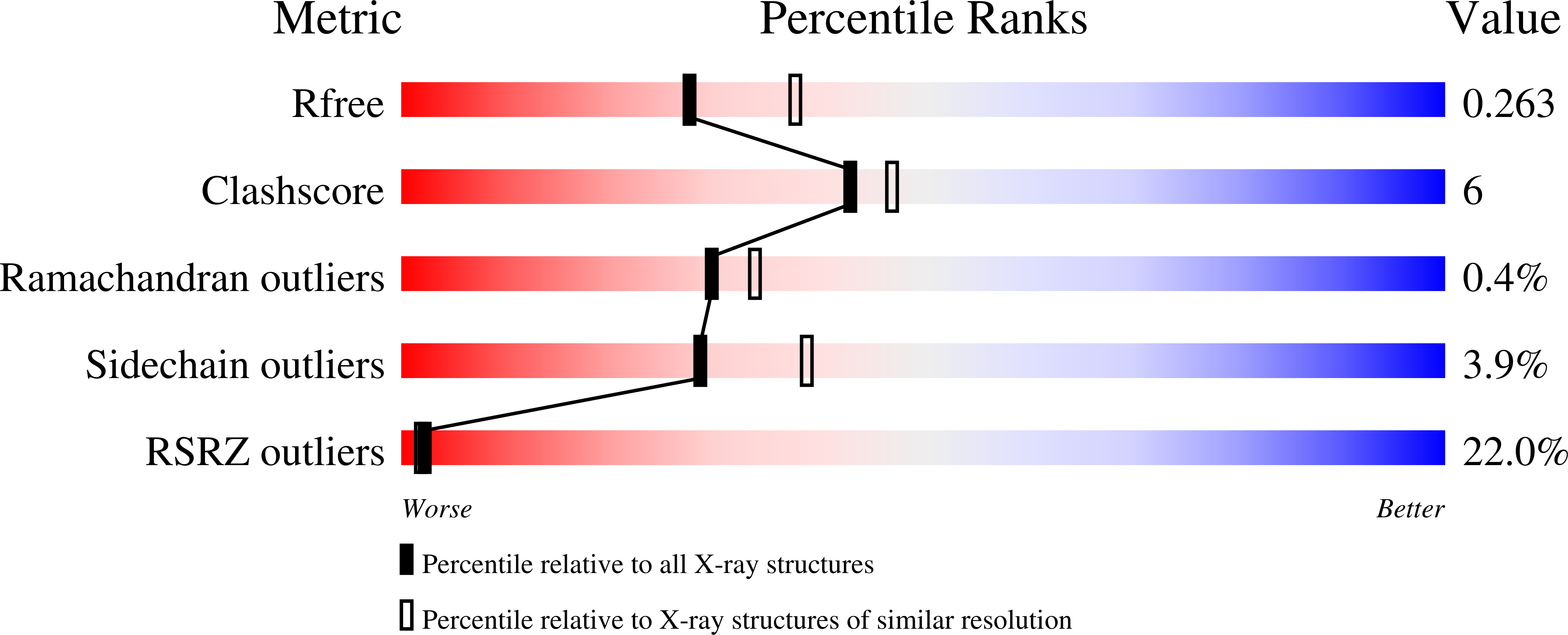
Resolution:
2.36 Å
R-Value Free:
0.26
R-Value Work:
0.24
R-Value Observed:
0.24
Space Group:
P 62 2 2