

Search Count: 434
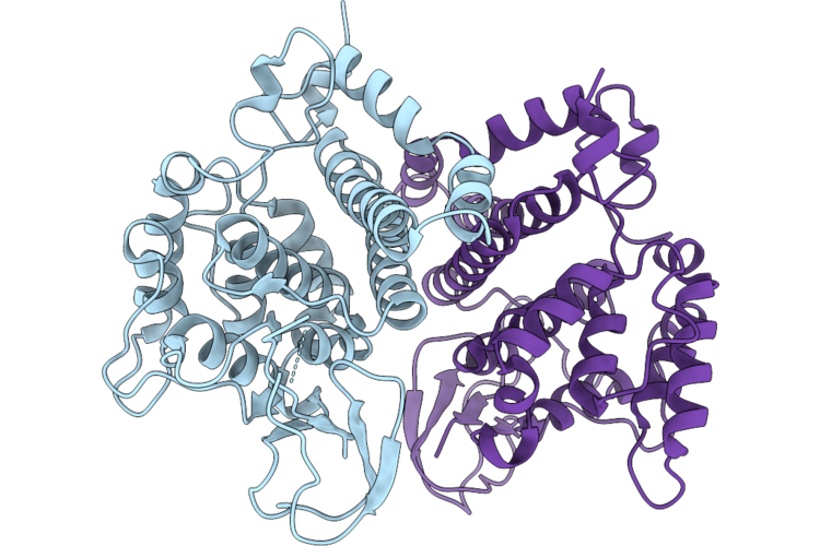 |
Crystal Structure Of Antitoxin Vp14460 (Poni)From Vibrio Parahaemolyticus
Organism: Vibrio parahaemolyticus
Method: X-RAY DIFFRACTION Resolution:2.32 Å Release Date: 2026-07-22 Classification: ANTITOXIN |
Organism: Vibrio parahaemolyticus
Method: X-RAY DIFFRACTION
Release Date: 2026-07-22
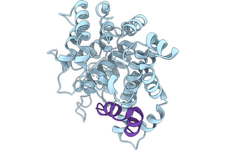 |
Crystal Structure Of Toxin-Antitoxin Complex Vp14460-Vp14465Peptide From Vibrio Parahaemolyticus
Organism: Vibrio parahaemolyticus
Method: X-RAY DIFFRACTION Resolution:2.50 Å Release Date: 2026-07-22 Classification: TOXIN |
Organism: Vibrio parahaemolyticus
Method: X-RAY DIFFRACTION
Release Date: 2026-07-22
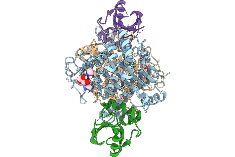 |
Crystal Structure Of The Effector-Immunity Vp14460-Vp14465 In Vibrio Parahaemolyticus
Organism: Vibrio parahaemolyticus
Method: X-RAY DIFFRACTION Resolution:1.97 Å Release Date: 2026-07-22 Classification: TOXIN Ligands: GOL, IMD |
Organism: Vibrio parahaemolyticus
Method: X-RAY DIFFRACTION
Release Date: 2026-07-22
Ligands: GOL, IMD
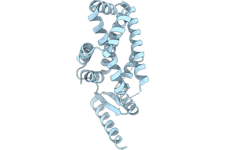 |
Crystal Structure Of A Designed Protein Three-Twist Knot
Organism: Helicobacter pylori
Method: X-RAY DIFFRACTION Resolution:2.28 Å Release Date: 2026-07-01 Classification: DE NOVO PROTEIN |
Organism: Helicobacter pylori
Method: X-RAY DIFFRACTION
Release Date: 2026-07-01
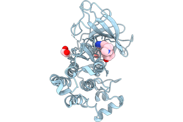 |
Crystal Structure Of Egfr Kinase (C797S) In Complex With Ln-B72
Organism: Homo sapiens
Method: X-RAY DIFFRACTION Resolution:2.95 Å Release Date: 2026-05-27 Classification: SIGNALING PROTEIN Ligands: A1ETY, EDO |
Organism: Homo sapiens
Method: X-RAY DIFFRACTION
Release Date: 2026-05-27
Ligands: A1ETY, EDO
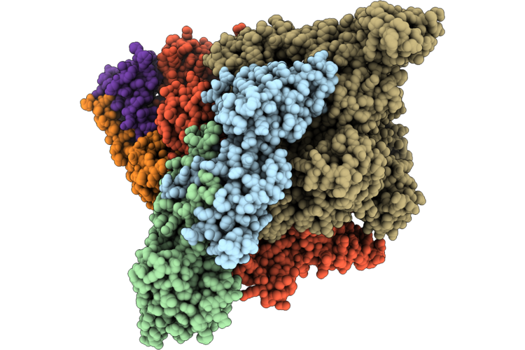 |
Cryo-Em Structure Of E. Coli Rna Polymerase In Complex With Ep1
Organism: Escherichia coli, Escherichia phage phisusp1
Method: ELECTRON MICROSCOPY Resolution:4.00 Å Release Date: 2026-04-29 Classification: TRANSCRIPTION |
Organism: Escherichia coli, Escherichia phage phisusp1
Method: ELECTRON MICROSCOPY
Release Date: 2026-04-29
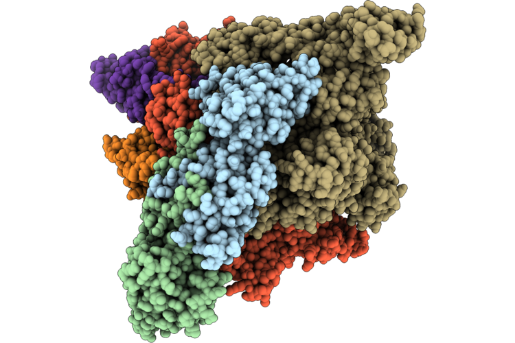 |
Cryo-Em Structure Of E. Coli Rna Polymerase In Complex With Pp1
Organism: Escherichia coli, Pseudomonas phage vb_paem_pao1_ab17
Method: ELECTRON MICROSCOPY Resolution:3.60 Å Release Date: 2026-04-29 Classification: TRANSCRIPTION |
Organism: Escherichia coli, Pseudomonas phage vb_paem_pao1_ab17
Method: ELECTRON MICROSCOPY
Release Date: 2026-04-29
 |
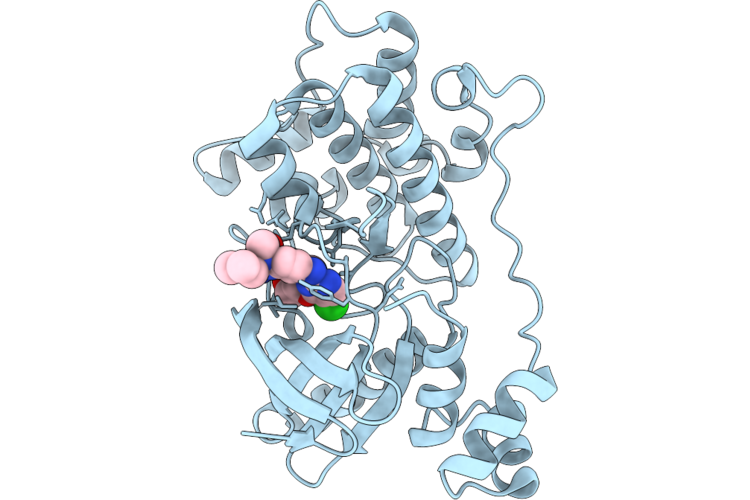
Cryo-Em Structure Of E. Coli Rna Polymerase In Complex With Vp1
Organism: Escherichia coli, Escherichia coli (strain k12), Vibrio phage vb_vpap_m3
Method: ELECTRON MICROSCOPY Resolution:3.30 Å Release Date: 2026-04-29 Classification: TRANSCRIPTION |
Organism: Escherichia coli, Escherichia coli (strain k12), Vibrio phage vb_vpap_m3
Method: ELECTRON MICROSCOPY
Release Date: 2026-04-29
 |
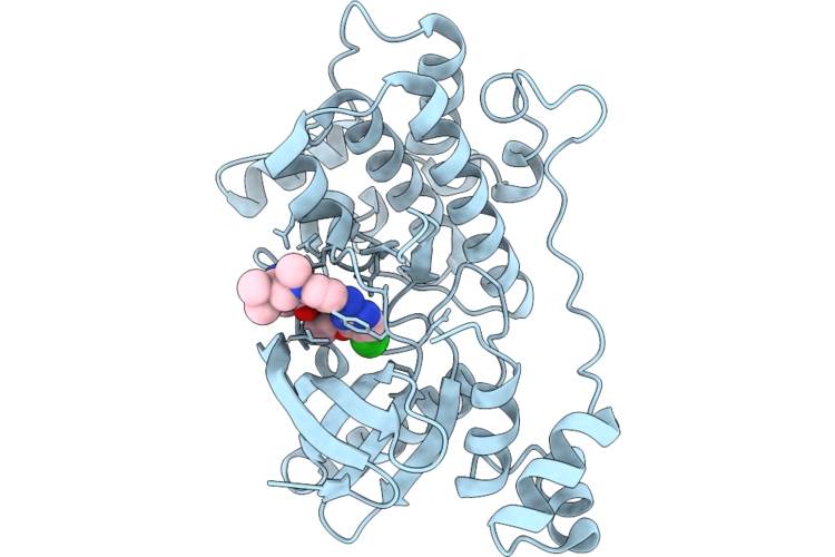
Crystal Structure Of The Nuak1-Mark3 Kinase Domain Chimera Bound With Small Molecule Inhibitor 2-Amino-N-(5-((5-Chloro-4-(((3R,3Ar,6R,6Ar)-6-Methoxyhexahydrofuro[3,2-B]Furan-3-Yl)Oxy)Pyrimidin-2-Yl)Amino)-2-((2-(Dimethylamino)Ethyl)(Methyl)Amino)Phenyl)Acetamide
Organism: Homo sapiens
Method: X-RAY DIFFRACTION Resolution:2.50 Å Release Date: 2026-04-22 Classification: TRANSFERASE Ligands: A1ER0 |
Organism: Homo sapiens
Method: X-RAY DIFFRACTION
Release Date: 2026-04-22
Ligands: A1ER0
 |
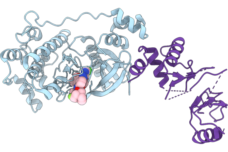
Crystal Structure Of The Nuak1-Mark3 Kinase Domain Chimera Bound With Small Molecule Inhibitor 4-((5-((5-Chloro-4-(((3R,3Ar,6R,6Ar)-6-Methoxyhexahydrofuro[3,2-B]Furan-3-Yl)Oxy)Pyrimidin-2-Yl)Amino)-2-((2-(Dimethylamino)Ethyl)(Methyl)Amino)Phenyl)Carbamoyl)-1-Methyl-3H-Pyrazol-1-Ium-3-Ide
Organism: Homo sapiens
Method: X-RAY DIFFRACTION Resolution:2.00 Å Release Date: 2026-04-22 Classification: CYTOSOLIC PROTEIN Ligands: A1ER9 |
Organism: Homo sapiens
Method: X-RAY DIFFRACTION
Release Date: 2026-04-22
Ligands: A1ER9
 |
Crystal Structure Of The Nuak1-Mark3 Kinase Domain Chimera Bound With Small Molecule Inhibitor N-(5-((5-Chloro-4-(((3As,6R,6Ar)-6-Methoxy-3A,5,6,6A-Tetrahydrofuro[3,2-B]Furan-3-Yl)Oxy)Pyrimidin-2-Yl)Amino)-2-(((2R,7Ar)-2-Fluorotetrahydro-1H-Pyrrolizin-7A(5H)-Yl)Methoxy)Phenyl)-1-Methyl-1H-Pyrazole-4-Carboxamide
Organism: Homo sapiens
Method: X-RAY DIFFRACTION Resolution:2.80 Å Release Date: 2026-04-22 Classification: CYTOSOLIC PROTEIN Ligands: A1ESF |
Organism: Homo sapiens
Method: X-RAY DIFFRACTION
Release Date: 2026-04-22
Ligands: A1ESF
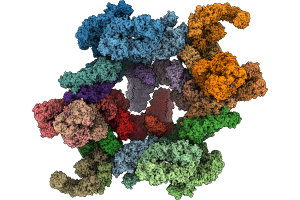 |
Cryo-Em Structure Of The Cytoplasmic Lattice(Cpl) From Mouse Oocyte
Organism: Mus musculus
Method: ELECTRON MICROSCOPY Release Date: 2026-02-25 Classification: STRUCTURAL PROTEIN Ligands: GDP, ADP |
Organism: Mus musculus
Method: ELECTRON MICROSCOPY
Release Date: 2026-02-25
Ligands: GDP, ADP
 |
Structure Of Wdr5 In Complex With Peptide 2
Organism: Homo sapiens, Synthetic construct
Method: X-RAY DIFFRACTION Resolution:1.70 Å Release Date: 2026-02-11 Classification: NUCLEAR PROTEIN |
Organism: Homo sapiens, Synthetic construct
Method: X-RAY DIFFRACTION
Release Date: 2026-02-11
 |
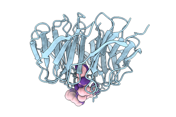
Structure Of Wdr5 In Complex With Peptide 2_R4A
Organism: Homo sapiens, Synthetic construct
Method: X-RAY DIFFRACTION Resolution:1.76 Å Release Date: 2026-02-11 Classification: NUCLEAR PROTEIN |
Organism: Homo sapiens, Synthetic construct
Method: X-RAY DIFFRACTION
Release Date: 2026-02-11
 |
Structure Of Wdr5 In Complex With Peptide 1
Organism: Homo sapiens, Synthetic construct
Method: X-RAY DIFFRACTION Resolution:1.48 Å Release Date: 2026-02-11 Classification: NUCLEAR PROTEIN |
Organism: Homo sapiens, Synthetic construct
Method: X-RAY DIFFRACTION
Release Date: 2026-02-11
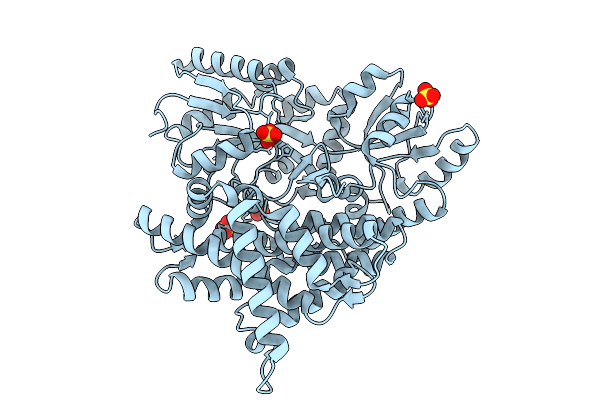 |
Structure Of Mbp Tagged H2A.W-H2B
Organism: Arabidopsis thaliana
Method: X-RAY DIFFRACTION Resolution:1.85 Å Release Date: 2026-01-28 Classification: NUCLEAR PROTEIN Ligands: SO4 |
Organism: Arabidopsis thaliana
Method: X-RAY DIFFRACTION
Release Date: 2026-01-28
Ligands: SO4
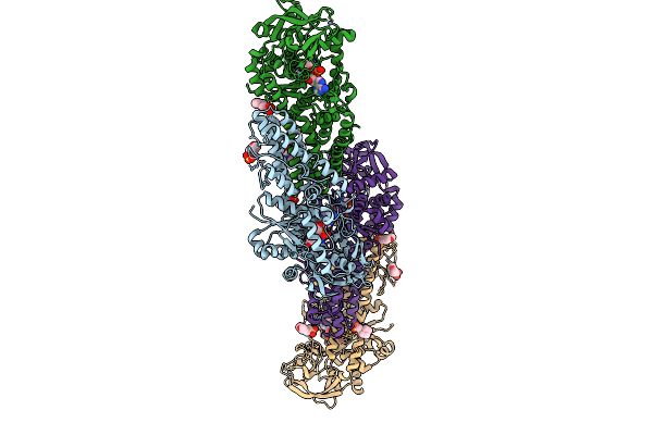 |
Pyrn C-Terminl Domain In Complex With L-Glutamyl-Sulfamoyl-Adenosine (Gsa)
Organism: Streptomyces candidus
Method: X-RAY DIFFRACTION Resolution:2.23 Å Release Date: 2025-12-31 Classification: LIGASE Ligands: MES, ZN, GSU |
Organism: Streptomyces candidus
Method: X-RAY DIFFRACTION
Release Date: 2025-12-31
Ligands: MES, ZN, GSU
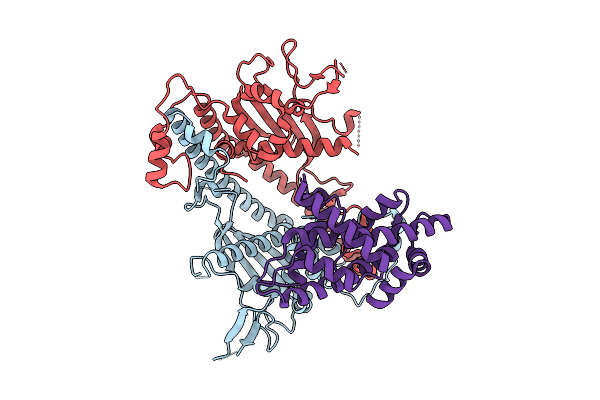 |
Structure Of Nap1 In Complex With H2A.Z-H2B
Organism: Caenorhabditis elegans
Method: X-RAY DIFFRACTION Resolution:2.80 Å Release Date: 2025-12-24 Classification: CHAPERONE |
Organism: Caenorhabditis elegans
Method: X-RAY DIFFRACTION
Release Date: 2025-12-24
 |
Crystal Structure Of Namat In Complex With Nad
Organism: Bacillus phage spbetal1
Method: X-RAY DIFFRACTION Resolution:2.30 Å Release Date: 2025-12-24 Classification: VIRAL PROTEIN Ligands: NAD, SO4, GOL |
Organism: Bacillus phage spbetal1
Method: X-RAY DIFFRACTION
Release Date: 2025-12-24
Ligands: NAD, SO4, GOL
 |
Crystal Structure Of Namat In Complex With Nicotinamide
Organism: Bacillus phage spbetal1
Method: X-RAY DIFFRACTION Resolution:2.40 Å Release Date: 2025-12-24 Classification: VIRAL PROTEIN Ligands: GOL, NCA, SO4 |
Organism: Bacillus phage spbetal1
Method: X-RAY DIFFRACTION
Release Date: 2025-12-24
Ligands: GOL, NCA, SO4