9W2M image
Deposition Date 2025-07-28
Release Date 2026-02-25
Last Version Date 2026-05-06
Entry Detail
PDB ID:
9W2M
Title:
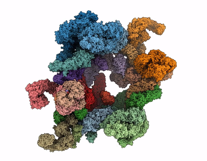
Cryo-EM structure of the Cytoplasmic lattice(CPL) from mouse oocyte
Biological Source:
Source Organism(s):
Mus musculus (Taxon ID: 10090)
Method Details:
Experimental Method:
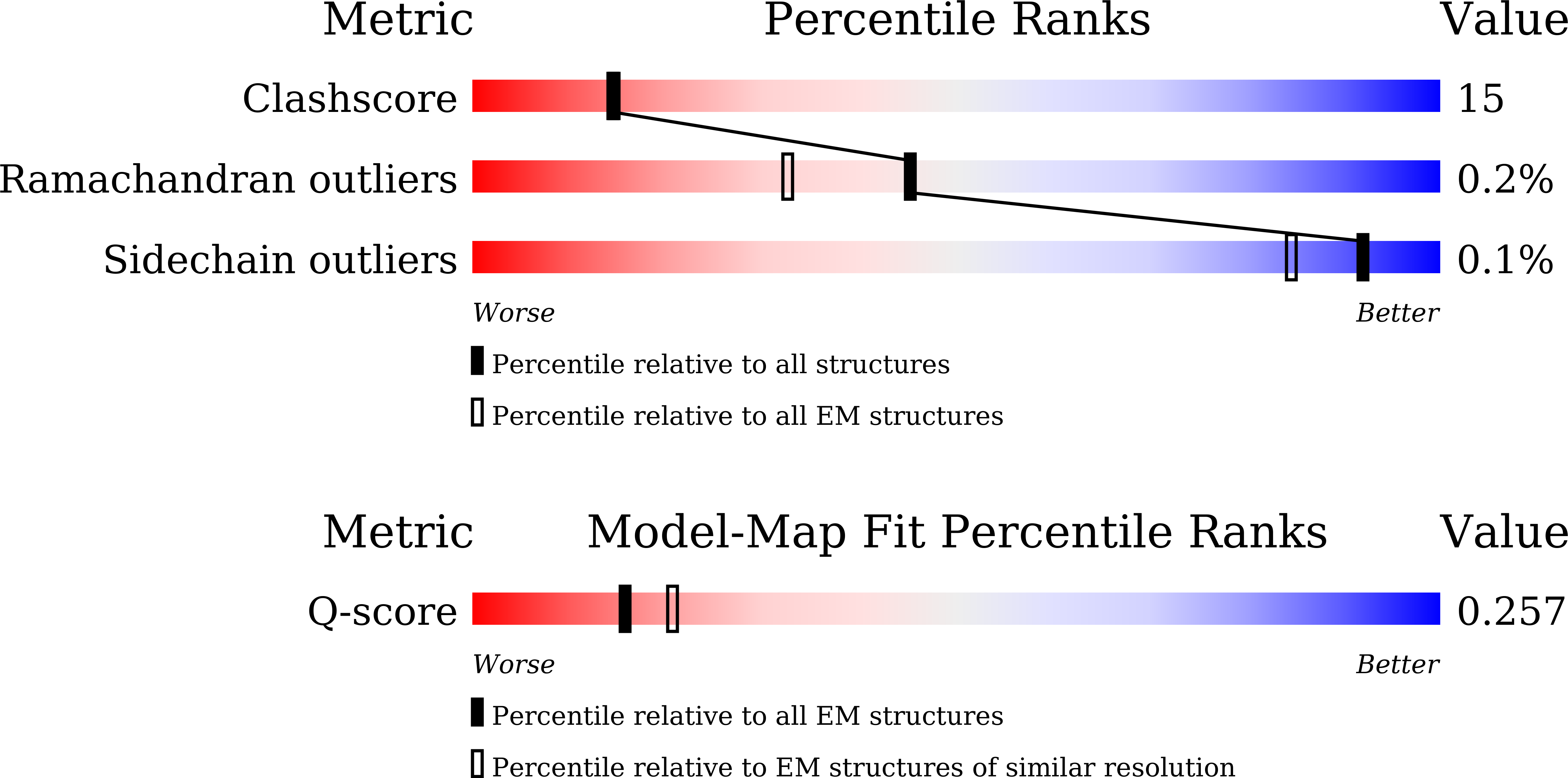
Resolution:
4.20 Å
Aggregation State:
FILAMENT
Reconstruction Method:
SINGLE PARTICLE
Macromolecular Entities
Structures with similar UniProt ID
Protein Blast
Polymer Type:polypeptide(L)
Molecule:Inactive protein-arginine dei
Gene (Uniprot):Padi6
Chain IDs:A (auth: 1), L (auth: Z), W (auth: z), X (auth: 2), Y (auth: 3), Z (auth: 4), AA (auth: 5), BA (auth: 6), CA (auth: 7), DA (auth: 8), EA (auth: 9), FA (auth: 12), WA (auth: 11), XA (auth: 19), YA (auth: 15), ZA (auth: 16), AB (auth: 14), BB (auth: 13), CB (auth: 17), DB (auth: 18)
Chain Length:682
Number of Molecules:20
Biological Source:Mus musculus
Structures with similar UniProt ID
Protein Blast
Polymer Type:polypeptide(L)
Molecule:Isoform 4 of NACHT, LRR and P
Gene (Uniprot):Nlrp5
Chain IDs:KA (auth: A), MA (auth: F), SA (auth: a), UA (auth: f)
Chain Length:963
Number of Molecules:4
Biological Source:Mus musculus
Structures with similar UniProt ID
Protein Blast
Polymer Type:polypeptide(L)
Molecule:Transducin-like enhancer prot
Gene (Uniprot):Tle6
Chain IDs:LA (auth: B), NA (auth: G), TA (auth: b), VA (auth: g)
Chain Length:436
Number of Molecules:4
Biological Source:Mus musculus
Structures with similar UniProt ID
Protein Blast
Polymer Type:polypeptide(L)
Molecule:Oocyte-expressed protein homo
Gene (Uniprot):Ooep
Chain IDs:B (auth: C), E (auth: H), M (auth: c), P (auth: h)
Chain Length:125
Number of Molecules:4
Biological Source:Mus musculus
Structures with similar UniProt ID
Protein Blast
Polymer Type:polypeptide(L)
Molecule:KH domain-containing protein
Gene (Uniprot):Khdc3
Chain IDs:C (auth: D), N (auth: d)
Chain Length:128
Number of Molecules:2
Biological Source:Mus musculus
Structures with similar UniProt ID
Protein Blast
Polymer Type:polypeptide(L)
Molecule:NLR family, pyrin domain cont
Gene (Uniprot):Nlrp4f
Chain IDs:D (auth: E), F (auth: I), O (auth: e), Q (auth: i)
Chain Length:933
Number of Molecules:4
Biological Source:Mus musculus
Protein Blast
Polymer Type:polypeptide(L)
Molecule:FBXW24
Chain IDs:G (auth: J), R (auth: j)
Chain Length:466
Number of Molecules:2
Biological Source:Mus musculus
Structures with similar UniProt ID
Protein Blast
Polymer Type:polypeptide(L)
Molecule:S-phase kinase-associated pro
Gene (Uniprot):Skp1
Chain IDs:H (auth: K), S (auth: k)
Chain Length:163
Number of Molecules:2
Biological Source:Mus musculus
Structures with similar UniProt ID
Protein Blast
Polymer Type:polypeptide(L)
Molecule:Tubulin beta-2A chain
Gene (Uniprot):Tubb2a
Chain IDs:IA (auth: L), QA (auth: l)
Chain Length:445
Number of Molecules:2
Biological Source:Mus musculus
Structures with similar UniProt ID
Protein Blast
Polymer Type:polypeptide(L)
Molecule:Tubulin beta-2B chain
Chain IDs:JA (auth: M), RA (auth: m)
Chain Length:445
Number of Molecules:2
Biological Source:Mus musculus
Structures with similar UniProt ID
Protein Blast
Polymer Type:polypeptide(L)
Molecule:NACHT, LRR and PYD domains-co
Gene (Uniprot):Nlrp14
Chain IDs:GA (auth: N), OA (auth: n)
Chain Length:966
Number of Molecules:2
Biological Source:Mus musculus
Structures with similar UniProt ID
Protein Blast
Polymer Type:polypeptide(L)
Molecule:Ubiquitin-conjugating enzyme
Gene (Uniprot):Ube2d3
Chain IDs:I (auth: O), T (auth: o)
Chain Length:147
Number of Molecules:2
Biological Source:Mus musculus
Structures with similar UniProt ID
Protein Blast
Polymer Type:polypeptide(L)
Molecule:E3 ubiquitin-protein ligase U
Gene (Uniprot):Uhrf1
Chain IDs:J (auth: Q), U (auth: q), HA (auth: P), PA (auth: p)
Chain Length:782
Number of Molecules:4
Biological Source:Mus musculus
Structures with similar UniProt ID
Protein Blast
Polymer Type:polypeptide(L)
Molecule:Zinc finger BED domain-contai
Gene (Uniprot):Zbed3
Chain IDs:K (auth: R), V (auth: r)
Chain Length:55
Number of Molecules:2
Biological Source:Mus musculus
Primary Citation
Molecular basis of oocyte cytoplasmic lattice assembly.
Nature ? ? ? (2026)
PMID: 41845018 DOI: 10.1038/s41586-026-10360-7

Abstact

Mammalian oocytes are filled with fibric structures called cytoplasmic lattice (CPL) that are essential for oocyte maturation and early embryonic development(1-3). CPL comprises subcortical maternal complex (SCMC) and multiple components, including PADI6(2,4,5). Although it was first discovered in the 1960s, the molecular architecture and assembly mechanisms of CPL remain poorly understood. Here we present the cryo-electron microscopy structure of CPL isolated from mouse oocytes. Our analysis identified 14 constitutive protein subunits and revealed that CPL is composed of repeating units comprising U-shaped basket (UB) and adapter ring (AR) features, forming a filamentous architecture. The AR adopts a two-fold symmetric conformation, containing two NLRP4F, four SCMC and two ZBED3 subunits circularized via two distinct interaction clusters. The UB is anchored by PADI6, a didecamer composed of ten homodimers assembled by two back-to-back pentamers, each forming the lateral side of the UB. The underfoot base and up and down sides of the UB are formed by multiple central-symmetric assemblies (UBE2D3-UHRF1-NLRP14) and (TUBB2B-TUBB2A-FBXW24-SKP1), respectively, associating with the PADI6 pentamers to construct the intact UB structure. Two SCMC dimers within each AR connect the up and down sides of two adjacent UBs with an extensive protein-protein interaction network, and thus maintain the repetitive connection between the neighbouring CPL units. Our work unveils the architectural principles underlying the assembly of this large, periodic CPL filament, offering a molecular basis for understanding the functions of CPL in early mammalian embryogenesis and female reproductive disorders.

Legend

Protein

Chemical

Disease

Primary Citation of related structures
Feedback Form
Name
Email
Institute
Feedback