

Search Count: 615
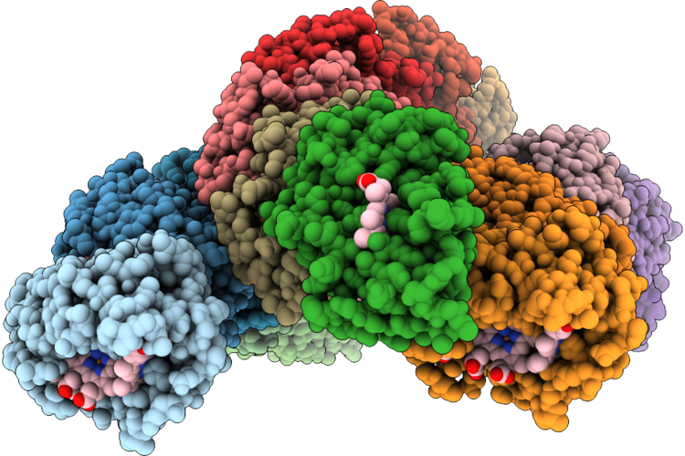 |
A Bundled Antiparallel Cytochrome Nanowire Produced By Desulfuromonas Soudanensis Wtl
Organism: Desulfuromonas soudanensis
Method: ELECTRON MICROSCOPY Resolution:3.11 Å Release Date: 2026-08-26 Classification: ELECTRON TRANSPORT Ligands: HEC, CA |
Organism: Desulfuromonas soudanensis
Method: ELECTRON MICROSCOPY
Release Date: 2026-08-26
Ligands: HEC, CA
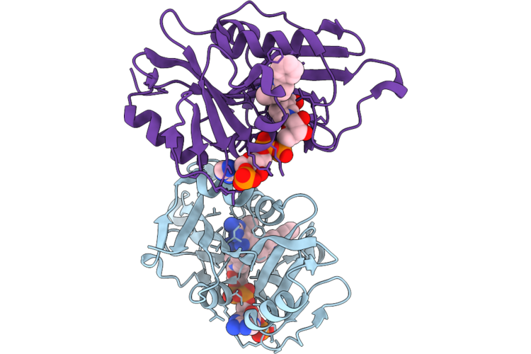 |
Crystal Structure Of Candida Auris Dihydrofolate Reductase In Complex With Inhibitor 1051 (S-Form) And Nadph
Organism: Candidozyma auris
Method: X-RAY DIFFRACTION Resolution:2.42 Å Release Date: 2026-07-22 Classification: OXIDOREDUCTASE Ligands: NDP, A1CTC |
Organism: Candidozyma auris
Method: X-RAY DIFFRACTION
Release Date: 2026-07-22
Ligands: NDP, A1CTC
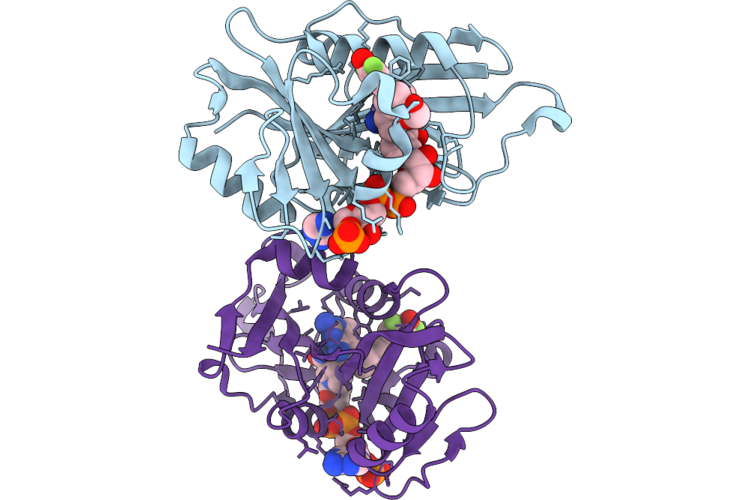 |
Crystal Structure Of Candida Auris Dihydrofolate Reductase In Complex With Inhibitor 1283 And Nadph
Organism: Candidozyma auris
Method: X-RAY DIFFRACTION Resolution:2.13 Å Release Date: 2026-07-22 Classification: OXIDOREDUCTASE Ligands: NDP, A1CTB |
Organism: Candidozyma auris
Method: X-RAY DIFFRACTION
Release Date: 2026-07-22
Ligands: NDP, A1CTB
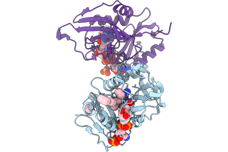 |
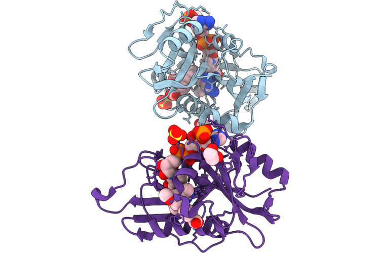
Crystal Structure Of Candida Auris Dihydrofolate Reductase In Complex With Inhibitor 1051 (R-Form) And Nadph
Organism: Candidozyma auris
Method: X-RAY DIFFRACTION Resolution:1.90 Å Release Date: 2026-07-22 Classification: OXIDOREDUCTASE Ligands: NDP, A1CTA, EDO, SO4 |
Organism: Candidozyma auris
Method: X-RAY DIFFRACTION
Release Date: 2026-07-22
Ligands: NDP, A1CTA, EDO, SO4
 |
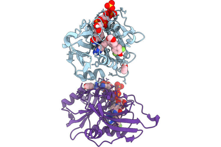
Crystal Structure Of Candida Auris Dihydrofolate Reductase In Complex With Inhibitor 1172 And Nadph
Organism: Candidozyma auris
Method: X-RAY DIFFRACTION Resolution:1.98 Å Release Date: 2026-07-22 Classification: OXIDOREDUCTASE Ligands: NDP, G6Y, EDO, SO4 |
Organism: Candidozyma auris
Method: X-RAY DIFFRACTION
Release Date: 2026-07-22
Ligands: NDP, G6Y, EDO, SO4
 |
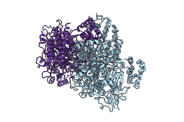
Crystal Structure Of Candida Auris Dihydrofolate Reductase In Complex With Inhibitor 1282 And Nadph
Organism: Candidozyma auris
Method: X-RAY DIFFRACTION Resolution:1.65 Å Release Date: 2026-07-22 Classification: OXIDOREDUCTASE Ligands: NDP, A1CS9, EDO, SO4 |
Organism: Candidozyma auris
Method: X-RAY DIFFRACTION
Release Date: 2026-07-22
Ligands: NDP, A1CS9, EDO, SO4
 |
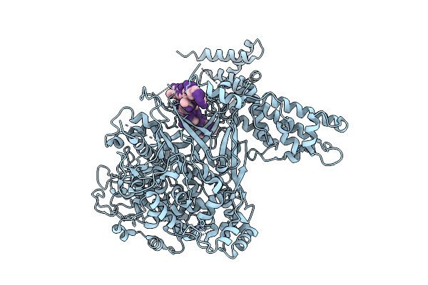
Single Stranded Dna-Binding Protein (Icp8) From Herpes Simplex Virus-1, Apo Form. Mutations: C254S, C455S, K769A, E770A
Organism: Human alphaherpesvirus 1
Method: X-RAY DIFFRACTION Resolution:2.75 Å Release Date: 2026-04-15 Classification: DNA BINDING PROTEIN Ligands: ZN |
Organism: Human alphaherpesvirus 1
Method: X-RAY DIFFRACTION
Release Date: 2026-04-15
Ligands: ZN
 |
Poly Dt Bound Form Of Single Stranded Dna-Binding Protein(Icp8) From Herpes Simplex Virus-1. Mutations: C254S, C455S
Organism: Human alphaherpesvirus 1, Synthetic construct
Method: X-RAY DIFFRACTION Resolution:3.10 Å Release Date: 2026-04-15 Classification: DNA BINDING PROTEIN/DNA Ligands: ZN |
Organism: Human alphaherpesvirus 1, Synthetic construct
Method: X-RAY DIFFRACTION
Release Date: 2026-04-15
Ligands: ZN
 |
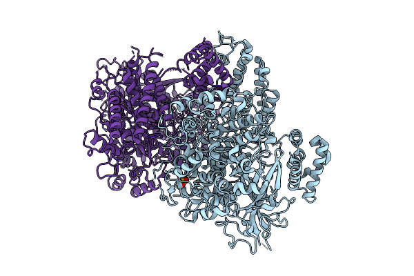
Poly Da Bound Form Of Single Stranded Dna-Binding Protein(Icp8) From Herpes Simplex Virus-1. Mutations: C254S, C455S
Organism: Human alphaherpesvirus 1, Synthetic construct
Method: X-RAY DIFFRACTION Resolution:3.00 Å Release Date: 2026-04-15 Classification: DNA BINDING PROTEIN/DNA |
Organism: Human alphaherpesvirus 1, Synthetic construct
Method: X-RAY DIFFRACTION
Release Date: 2026-04-15
 |
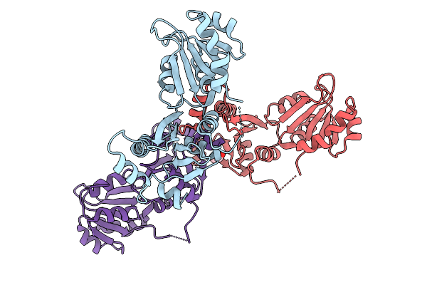
Single Stranded Dna-Binding Protein (Icp8) From Herpes Simplex Virus-1, Apo Form.Mutations: K166A, E167A, C254S, C455S
Organism: Human alphaherpesvirus 1
Method: X-RAY DIFFRACTION Resolution:2.75 Å Release Date: 2026-04-15 Classification: DNA BINDING PROTEIN Ligands: ZN, SO4, GOL |
Organism: Human alphaherpesvirus 1
Method: X-RAY DIFFRACTION
Release Date: 2026-04-15
Ligands: ZN, SO4, GOL
 |
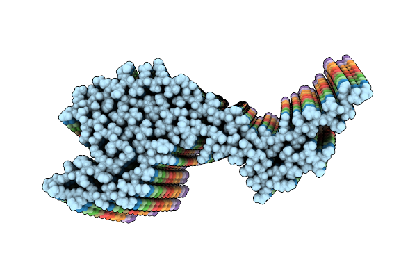
Crystal Structure Of The N-Terminal Domain Of The A Subunit Of The Bacillus Cereus Geri Germinant Receptor
Organism: Bacillus cereus atcc 14579
Method: X-RAY DIFFRACTION Resolution:2.80 Å Release Date: 2026-03-25 Classification: PROTEIN TRANSPORT Ligands: ACT |
Organism: Bacillus cereus atcc 14579
Method: X-RAY DIFFRACTION
Release Date: 2026-03-25
Ligands: ACT
 |
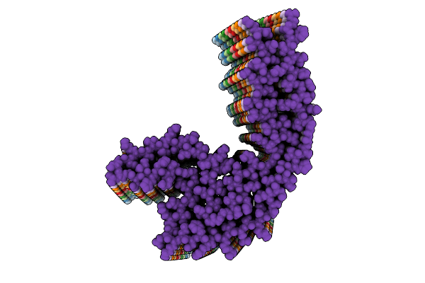
S305I Frontotemporal Lobar Degeneration (Ftld) Type I Tau Filament
Organism: Homo sapiens
Method: ELECTRON MICROSCOPY Release Date: 2026-03-11 Classification: PROTEIN FIBRIL |
Organism: Homo sapiens
Method: ELECTRON MICROSCOPY
Release Date: 2026-03-11
 |
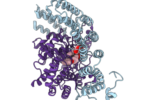
S305I Frontotemporal Lobar Degeneration (Ftld) Type Ii Tau Filament
Organism: Homo sapiens
Method: ELECTRON MICROSCOPY Release Date: 2026-03-11 Classification: PROTEIN FIBRIL |
Organism: Homo sapiens
Method: ELECTRON MICROSCOPY
Release Date: 2026-03-11
 |
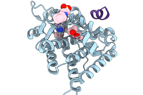
Co-Crystal Structure Of Rat Protein Farnesyltransferase Complexed With A-176120
Organism: Rattus norvegicus
Method: X-RAY DIFFRACTION Resolution:2.31 Å Release Date: 2026-03-04 Classification: TRANSFERASE Ligands: GOL, ZN, A1JZU |
Organism: Rattus norvegicus
Method: X-RAY DIFFRACTION
Release Date: 2026-03-04
Ligands: GOL, ZN, A1JZU
 |
Mineralocorticoid Receptor In Complex With Acylurea Antagonists.
Organism: Homo sapiens
Method: X-RAY DIFFRACTION Resolution:1.46 Å Release Date: 2026-02-18 Classification: TRANSCRIPTION Ligands: MPD, NHE, A1IJ6 |
Organism: Homo sapiens
Method: X-RAY DIFFRACTION
Release Date: 2026-02-18
Ligands: MPD, NHE, A1IJ6
 |
Crystal Structure Of The Daba Transaminase Ectb From The Halophilic And Cold-Adapted Marinobacter Sp. Ck1 -Mutant K264A
Organism: Marinobacter sp. ck-1
Method: X-RAY DIFFRACTION Resolution:1.55 Å Release Date: 2026-02-18 Classification: TRANSFERASE Ligands: PLP, EDO, SO4, NA |
Organism: Marinobacter sp. ck-1
Method: X-RAY DIFFRACTION
Release Date: 2026-02-18
Ligands: PLP, EDO, SO4, NA
 |
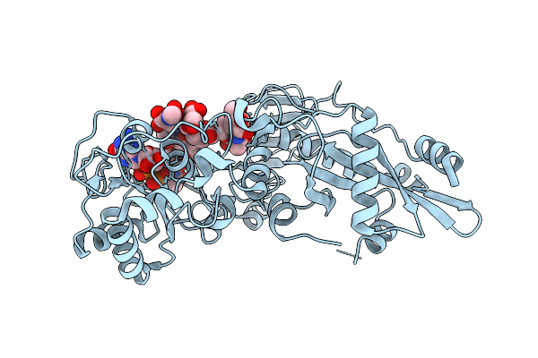
Crystal Structure Of The Daba Transaminase Ectb From The Halophilic And Cold-Adapted Marinobacter Sp. Ck1
Organism: Marinobacter sp. ck-1
Method: X-RAY DIFFRACTION Resolution:2.00 Å Release Date: 2026-02-18 Classification: TRANSFERASE Ligands: PMP, GOL |
Organism: Marinobacter sp. ck-1
Method: X-RAY DIFFRACTION
Release Date: 2026-02-18
Ligands: PMP, GOL
 |
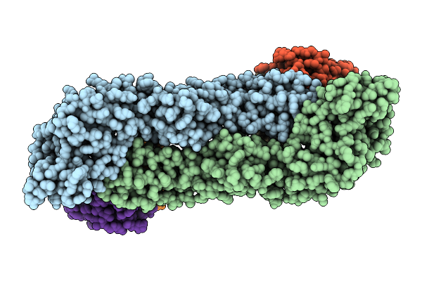
Crystal Structure Of Arabidopsis Thaliana Fut11 Complexed To Gdp And G0
Organism: Arabidopsis thaliana
Method: X-RAY DIFFRACTION Resolution:1.41 Å Release Date: 2026-01-28 Classification: TRANSFERASE Ligands: GDP |
Organism: Arabidopsis thaliana
Method: X-RAY DIFFRACTION
Release Date: 2026-01-28
Ligands: GDP
 |
Cryo-Em Structure Of Modified Zika Virus E Protein Dimer Complexed With A Neutralizing Antibody Smzab2 Fab
Organism: Homo sapiens, Zika virus
Method: ELECTRON MICROSCOPY Release Date: 2026-01-21 Classification: VIRAL PROTEIN/IMMUNE SYSTEM |
Organism: Homo sapiens, Zika virus
Method: ELECTRON MICROSCOPY
Release Date: 2026-01-21
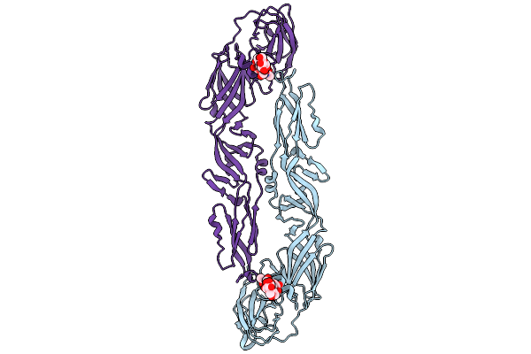 |
Cryo-Em Structure Of Modified Jev Virus E Protein Dimer
Organism: Japanese encephalitis virus
Method: ELECTRON MICROSCOPY Resolution:4.18 Å Release Date: 2026-01-21 Classification: VIRAL PROTEIN |
Organism: Japanese encephalitis virus
Method: ELECTRON MICROSCOPY
Release Date: 2026-01-21