

Search Count: 9,669
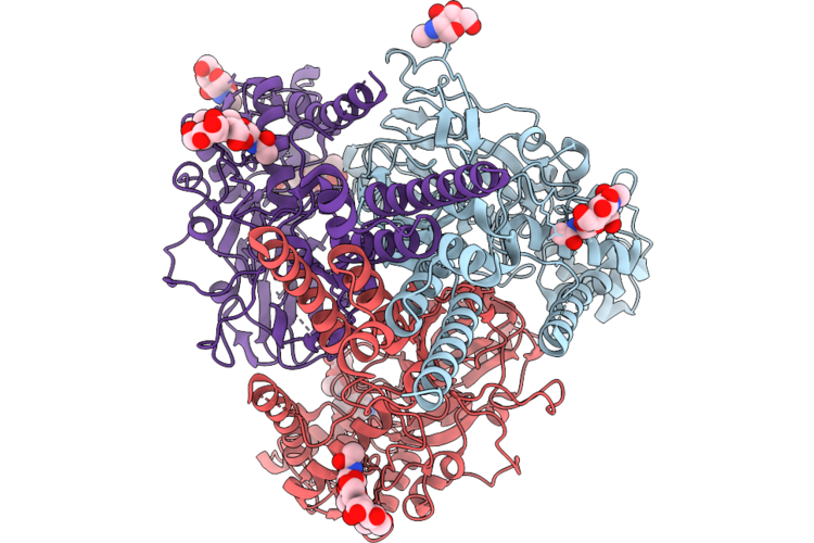 |
Heterotrimeric Enac Channel Complex Ppk1/Ppk26/Ppk26
Organism: Drosophila melanogaster
Method: ELECTRON MICROSCOPY Resolution:2.86 Å Release Date: 2026-08-19 Classification: NEUROPEPTIDE Ligands: NAG |
Organism: Drosophila melanogaster
Method: ELECTRON MICROSCOPY
Release Date: 2026-08-19
Ligands: NAG
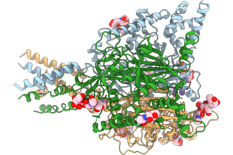 |
Heterotrimeric Enac Channel Complex Ppk1/Ppk26/Ppk26 With Vulnusin
Organism: Drosophila melanogaster
Method: ELECTRON MICROSCOPY Resolution:2.86 Å Release Date: 2026-08-19 Classification: NEUROPEPTIDE Ligands: NAG, NH4 |
Organism: Drosophila melanogaster
Method: ELECTRON MICROSCOPY
Release Date: 2026-08-19
Ligands: NAG, NH4
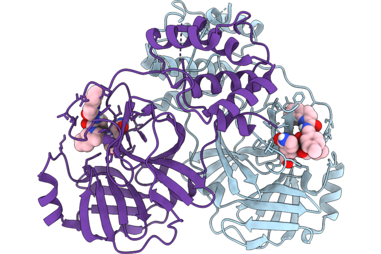 |
Crystal Structure Of Munia Coronavirus Hku13 Main Proteinase In Complex With A Broad-Spectrum Inhibitor
Organism: Munia coronavirus hku13-3514
Method: X-RAY DIFFRACTION Resolution:2.25 Å Release Date: 2026-08-19 Classification: VIRAL PROTEIN Ligands: A1EYZ |
Organism: Munia coronavirus hku13-3514
Method: X-RAY DIFFRACTION
Release Date: 2026-08-19
Ligands: A1EYZ
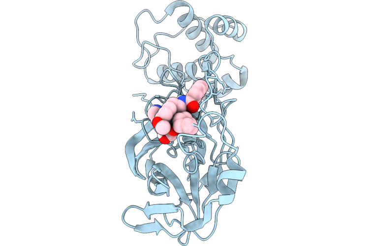 |
Crystal Structure Of Sars Coronavirus Main Proteinase In Complex With A Broad-Spectrum Inhibitor
Organism: Sars coronavirus urbani
Method: X-RAY DIFFRACTION Resolution:2.39 Å Release Date: 2026-08-19 Classification: VIRAL PROTEIN Ligands: A1EYZ |
Organism: Sars coronavirus urbani
Method: X-RAY DIFFRACTION
Release Date: 2026-08-19
Ligands: A1EYZ
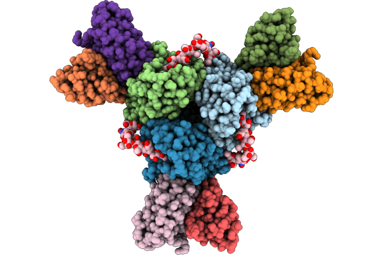 |
Cryoem Structure Of H5N1 A/Texas/37/2024 Ha Bound To Fab H91
Organism: Mus musculus, Influenza a virus
Method: ELECTRON MICROSCOPY Release Date: 2026-08-19 Classification: VIRAL PROTEIN Ligands: NAG |
Organism: Mus musculus, Influenza a virus
Method: ELECTRON MICROSCOPY
Release Date: 2026-08-19
Ligands: NAG
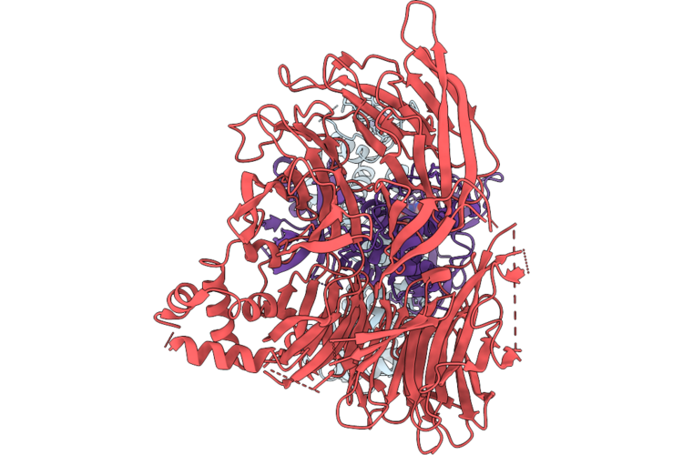 |
Structure Of Crbn/Ddb1Db-Kat2A-Compound4 Ternary Complex
Organism: Homo sapiens
Method: ELECTRON MICROSCOPY Resolution:2.90 Å Release Date: 2026-08-12 Classification: TRANSFERASE Ligands: ZN, A1C5C |
Organism: Homo sapiens
Method: ELECTRON MICROSCOPY
Release Date: 2026-08-12
Ligands: ZN, A1C5C
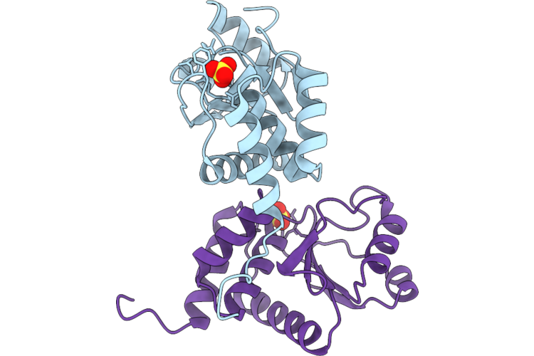 |
Crystal Structure Of Bacterial Dusp From Candidatus Chlorohelix Allophototropha
Organism: Candidatus chlorohelix allophototropha
Method: X-RAY DIFFRACTION Resolution:2.40 Å Release Date: 2026-08-12 Classification: HYDROLASE Ligands: SO4, CL |
Organism: Candidatus chlorohelix allophototropha
Method: X-RAY DIFFRACTION
Release Date: 2026-08-12
Ligands: SO4, CL
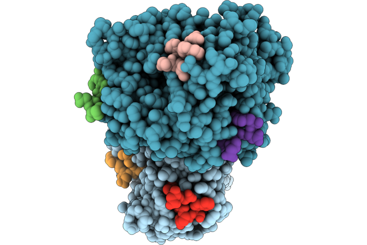 |
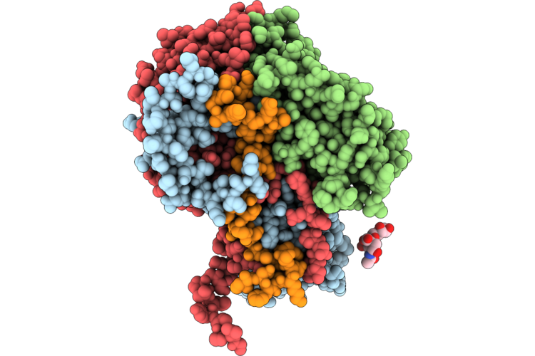
The Role Of Llps In Yeast Clathrin-Mediated Endocytosis And Trafficking
Organism: Saccharomyces
Method: X-RAY DIFFRACTION Resolution:1.70 Å Release Date: 2026-08-12 Classification: STRUCTURAL PROTEIN |
Organism: Saccharomyces
Method: X-RAY DIFFRACTION
Release Date: 2026-08-12
 |
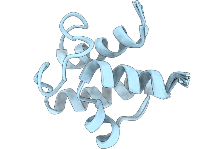
Cryoem Structure Of Ev-D68 Strain Fermon In Complex With Mfsd6-R196-L226
Organism: Enterovirus d68, Homo sapiens
Method: ELECTRON MICROSCOPY Resolution:2.00 Å Release Date: 2026-08-12 Classification: VIRAL PROTEIN |
Organism: Enterovirus d68, Homo sapiens
Method: ELECTRON MICROSCOPY
Release Date: 2026-08-12
 |
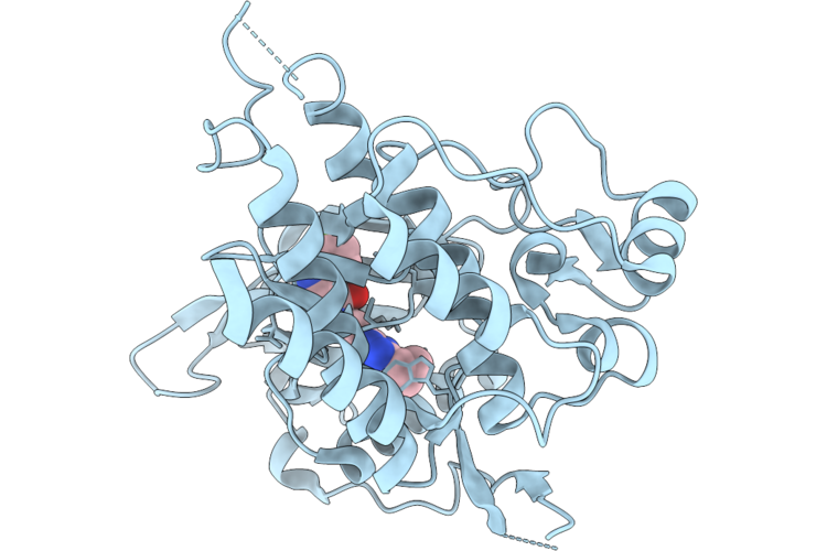
Solution Structure Of Holo Acyl Carrier Protein From Escherichia Coli
Organism: Escherichia coli (strain k12)
Method: SOLUTION NMR Release Date: 2026-08-12 Classification: PROTEIN TRANSPORT |
Organism: Escherichia coli (strain k12)
Method: SOLUTION NMR
Release Date: 2026-08-12
 |
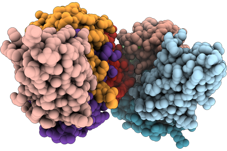
Crystal Structure Of 6-Fluoropyrazolo[1,5-A]Pyridine Derivative Bound To Kit
Organism: Homo sapiens
Method: X-RAY DIFFRACTION Resolution:2.63 Å Release Date: 2026-08-05 Classification: TRANSFERASE Ligands: A1DJF |
Organism: Homo sapiens
Method: X-RAY DIFFRACTION
Release Date: 2026-08-05
Ligands: A1DJF
 |
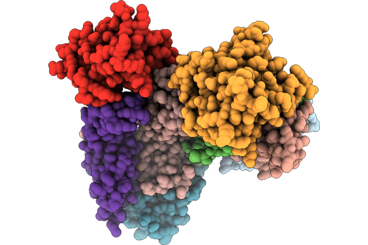
Crystal Structure Of A De Novo-Designed Vhh Targeting S100A4
Organism: Synthetic construct, Homo sapiens
Method: X-RAY DIFFRACTION Resolution:1.82 Å Release Date: 2026-08-05 Classification: DE NOVO PROTEIN Ligands: CA, PEG, GOL |
Organism: Synthetic construct, Homo sapiens
Method: X-RAY DIFFRACTION
Release Date: 2026-08-05
Ligands: CA, PEG, GOL
 |
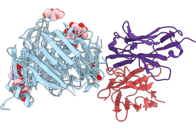
Crystal Structure Of A De Novo-Designed Binder Targeting Vegfa
Organism: Synthetic construct, Homo sapiens
Method: X-RAY DIFFRACTION Resolution:2.30 Å Release Date: 2026-08-05 Classification: DE NOVO PROTEIN Ligands: GOL |
Organism: Synthetic construct, Homo sapiens
Method: X-RAY DIFFRACTION
Release Date: 2026-08-05
Ligands: GOL
 |
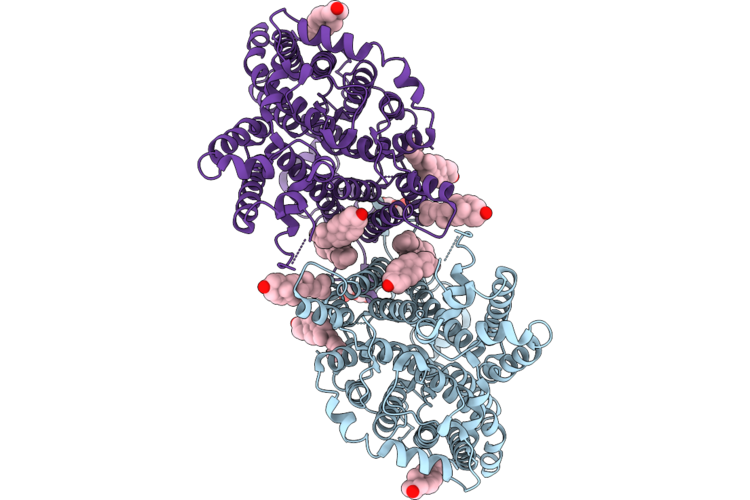
Cryo-Em Structure Of N19 Fab : D0D-S1A
Organism: Porcine epidemic diarrhea virus, Sus scrofa
Method: ELECTRON MICROSCOPY Resolution:2.30 Å Release Date: 2026-08-05 Classification: VIRAL PROTEIN/IMMUNE SYSTEM Ligands: NAG |
Organism: Porcine epidemic diarrhea virus, Sus scrofa
Method: ELECTRON MICROSCOPY
Release Date: 2026-08-05
Ligands: NAG
 |
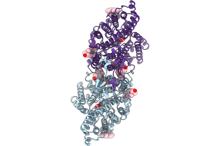
Cryo-Em Structure Of Human Nbcn2
Organism: Homo sapiens
Method: ELECTRON MICROSCOPY Resolution:2.85 Å Release Date: 2026-08-05 Classification: MEMBRANE PROTEIN Ligands: CLR, NAG |
Organism: Homo sapiens
Method: ELECTRON MICROSCOPY
Release Date: 2026-08-05
Ligands: CLR, NAG
 |
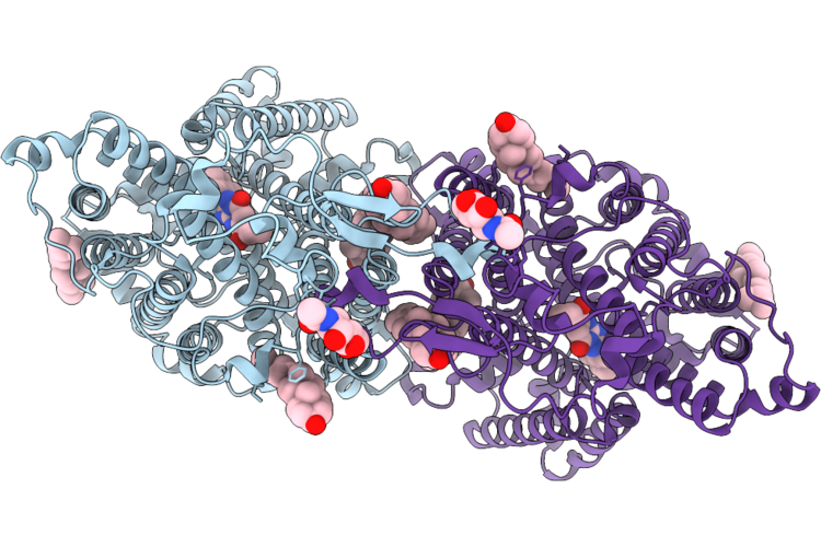
Cryo-Em Structure Of Human Nbcn2 Bound To Carbonate
Organism: Homo sapiens
Method: ELECTRON MICROSCOPY Resolution:2.45 Å Release Date: 2026-08-05 Classification: MEMBRANE PROTEIN Ligands: CLR, CO3, NA |
Organism: Homo sapiens
Method: ELECTRON MICROSCOPY
Release Date: 2026-08-05
Ligands: CLR, CO3, NA
 |
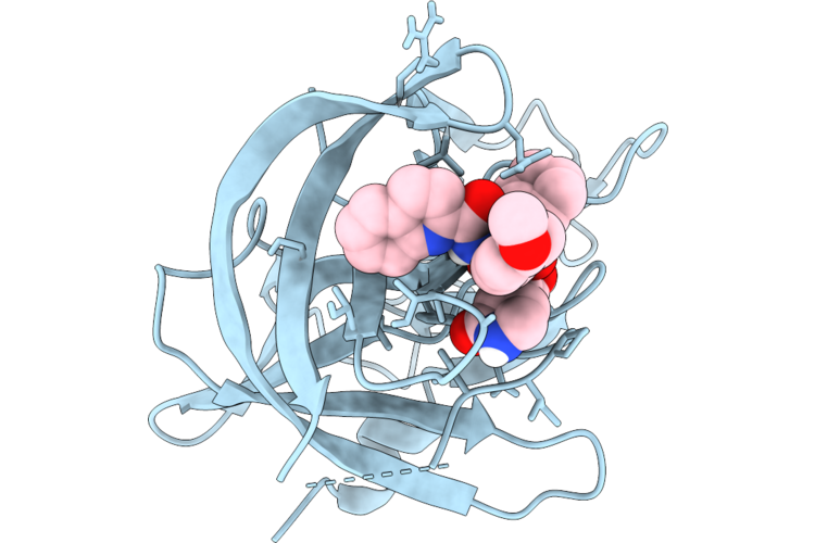
Cryo-Em Structure Of Human Nbcn2 Bound To Compound 38J
Organism: Homo sapiens
Method: ELECTRON MICROSCOPY Resolution:3.24 Å Release Date: 2026-08-05 Classification: MEMBRANE PROTEIN Ligands: CLR, A1BLW, NAG |
Organism: Homo sapiens
Method: ELECTRON MICROSCOPY
Release Date: 2026-08-05
Ligands: CLR, A1BLW, NAG
 |
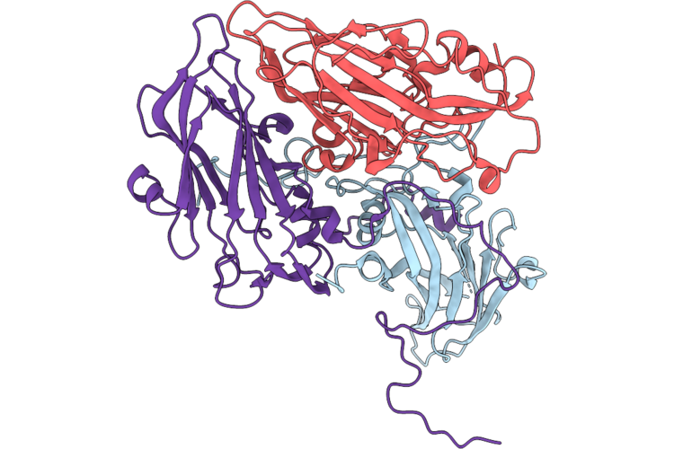
Crystal Structure Of Norovirus 3C-Like Protease In Complex With A Broad-Spectrum Inhibitor
Organism: Norovirus (strain human/nov/united states/norwalk/1968/gi)
Method: X-RAY DIFFRACTION Resolution:1.76 Å Release Date: 2026-08-05 Classification: VIRAL PROTEIN Ligands: A1EYZ |
Organism: Norovirus (strain human/nov/united states/norwalk/1968/gi)
Method: X-RAY DIFFRACTION
Release Date: 2026-08-05
Ligands: A1EYZ
 |
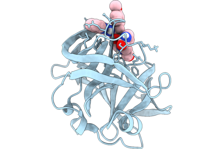
Cryoem Structure Of Ev-D68 Strain Fermon Procapsid
Organism: Enterovirus d68
Method: ELECTRON MICROSCOPY Resolution:3.21 Å Release Date: 2026-08-05 Classification: VIRAL PROTEIN |
Organism: Enterovirus d68
Method: ELECTRON MICROSCOPY
Release Date: 2026-08-05
 |
Crystal Structure Of Enterovirus A71 3C Protease In Complex With A Broad-Spectrum Inhibitor
Organism: Human enterovirus 71 (strain brcr)
Method: X-RAY DIFFRACTION Resolution:1.59 Å Release Date: 2026-08-05 Classification: VIRAL PROTEIN Ligands: A1EYZ |
Organism: Human enterovirus 71 (strain brcr)
Method: X-RAY DIFFRACTION
Release Date: 2026-08-05
Ligands: A1EYZ