

Search Count: 652
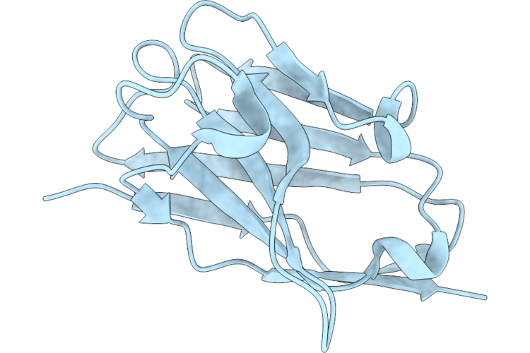 |
Structure Of Nanobody Sm8 Against Fenitrothion
Organism: Vicugna pacos
Method: X-RAY DIFFRACTION Resolution:1.73 Å Release Date: 2026-07-29 Classification: IMMUNE SYSTEM |
Organism: Vicugna pacos
Method: X-RAY DIFFRACTION
Release Date: 2026-07-29
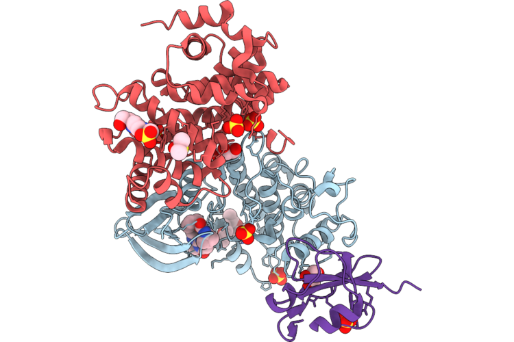 |
Crystal Structure Of Selective Inhibitor 16 Bound At The Active Site Of Cdk1
Organism: Homo sapiens
Method: X-RAY DIFFRACTION Resolution:2.40 Å Release Date: 2026-07-08 Classification: SIGNALING PROTEIN Ligands: A1DEQ, A1DJL, GOL, SO4, DMS, EPE |
Organism: Homo sapiens
Method: X-RAY DIFFRACTION
Release Date: 2026-07-08
Ligands: A1DEQ, A1DJL, GOL, SO4, DMS, EPE
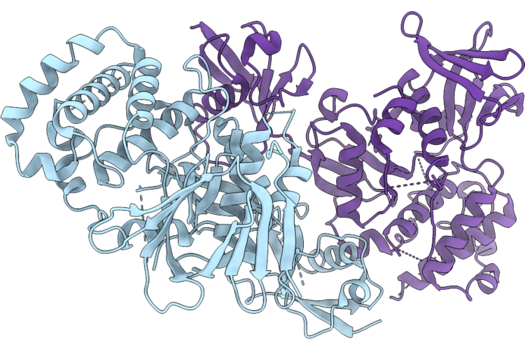 |
Kindlin-3/Integrin Beta2 Cytoplasmic Tail Complex
Organism: Homo sapiens
Method: X-RAY DIFFRACTION Resolution:2.40 Å Release Date: 2026-07-08 Classification: CELL ADHESION |
Organism: Homo sapiens
Method: X-RAY DIFFRACTION
Release Date: 2026-07-08
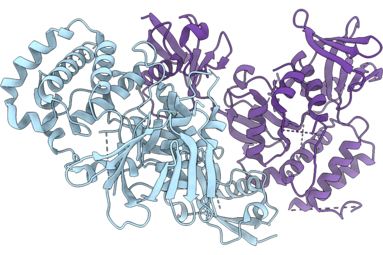 |
Kindlin-3/Integrin Beta3 Cytoplasmic Tail Complex
Organism: Homo sapiens
Method: X-RAY DIFFRACTION Resolution:2.20 Å Release Date: 2026-07-08 Classification: CELL ADHESION |
Organism: Homo sapiens
Method: X-RAY DIFFRACTION
Release Date: 2026-07-08
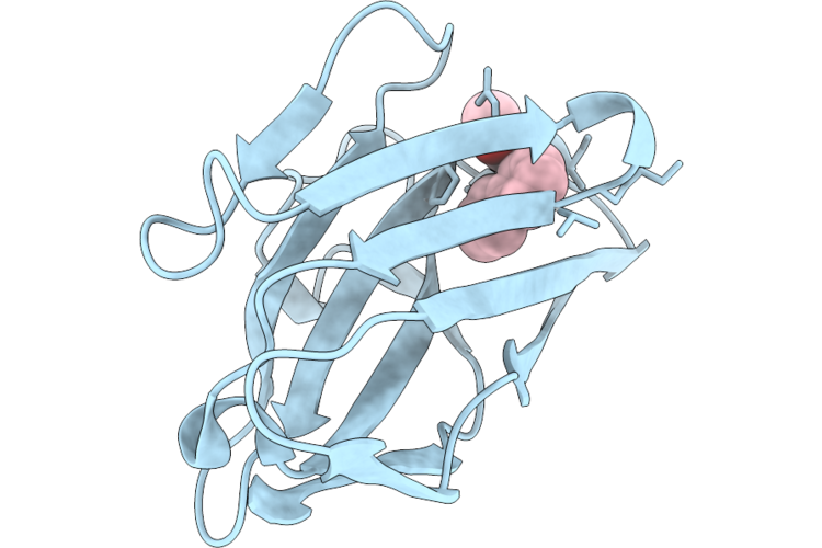 |
Fundamental Structural Studies Of Metanaphthene And Introduction Of Disulfide Bonds
Organism: Synthetic construct
Method: X-RAY DIFFRACTION Resolution:1.45 Å Release Date: 2026-07-01 Classification: IMMUNE SYSTEM Ligands: A1ES9 |
Organism: Synthetic construct
Method: X-RAY DIFFRACTION
Release Date: 2026-07-01
Ligands: A1ES9
 |
Cryo-Em Structure Of Human Protease-Activated Receptor 4 In Complex With Gq Heterotrimers And Scfv16 Bound To Tethered Ligand
Organism: Homo sapiens, Escherichia coli k-12
Method: ELECTRON MICROSCOPY Release Date: 2026-06-10 Classification: MEMBRANE PROTEIN/IMMUNE SYSTEM |
Organism: Homo sapiens, Escherichia coli k-12
Method: ELECTRON MICROSCOPY
Release Date: 2026-06-10
 |
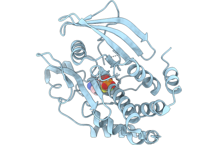
Structure Of Ptp1B Complexed With Difluoromethylphosphonate Inhibitor Compound 2
Organism: Homo sapiens
Method: X-RAY DIFFRACTION Resolution:1.73 Å Release Date: 2026-05-27 Classification: HYDROLASE/INHIBITOR Ligands: A1C3A |
Organism: Homo sapiens
Method: X-RAY DIFFRACTION
Release Date: 2026-05-27
Ligands: A1C3A
 |
Structure Of Ptp1B Complexed With Difluoromethylphosphonate Inhibitor Compound 10
Organism: Homo sapiens
Method: X-RAY DIFFRACTION Resolution:2.51 Å Release Date: 2026-05-27 Classification: HYDROLASE/INHIBITOR Ligands: A1C3B |
Organism: Homo sapiens
Method: X-RAY DIFFRACTION
Release Date: 2026-05-27
Ligands: A1C3B
 |
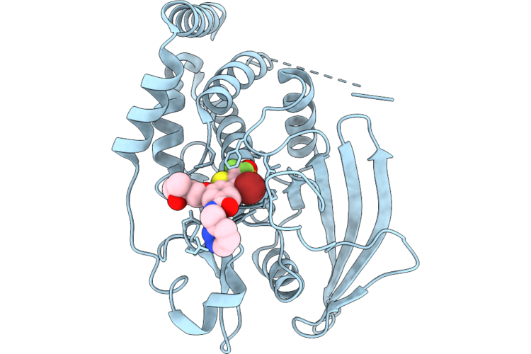
Structure Of Ptp1B Complexed With Difluoromethylphosphonate Inhibitor Compound 15
Organism: Homo sapiens
Method: X-RAY DIFFRACTION Resolution:2.15 Å Release Date: 2026-05-27 Classification: HYDROLASE/INHIBITOR Ligands: A1C3C, DMS |
Organism: Homo sapiens
Method: X-RAY DIFFRACTION
Release Date: 2026-05-27
Ligands: A1C3C, DMS
 |
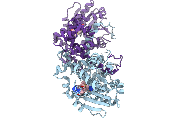
Structure Of Ptp1B Complexed With Difluoromethylphosphonate Inhibitor Compound 30
Organism: Homo sapiens
Method: X-RAY DIFFRACTION Resolution:1.94 Å Release Date: 2026-05-27 Classification: HYDROLASE/INHIBITOR Ligands: A1C3D |
Organism: Homo sapiens
Method: X-RAY DIFFRACTION
Release Date: 2026-05-27
Ligands: A1C3D
 |
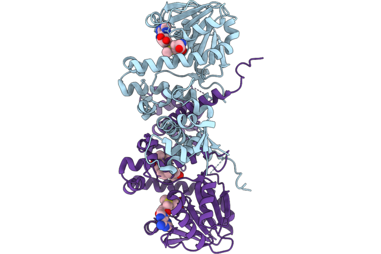
Crystal Structure Of Schomt2-Sam From Schisandra Chinensis
Organism: Schisandra chinensis
Method: X-RAY DIFFRACTION Resolution:1.63 Å Release Date: 2026-05-13 Classification: TRANSFERASE Ligands: SAM |
Organism: Schisandra chinensis
Method: X-RAY DIFFRACTION
Release Date: 2026-05-13
Ligands: SAM
 |
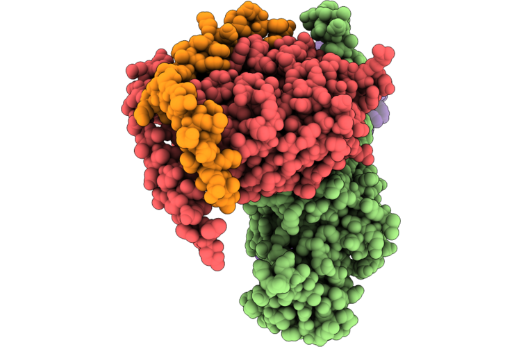
Crystal Structure Of Schomt2-Sam-Gom From Schisandra Chinensis
Organism: Schisandra chinensis
Method: X-RAY DIFFRACTION Resolution:2.43 Å Release Date: 2026-05-13 Classification: TRANSFERASE Ligands: SAM, A1E80 |
Organism: Schisandra chinensis
Method: X-RAY DIFFRACTION
Release Date: 2026-05-13
Ligands: SAM, A1E80
 |
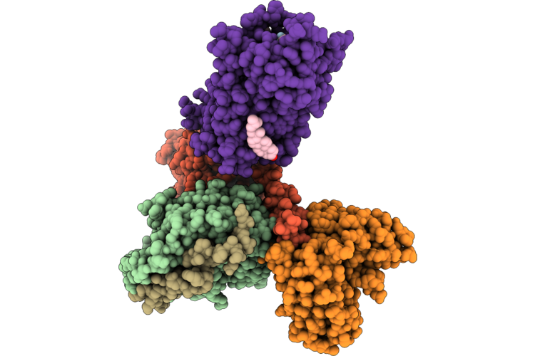
Gi Bound Kappa-Opioid Receptor In Complex With Beta01
Organism: Homo sapiens, Mus musculus, Bos taurus, Escherichia coli, Synthetic construct
Method: ELECTRON MICROSCOPY Release Date: 2026-05-06 Classification: SIGNALING PROTEIN |
Organism: Homo sapiens, Mus musculus, Bos taurus, Escherichia coli, Synthetic construct
Method: ELECTRON MICROSCOPY
Release Date: 2026-05-06
 |
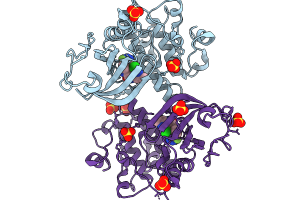
Gi Bound Kappa-Opioid Receptor In Complex With Difelikefalin
Organism: Rattus norvegicus, Bos taurus, Homo sapiens, Synthetic construct
Method: ELECTRON MICROSCOPY Release Date: 2026-05-06 Classification: SIGNALING PROTEIN/IMMUNE SYSTEM Ligands: CLR |
Organism: Rattus norvegicus, Bos taurus, Homo sapiens, Synthetic construct
Method: ELECTRON MICROSCOPY
Release Date: 2026-05-06
Ligands: CLR
 |
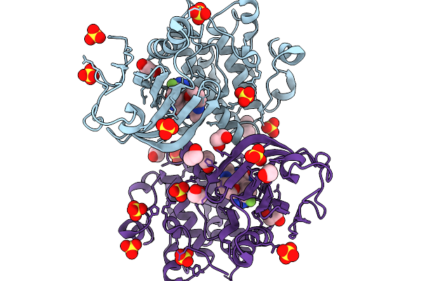
Crystal Structure Of Human Pkmyt1 Protein Kinase Domain With Naphthyridinone Inhibitor Compound 11
Organism: Homo sapiens
Method: X-RAY DIFFRACTION Resolution:1.74 Å Release Date: 2026-03-25 Classification: TRANSFERASE Ligands: A1E5A, SO4 |
Organism: Homo sapiens
Method: X-RAY DIFFRACTION
Release Date: 2026-03-25
Ligands: A1E5A, SO4
 |
Crystal Structure Of Human Pkmyt1 Protein Kinase Domain With Naphthyridinone Inhibitor Compound 16
Organism: Homo sapiens
Method: X-RAY DIFFRACTION Resolution:1.59 Å Release Date: 2026-03-25 Classification: TRANSFERASE Ligands: A1E5J, DMS, EDO, SO4 |
Organism: Homo sapiens
Method: X-RAY DIFFRACTION
Release Date: 2026-03-25
Ligands: A1E5J, DMS, EDO, SO4
 |
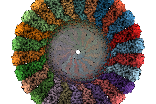
Cryo-Em Structure Of The Erlin1/2 Complex Purified Using Gdn And Chs
Organism: Homo sapiens
Method: ELECTRON MICROSCOPY Resolution:2.70 Å Release Date: 2026-03-25 Classification: MEMBRANE PROTEIN |
Organism: Homo sapiens
Method: ELECTRON MICROSCOPY
Release Date: 2026-03-25
 |
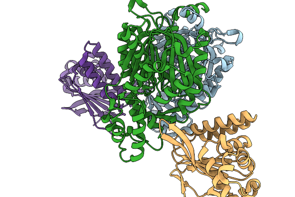
Cryo-Em Structure Of The Erlin1/2 Complex Purified Using Ddm And Gdn
Organism: Homo sapiens
Method: ELECTRON MICROSCOPY Resolution:2.60 Å Release Date: 2026-03-25 Classification: MEMBRANE PROTEIN |
Organism: Homo sapiens
Method: ELECTRON MICROSCOPY
Release Date: 2026-03-25
 |
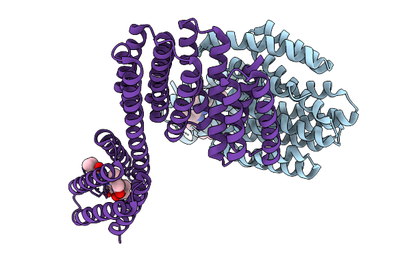
Crystal Structure Of The Chs-Chil Complex
Organism: Arabidopsis thaliana
Method: X-RAY DIFFRACTION Resolution:1.91 Å Release Date: 2026-03-04 Classification: TRANSFERASE |
Organism: Arabidopsis thaliana
Method: X-RAY DIFFRACTION
Release Date: 2026-03-04
 |
Crystal Structrue Of Bvfp-Pr1-88.4
Organism: Homo sapiens
Method: X-RAY DIFFRACTION Resolution:1.80 Å Release Date: 2026-01-28 Classification: DE NOVO PROTEIN Ligands: BLA |
Organism: Homo sapiens
Method: X-RAY DIFFRACTION
Release Date: 2026-01-28
Ligands: BLA