
Search Count: 2,996
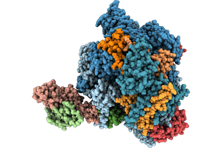 |
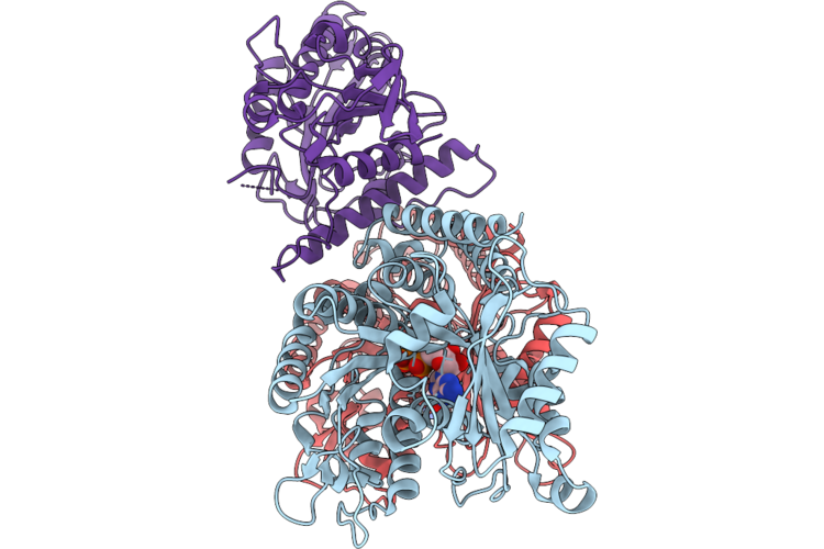
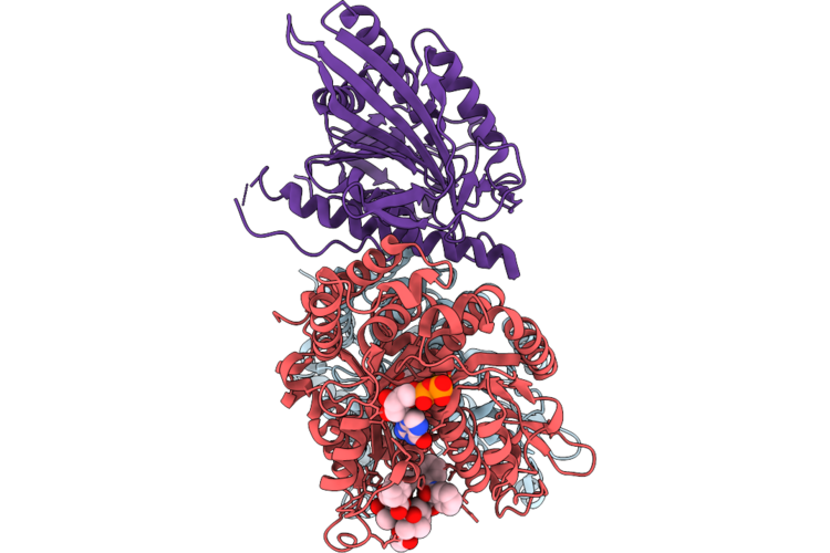
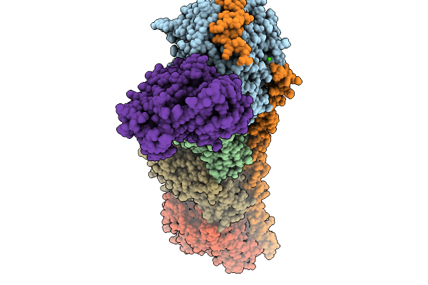
Structure Of Mammalian Rna Polymerase Ii Stalled By A Cisplatin-Induced Inter-Strand Crosslink Lesion At The +2 Position.
Organism: Synthetic construct, Sus scrofa
Method: ELECTRON MICROSCOPY Resolution:3.04 Å Release Date: 2026-06-24 Classification: TRANSCRIPTION Ligands: ZN, CPT, MG |
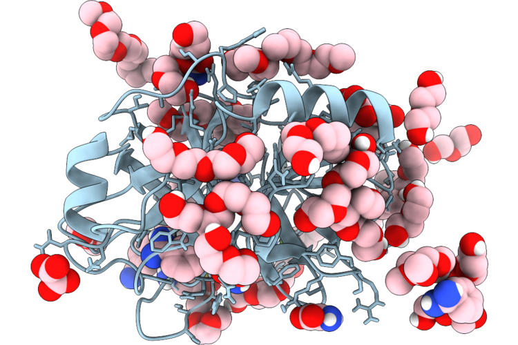 |
The Structure Of Porcine Trypsin In Complex With Crystallization Additives I
Organism: Sus scrofa
Method: X-RAY DIFFRACTION Resolution:1.28 Å Release Date: 2026-05-27 Classification: HYDROLASE Ligands: CA, BEN, PG5, PEG, PG4, OXL, A1CHW, OXM, PG6 |
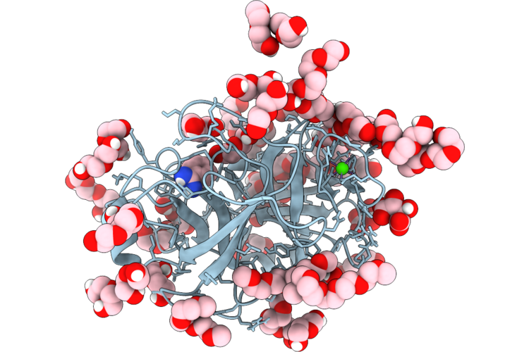 |
Porcine Trypsin Grown From Peg And Complexed With Crystallization Additives Ii
Organism: Sus scrofa
Method: X-RAY DIFFRACTION Resolution:1.27 Å Release Date: 2026-05-27 Classification: HYDROLASE Ligands: CA, TLA, PEG, PG4, PG6, PG5, BEN |
 |
Cryo-Em Reconstruction Of Undecorated Gdp Microtubule
Organism: Sus scrofa
Method: ELECTRON MICROSCOPY Resolution:2.90 Å Release Date: 2026-05-06 Classification: STRUCTURAL PROTEIN Ligands: GTP, MG, GDP |
 |
Giraffe Kif5A Motor Domain In Nucleotide Free State Bound To Microtubule
Organism: Giraffa camelopardalis, Sus scrofa
Method: ELECTRON MICROSCOPY Resolution:4.40 Å Release Date: 2026-04-29 Classification: MOTOR PROTEIN Ligands: GTP, GDP |
 |
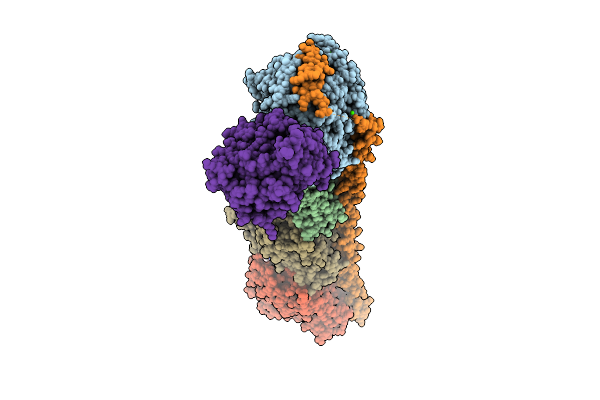
Complex Of Fmdv O/18074 And Porcine-Derived Neutralizing Monoclonal Antibody Po18-10
Organism: Sus scrofa, Foot-and-mouth disease virus o
Method: ELECTRON MICROSCOPY Resolution:2.27 Å Release Date: 2026-04-22 Classification: VIRUS |
 |
Kif1A R350G Bound To Microtubules In Two-Heads-Bound State With Amp-Pnp
Organism: Homo sapiens, Sus scrofa
Method: ELECTRON MICROSCOPY Release Date: 2026-04-22 Classification: MOTOR PROTEIN Ligands: GTP, MG, GDP, TA1, ANP |
 |
Kif1A R350G Bound To Microtubules In The Apo State
Organism: Homo sapiens, Sus scrofa
Method: ELECTRON MICROSCOPY Resolution:3.29 Å Release Date: 2026-04-22 Classification: MOTOR PROTEIN Ligands: GTP, MG, GDP, TA1 |
 |
Kif1A R350W Bound To Microtubules In The Apo State
Organism: Homo sapiens, Sus scrofa
Method: ELECTRON MICROSCOPY Release Date: 2026-04-22 Classification: MOTOR PROTEIN Ligands: GTP, MG, GDP, TA1 |
 |
Kif1A R350W Bound To Microtubules In Two-Heads-Bound State With Amp-Pnp
Organism: Homo sapiens, Sus scrofa
Method: ELECTRON MICROSCOPY Release Date: 2026-04-22 Classification: MOTOR PROTEIN Ligands: MG, ANP, GTP, GDP, TA1 |
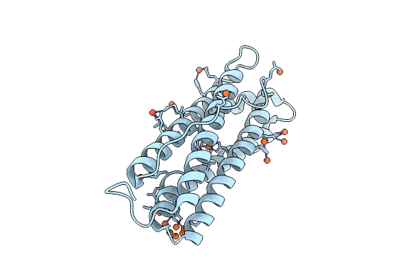 |
Crystal Structure Of Ferritin From Pig Spleen
Organism: Sus scrofa
Method: X-RAY DIFFRACTION Resolution:2.31 Å Release Date: 2026-04-15 Classification: METAL BINDING PROTEIN Ligands: FE |
 |
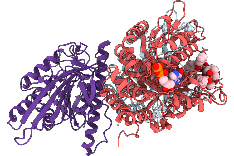
Crystal Structure Of The Post-Reactive State Of Porcine Oas1 In Complex With Dsrna And Products 25A2 And Ppi Bound To The Catalytic Center.
Organism: Sus scrofa, Synthetic construct
Method: X-RAY DIFFRACTION Resolution:1.80 Å Release Date: 2026-04-08 Classification: TRANSFERASE/RNA Ligands: MG, MN, 25L, POP, EDO |
 |
Crystal Structure Of The Post-Reactive State Of Porcine Oas1 In Complex With Dsrna, Catalytic Center Bound Ppi, And Dissociated 25A2.
Organism: Sus scrofa, Synthetic construct
Method: X-RAY DIFFRACTION Resolution:2.74 Å Release Date: 2026-04-08 Classification: TRANSFERASE/RNA Ligands: MG, POP, 25L |
 |
Crystal Structure Of The Pre-Reactive State Of Porcine Oas1 In Complex With Dsrna, Two Apcpp Substrate Analogs, Three Catalytic Mn2+ Ions.
Organism: Sus scrofa, Synthetic construct
Method: X-RAY DIFFRACTION Resolution:1.60 Å Release Date: 2026-04-08 Classification: TRANSFERASE/RNA Ligands: APC, MN, EDO |
 |
Tubulin-Rb3_Sld In Complex With Compound Qw-5-70
Organism: Rattus norvegicus, Sus scrofa
Method: X-RAY DIFFRACTION Resolution:2.51 Å Release Date: 2026-04-08 Classification: CELL CYCLE Ligands: GTP, GDP, A1B97, SO4 |
 |
Tubulin-Rb3-Ttl In Complex With C38
Organism: Rattus norvegicus, Gallus gallus, Sus scrofa
Method: X-RAY DIFFRACTION Resolution:2.93 Å Release Date: 2026-04-01 Classification: CELL CYCLE Ligands: GTP, MG, CA, MES, A1L80, GDP, ACP |
 |
Crystal Structure Of Tubulin-Rb3-Ttl In Complex With X8
Organism: Rattus norvegicus, Gallus gallus, Sus scrofa
Method: X-RAY DIFFRACTION Resolution:2.54 Å Release Date: 2026-03-25 Classification: CELL CYCLE Ligands: GTP, MG, CA, MES, GDP, A1L8Z |
 |
Asfv P15 In Complex With Fab 4E2
Organism: African swine fever virus (strain badajoz 1971 vero-adapted), Sus scrofa
Method: ELECTRON MICROSCOPY Resolution:3.75 Å Release Date: 2026-03-18 Classification: VIRAL PROTEIN |
 |
State 2 Map 3 Rna Pol Ii Activated Elongation Complex With Setd2 Bound To Proximal Upstream H3
Organism: Homo sapiens, Synthetic construct, Sus scrofa
Method: ELECTRON MICROSCOPY Release Date: 2026-03-18 Classification: TRANSCRIPTION Ligands: ZN, MG |
 |
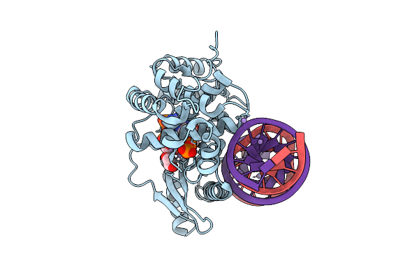
Native Glua1/Glua4-Cnih3 Complex In Resting State
Organism: Sus scrofa
Method: ELECTRON MICROSCOPY Release Date: 2026-03-04 Classification: MEMBRANE PROTEIN Ligands: DQC |