

Search Count: 2,271
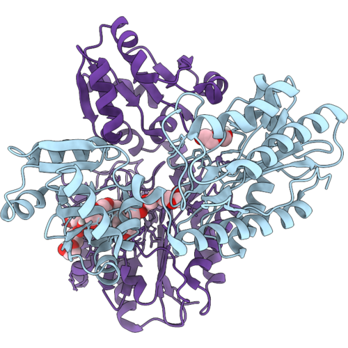 |
Crystal Structure Of Aminopeptidase P From Lactococcus Lactis With A Dinuclear Manganese Active Site At 2.73 Angstrom Resolution
Organism: Lactococcus lactis subsp. lactis
Method: X-RAY DIFFRACTION Resolution:2.73 Å Release Date: 2026-11-04 Classification: HYDROLASE Ligands: MN, PEG, 12P, PG4 |
Organism: Lactococcus lactis subsp. lactis
Method: X-RAY DIFFRACTION
Release Date: 2026-11-04
Ligands: MN, PEG, 12P, PG4
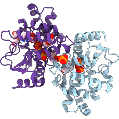 |
Crystal Structure Of A Cytidylate Kinase From Mycobacterium Tuberculosis (Cmp Complex)
Organism: Mycobacterium tuberculosis h37rv
Method: X-RAY DIFFRACTION Resolution:1.95 Å Release Date: 2026-10-28 Classification: TRANSFERASE Ligands: PO4, PEG, PG4, C5P |
Organism: Mycobacterium tuberculosis h37rv
Method: X-RAY DIFFRACTION
Release Date: 2026-10-28
Ligands: PO4, PEG, PG4, C5P
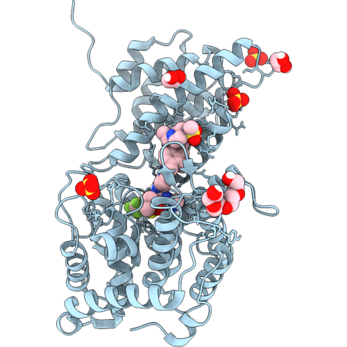 |
Menin Mutant - M327I In Complex With Ziftomenib
Organism: Homo sapiens
Method: X-RAY DIFFRACTION Resolution:1.60 Å Release Date: 2026-10-28 Classification: PROTEIN BINDING Ligands: K5O, SO4, PG4, EDO, DMS |
Organism: Homo sapiens
Method: X-RAY DIFFRACTION
Release Date: 2026-10-28
Ligands: K5O, SO4, PG4, EDO, DMS
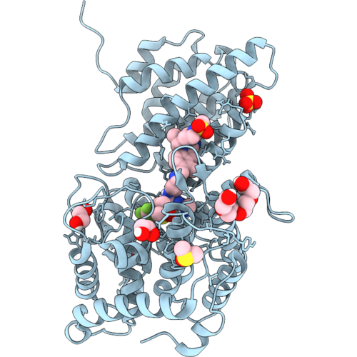 |
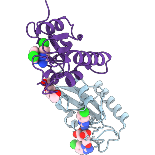
Menin Mutant - G331D In Complex With Ziftomenib
Organism: Homo sapiens
Method: X-RAY DIFFRACTION Resolution:1.45 Å Release Date: 2026-10-28 Classification: PROTEIN BINDING Ligands: K5O, DMS, EDO, SO4, PG4 |
Organism: Homo sapiens
Method: X-RAY DIFFRACTION
Release Date: 2026-10-28
Ligands: K5O, DMS, EDO, SO4, PG4
 |
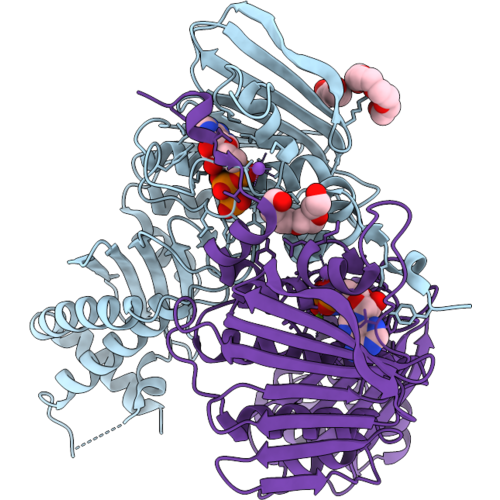
Crystal Structure Of Human Pin1 Bound To Compound 155
Organism: Homo sapiens
Method: X-RAY DIFFRACTION Resolution:1.60 Å Release Date: 2026-10-28 Classification: ISOMERASE Ligands: VGD, PG4, FLC |
Organism: Homo sapiens
Method: X-RAY DIFFRACTION
Release Date: 2026-10-28
Ligands: VGD, PG4, FLC
 |
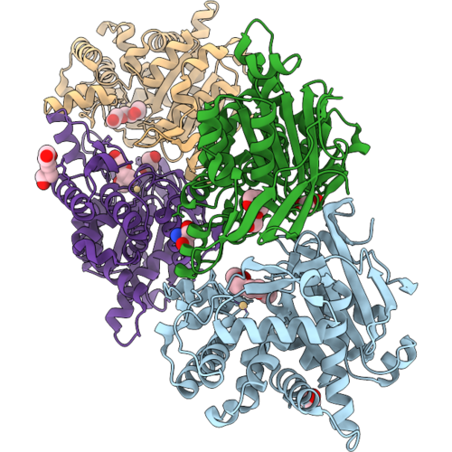
Crystal Structure Of The E48Q Variant Of The Aquifex Aeolicus Gyrb Atpase Domain Complexed With Amppnp
Organism: Aquifex aeolicus vf5
Method: X-RAY DIFFRACTION Resolution:1.70 Å Release Date: 2026-10-21 Classification: HYDROLASE Ligands: MG, ANP, PEG, NA, PG4 |
Organism: Aquifex aeolicus vf5
Method: X-RAY DIFFRACTION
Release Date: 2026-10-21
Ligands: MG, ANP, PEG, NA, PG4
 |
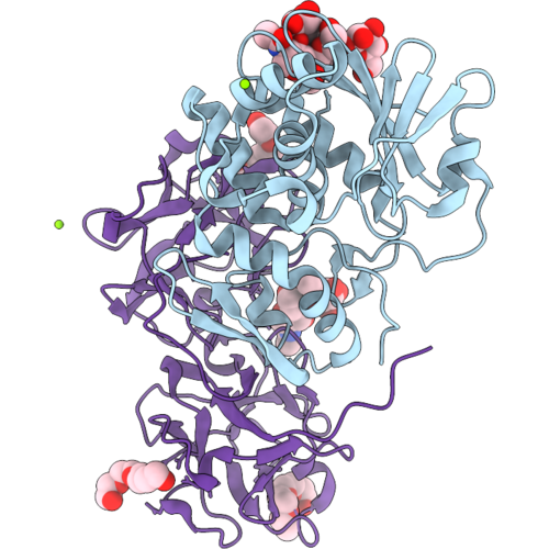
Class 3 L-Asparaginase From Colletotrichum Fructicola With Unmodified Active Site Ser51
Organism: Colletotrichum fructicola
Method: X-RAY DIFFRACTION Resolution:1.71 Å Release Date: 2026-10-21 Classification: HYDROLASE Ligands: PG4, EDO, CL, CD, PGE, TRS |
Organism: Colletotrichum fructicola
Method: X-RAY DIFFRACTION
Release Date: 2026-10-21
Ligands: PG4, EDO, CL, CD, PGE, TRS
 |
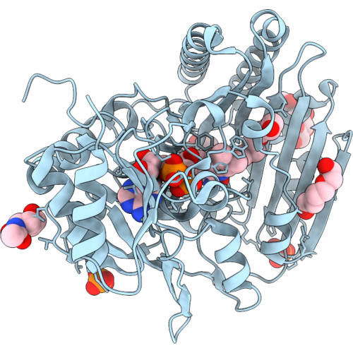
The Structure Of The Monoclinic Crystal Form Of The Type Ii Ribosome Inactivating Protein From Winter Aconite Eranthis Hyemalis.
Organism: Eranthis hyemalis
Method: X-RAY DIFFRACTION Resolution:1.40 Å Release Date: 2026-10-21 Classification: TOXIN Ligands: MG, PG4, CL |
Organism: Eranthis hyemalis
Method: X-RAY DIFFRACTION
Release Date: 2026-10-21
Ligands: MG, PG4, CL
 |
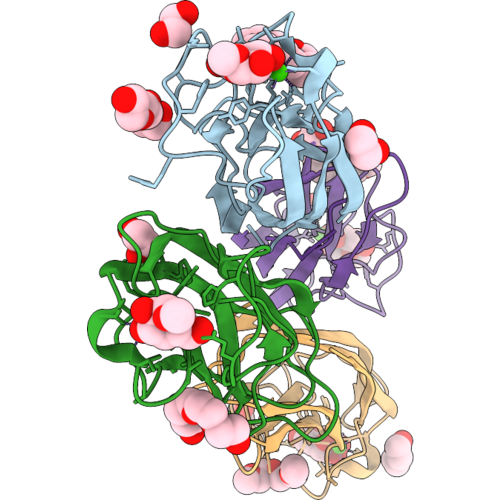
Crystal Structure Of Ncbbe14
Organism: Neurospora crassa
Method: X-RAY DIFFRACTION Resolution:2.80 Å Release Date: 2026-10-21 Classification: FLAVOPROTEIN Ligands: FAD, NAG, PO4, PG4, PGE |
Organism: Neurospora crassa
Method: X-RAY DIFFRACTION
Release Date: 2026-10-21
Ligands: FAD, NAG, PO4, PG4, PGE
 |
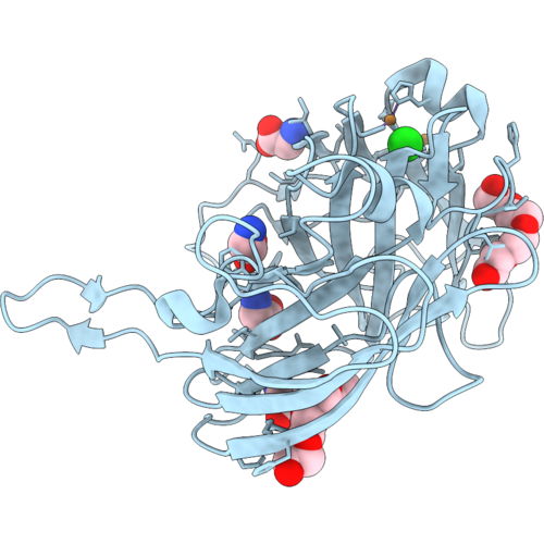
Crystal Structure Of Pseudomonas Aeruginosa Lectin 1 Complexed With Methyl Alpha-D-Galactopyranoside At 2 A Resolution
Organism: Pseudomonas aeruginosa pao1
Method: X-RAY DIFFRACTION Resolution:2.00 Å Release Date: 2026-10-14 Classification: SUGAR BINDING PROTEIN Ligands: AMG, P6G, EDO, PEG, CA, PG4, PG5 |
Organism: Pseudomonas aeruginosa pao1
Method: X-RAY DIFFRACTION
Release Date: 2026-10-14
Ligands: AMG, P6G, EDO, PEG, CA, PG4, PG5
 |
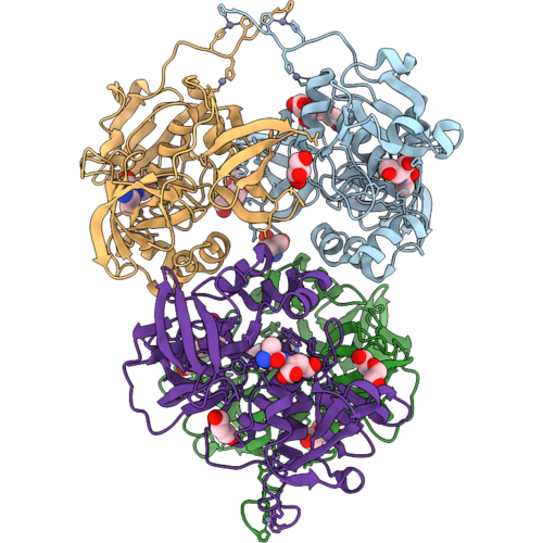
Crystal Structure Of A Tower-Slac, A Laccase From Streptomyces Coelicolor With A Tower Loop Mutation
Organism: Streptomyces coelicolor
Method: X-RAY DIFFRACTION Resolution:2.85 Å Release Date: 2026-10-14 Classification: OXIDOREDUCTASE Ligands: CU, OH, GLY, PG4, CL |
Organism: Streptomyces coelicolor
Method: X-RAY DIFFRACTION
Release Date: 2026-10-14
Ligands: CU, OH, GLY, PG4, CL
 |
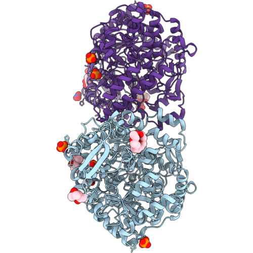
Crystal Structure Of The Nlpc/P60 Peptidase Ykfc From Bacillus Subtilis With Covalently Bound Dipeptide
Organism: Bacillus subtilis subsp. subtilis str. 168
Method: X-RAY DIFFRACTION Resolution:2.34 Å Release Date: 2026-10-14 Classification: HYDROLASE Ligands: ZN, PG4, TRS, PEG, ALA, DGL, GOL |
Organism: Bacillus subtilis subsp. subtilis str. 168
Method: X-RAY DIFFRACTION
Release Date: 2026-10-14
Ligands: ZN, PG4, TRS, PEG, ALA, DGL, GOL
 |
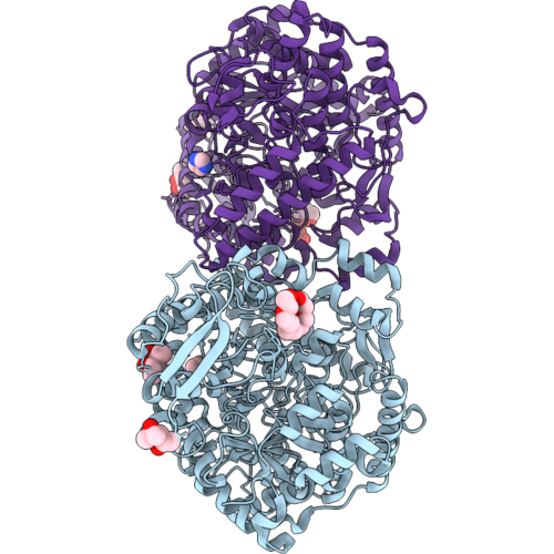
Crystal Structure Of A Formate Acetyltransferase (Pfl) From Megasphaera Lornae With An Acetylated Active Site Cysteine
Organism: Megasphaera lornae
Method: X-RAY DIFFRACTION Resolution:1.86 Å Release Date: 2026-10-07 Classification: TRANSFERASE Ligands: MPD, 1PE, PO4, CL, NO3, PG4 Modified Residue(s): SCY |
Organism: Megasphaera lornae
Method: X-RAY DIFFRACTION
Release Date: 2026-10-07
Ligands: MPD, 1PE, PO4, CL, NO3, PG4
 |
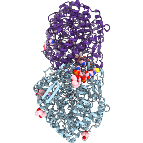
Crystal Structure Of A Formate Acetyltransferase (Pfl) From Megasphaera Lornae With An Acetylated Active Site Cysteine And Flipped Catalytic Loop
Organism: Megasphaera lornae
Method: X-RAY DIFFRACTION Resolution:1.93 Å Release Date: 2026-10-07 Classification: TRANSFERASE Ligands: MPD, IMD, 1PE, CL, PG4 Modified Residue(s): SCY |
Organism: Megasphaera lornae
Method: X-RAY DIFFRACTION
Release Date: 2026-10-07
Ligands: MPD, IMD, 1PE, CL, PG4
 |
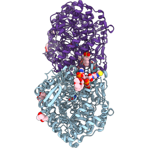
Crystal Structure Of A Formate Acetyltransferase (Pfl) From Megasphaera Lornae In Complex With Coa And Pyruvate
Organism: Megasphaera lornae
Method: X-RAY DIFFRACTION Resolution:1.75 Å Release Date: 2026-10-07 Classification: TRANSFERASE Ligands: MPD, IMD, COA, 1PE, CL, PYR, PG4 |
Organism: Megasphaera lornae
Method: X-RAY DIFFRACTION
Release Date: 2026-10-07
Ligands: MPD, IMD, COA, 1PE, CL, PYR, PG4
 |
Crystal Structure Of A Formate Acetyltransferase (Pfl) From Megasphaera Lornae In Complex With Coa And Oxamate
Organism: Megasphaera lornae
Method: X-RAY DIFFRACTION Resolution:1.99 Å Release Date: 2026-10-07 Classification: TRANSFERASE Ligands: MPO, OXM, MPD, COA, 1PE, CL, PG4 |
Organism: Megasphaera lornae
Method: X-RAY DIFFRACTION
Release Date: 2026-10-07
Ligands: MPO, OXM, MPD, COA, 1PE, CL, PG4
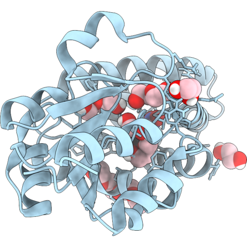 |
Crystal Structure Of Esnar1.3 (K39A) In Complex With 2,4-Dinitrobromobenzene
Organism: Synthetic construct
Method: X-RAY DIFFRACTION Resolution:2.10 Å Release Date: 2026-10-07 Classification: BIOSYNTHETIC PROTEIN Ligands: A1JRE, PGE, EDO, PEG, PG4 |
Organism: Synthetic construct
Method: X-RAY DIFFRACTION
Release Date: 2026-10-07
Ligands: A1JRE, PGE, EDO, PEG, PG4
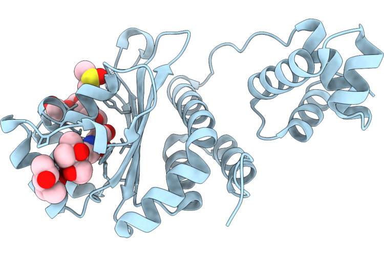 |
Crystal Structure Of Mrtr Bound To 3O-C8 Homoserine Lactone And Tetraethylene Glycol
Organism: Mesorhizobium tianshanense
Method: X-RAY DIFFRACTION Resolution:1.22 Å Release Date: 2026-09-23 Classification: TRANSCRIPTION Ligands: MPD, MRD, LAE, DMS, PG4 |
Organism: Mesorhizobium tianshanense
Method: X-RAY DIFFRACTION
Release Date: 2026-09-23
Ligands: MPD, MRD, LAE, DMS, PG4
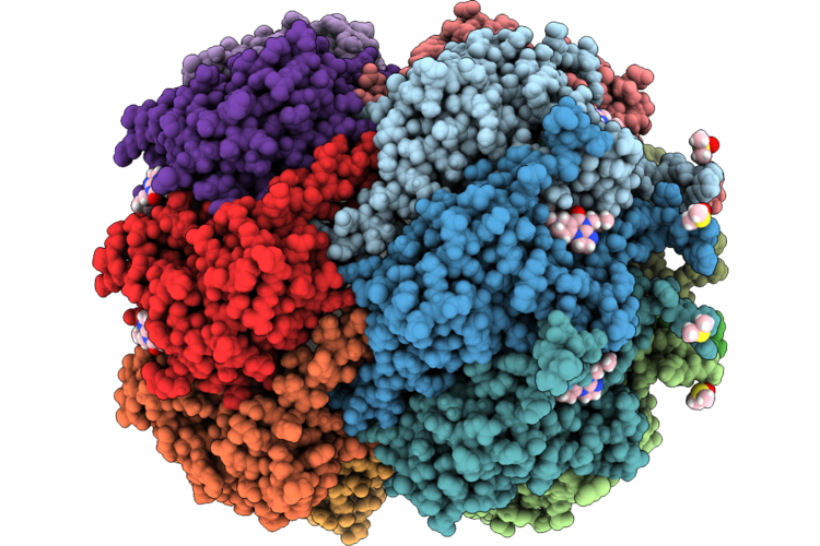 |
Crystal Structure Of M. Tuberculosis Clpp1P2 Bound To Onc201
Organism: Mycobacterium tuberculosis h37rv
Method: X-RAY DIFFRACTION Resolution:3.11 Å Release Date: 2026-09-23 Classification: ANTIBIOTIC Ligands: BEZ, LEU, A1C98, DMS, AE3, PEG, PG4, PGE, PG0 |
Organism: Mycobacterium tuberculosis h37rv
Method: X-RAY DIFFRACTION
Release Date: 2026-09-23
Ligands: BEZ, LEU, A1C98, DMS, AE3, PEG, PG4, PGE, PG0
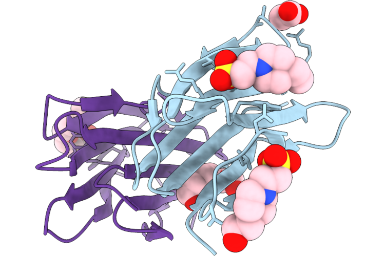 |
Crystal Structure Of Honey Truffle Active Component 1 Through 4 With Hepes Bound (Orthorhombic P Form)
Organism: Mattirolomyces terfezioides
Method: X-RAY DIFFRACTION Resolution:1.25 Å Release Date: 2026-09-23 Classification: PROTEIN BINDING Ligands: EPE, ACT, PG4 |
Organism: Mattirolomyces terfezioides
Method: X-RAY DIFFRACTION
Release Date: 2026-09-23
Ligands: EPE, ACT, PG4