
Search Count: 32,633
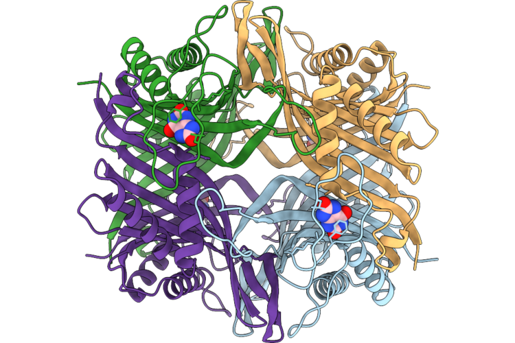 |
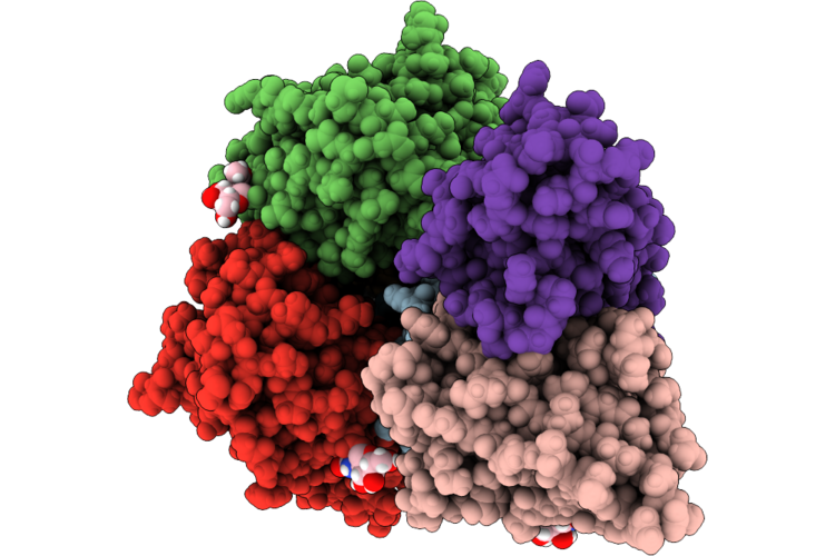
Structure Of Mouse Urate Oxidase In Complex With Uric Acid
Organism: Mus musculus
Method: ELECTRON MICROSCOPY Resolution:2.42 Å Release Date: 2026-07-08 Classification: OXIDOREDUCTASE Ligands: URC |
 |
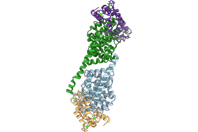
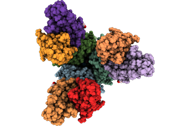
Structure Of Mouse Urate Oxidase In The Apo State
Organism: Mus musculus
Method: ELECTRON MICROSCOPY Resolution:3.00 Å Release Date: 2026-07-08 Classification: OXIDOREDUCTASE |
 |
Structure Of The Porcine Reproductive And Respiratory Syndrome Virus-2 Glycoprotein Gp4 Antigenic Region 59-69 Bound To The Neutralizing Scfv#18
Organism: Mus musculus, Prrsv vr2332
Method: X-RAY DIFFRACTION Resolution:2.30 Å Release Date: 2026-07-08 Classification: IMMUNE SYSTEM Ligands: ACT, NI |
 |
Structure Of The Human Astrovirus Va1 Capsid Spike Bound To Antibody 7C8
Organism: Astrovirus va1, Mus musculus
Method: ELECTRON MICROSCOPY Release Date: 2026-07-08 Classification: VIRAL PROTEIN |
 |
Structure Of The Human Astrovirus Va1 Capsid Spike Bound To Antibody 2A2
Organism: Astrovirus va1, Mus musculus
Method: ELECTRON MICROSCOPY Release Date: 2026-07-08 Classification: VIRAL PROTEIN |
 |
Cryo-Et Structure Of Full-Length Membrane-Bound Ehd2 Complex
Organism: Mus musculus
Method: ELECTRON MICROSCOPY Release Date: 2026-07-08 Classification: STRUCTURAL PROTEIN Ligands: ATP, MG |
 |
Cryo-Et Structure Of N-Terminally Truncated Membrane-Bound Ehd2 Complex
Organism: Mus musculus
Method: ELECTRON MICROSCOPY Release Date: 2026-07-08 Classification: STRUCTURAL PROTEIN Ligands: ATP, MG |
 |
Pfripr Egf6 And 7 Bound To Monoclonal Antibody Rp.012
Organism: Homo sapiens, Plasmodium falciparum 3d7, Mus musculus
Method: X-RAY DIFFRACTION Resolution:2.20 Å Release Date: 2026-07-08 Classification: CELL ADHESION |
 |
Crystal Structure Of Inf2 Did
Organism: Mus musculus
Method: X-RAY DIFFRACTION Resolution:2.39 Å Release Date: 2026-07-08 Classification: PROTEIN BINDING |
 |
Complex Structure Of Inf2 Did And Calcium Bound Cam
Organism: Mus musculus, Rattus norvegicus
Method: X-RAY DIFFRACTION Resolution:2.11 Å Release Date: 2026-07-08 Classification: PROTEIN BINDING Ligands: CA |
 |
Complex Structure Of Inf2 Did R91G And Calcium Bound Cam
Organism: Mus musculus, Rattus norvegicus
Method: X-RAY DIFFRACTION Resolution:2.11 Å Release Date: 2026-07-08 Classification: PROTEIN BINDING Ligands: CA |
 |
Fcgriia In Complex With Iv.3 Fab
Organism: Homo sapiens, Mus musculus
Method: ELECTRON MICROSCOPY Release Date: 2026-07-01 Classification: IMMUNE SYSTEM |
 |
Structure Of Mc5Ar2 In Complex With Mc5A-Desarg (Monomer)
Organism: Mus musculus
Method: ELECTRON MICROSCOPY Resolution:3.42 Å Release Date: 2026-07-01 Classification: SIGNALING PROTEIN |
 |
Topbp1 Brct0-2 In Complex With Phosphorylated Htatsf1
Organism: Homo sapiens, Mus musculus
Method: X-RAY DIFFRACTION Resolution:2.99 Å Release Date: 2026-07-01 Classification: PEPTIDE BINDING PROTEIN |
 |
Cryoem Structure Of A Catalytically Inactive Cxc Chemokine-Degrading Protease Spycep From Streptococcus Pyogenes Complexed With An Anti-Pa-Domain Monoclonal Antibody
Organism: Streptococcus pyogenes, Mus musculus
Method: ELECTRON MICROSCOPY Release Date: 2026-07-01 Classification: PROTEIN BINDING |
 |
Kl-H15-3G11 Fab In Complex With Soluble A/Wedge-Tailed Shearwater/Western Australia/2576/1979 H15 Hemagglutinin Trimer
Organism: Influenza a virus (a/shearwater/west australia/2576/79(h15n9)), Mus musculus
Method: ELECTRON MICROSCOPY Release Date: 2026-07-01 Classification: VIRAL PROTEIN/Immune System Ligands: NAG |
 |
Kl-H15-6H4 Fab In Complex With Soluble A/Wedge-Tailed Shearwater/Western Australia/2576/1979 H15 Hemagglutinin Trimer
Organism: Influenza a virus (a/shearwater/west australia/2576/79(h15n9)), Mus musculus
Method: ELECTRON MICROSCOPY Release Date: 2026-07-01 Classification: VIRAL PROTEIN/Immune System Ligands: NAG |
 |
Kl-H15-6G8 Fab In Complex With Soluble A/Wedge-Tailed Shearwater/Western Australia/2576/1979 H15 Hemagglutinin Trimer
Organism: Influenza a virus (a/shearwater/west australia/2576/79(h15n9)), Mus musculus
Method: ELECTRON MICROSCOPY Release Date: 2026-07-01 Classification: VIRAL PROTEIN/Immune System Ligands: NAG |
 |
07-5G01 Fab In Complex With Soluble A/Wedge-Tailed Shearwater/Western Australia/2576/1979 H15 Hemagglutinin Trimer
Organism: Influenza a virus (a/shearwater/west australia/2576/79(h15n9)), Mus musculus
Method: ELECTRON MICROSCOPY Release Date: 2026-07-01 Classification: VIRAL PROTEIN/Immune System Ligands: NAG |
 |
Pcp Bound Kappa-Opioid Receptor In Complex With Gi1
Organism: Escherichia coli, Homo sapiens, Mus musculus
Method: ELECTRON MICROSCOPY Resolution:2.63 Å Release Date: 2026-07-01 Classification: SIGNALING PROTEIN Ligands: 1PC |