
Search Count: 1,461
All
Selected
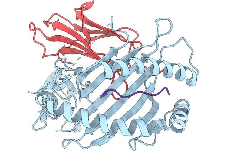 |
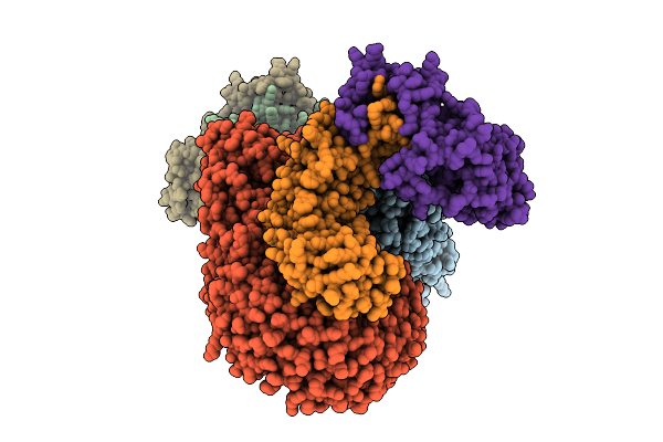
Conformational Flexibility In Hla-B8: Peptide Tuning Structural And Dynamical Changes
Organism: Homo sapiens, Human immunodeficiency virus 1
Method: X-RAY DIFFRACTION Resolution:1.43 Å Release Date: 2026-04-22 Classification: IMMUNE SYSTEM |
 |
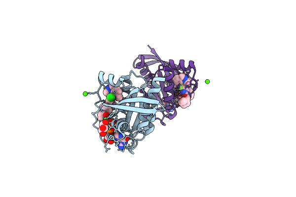
Organism: Homo sapiens
Method: X-RAY DIFFRACTION Resolution:2.28 Å Release Date: 2026-04-22 Classification: TRANSFERASE Ligands: A1ES7 |
 |
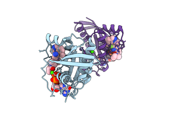
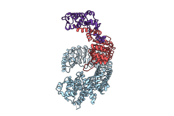
Organism: Homo sapiens
Method: X-RAY DIFFRACTION Resolution:1.50 Å Release Date: 2026-04-15 Classification: ONCOPROTEIN Ligands: CA, A1C6Y, GOL, GDP |
 |
Organism: Homo sapiens
Method: X-RAY DIFFRACTION Resolution:1.52 Å Release Date: 2026-04-15 Classification: ONCOPROTEIN Ligands: GDP, A1C60, CA |
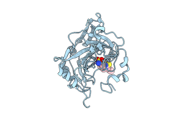 |
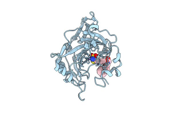
Crystal Structure Of Human Carbonic Anhydrase Vii In Complex With A Benzenesulfonamide Derivative Containing The Duloxetine Moiety.
Organism: Homo sapiens
Method: X-RAY DIFFRACTION Resolution:1.35 Å Release Date: 2026-04-15 Classification: LYASE Ligands: ZN, A1I9B |
 |
Crystal Structure Of Human Carbonic Anhydrase Vii In Complex With A Benzenesufonamide Derivative Containing The Duloxetine Moiety
Organism: Homo sapiens
Method: X-RAY DIFFRACTION Resolution:1.60 Å Release Date: 2026-04-15 Classification: LYASE Ligands: ZN, A1I9C |
 |
Organism: Escherichia phage t4
Method: ELECTRON MICROSCOPY Release Date: 2026-04-15 Classification: VIRAL PROTEIN |
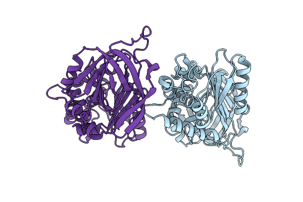 |
Crystal Structure Of The Recombinant A1-Antitrypsin F51L/M351V/M358V Triple Mutant
Organism: Homo sapiens
Method: X-RAY DIFFRACTION Resolution:2.60 Å Release Date: 2026-04-15 Classification: RECOMBINATION |
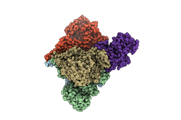 |
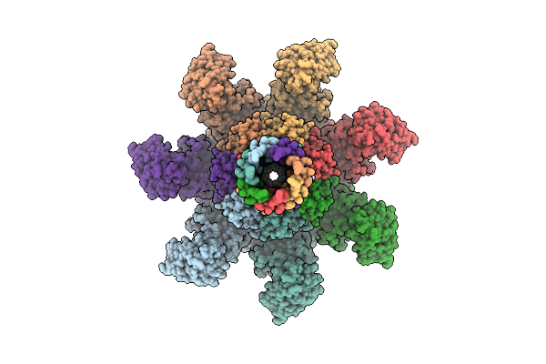
Structural Basis For The Assembly And Translocation Of The Vip1-Vip2 Insecticidal Binary Toxin From Bacillus Thuringiensis
Organism: Bacillus thuringiensis
Method: ELECTRON MICROSCOPY Resolution:3.05 Å Release Date: 2026-04-15 Classification: TOXIN Ligands: CA |
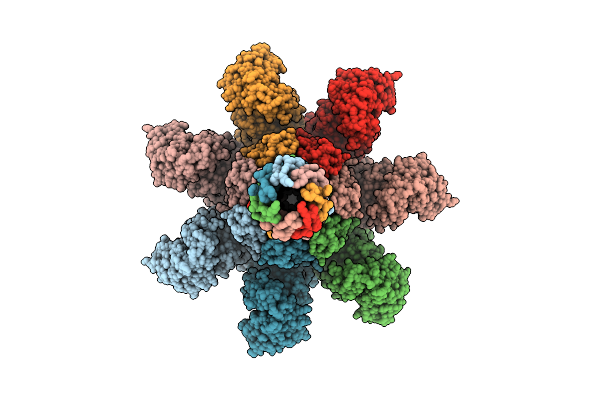 |
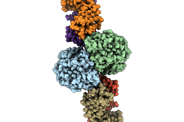
Structural Basis For The Assembly And Translocation Of The Vip1-Vip2 Insecticidal Binary Toxin From Bacillus Thuringiensis
Organism: Bacillus thuringiensis
Method: ELECTRON MICROSCOPY Resolution:2.40 Å Release Date: 2026-04-15 Classification: TOXIN Ligands: CA |
 |
Structural Basis For The Assembly And Translocation Of The Vip1-Vip2 Insecticidal Binary Toxin From Bacillus Thuringiensis
Organism: Bacillus thuringiensis
Method: ELECTRON MICROSCOPY Resolution:3.31 Å Release Date: 2026-04-15 Classification: TOXIN Ligands: CA |
 |
Organism: Mus musculus
Method: ELECTRON MICROSCOPY Resolution:3.50 Å Release Date: 2026-03-25 Classification: MEMBRANE PROTEIN |
 |
Organism: Mus musculus
Method: ELECTRON MICROSCOPY Resolution:3.20 Å Release Date: 2026-03-25 Classification: MEMBRANE PROTEIN |
 |
Organism: Mus musculus
Method: ELECTRON MICROSCOPY Release Date: 2026-03-25 Classification: MEMBRANE PROTEIN |
 |
Organism: Mus musculus
Method: ELECTRON MICROSCOPY Release Date: 2026-03-25 Classification: MEMBRANE PROTEIN |
 |
Organism: Escherichia coli k-12, Homo sapiens
Method: ELECTRON MICROSCOPY Resolution:3.49 Å Release Date: 2026-03-18 Classification: CYTOSOLIC PROTEIN |
 |
Organism: Mus musculus
Method: ELECTRON MICROSCOPY Release Date: 2026-03-18 Classification: CYTOSOLIC PROTEIN |
 |
Organism: Mus musculus
Method: ELECTRON MICROSCOPY Release Date: 2026-03-18 Classification: CYTOSOLIC PROTEIN |
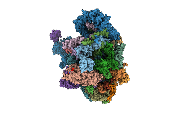 |
Assembly Intermediate Of Human Mitochondrial Ribosome Small Subunit Bound To Mettl15, Rbfa, And Mtif2 (State M2.1)
Organism: Homo sapiens
Method: ELECTRON MICROSCOPY Release Date: 2026-03-11 Classification: RIBOSOME Ligands: MG, ZN, FES, ATP, GDP, NAD, SPM |
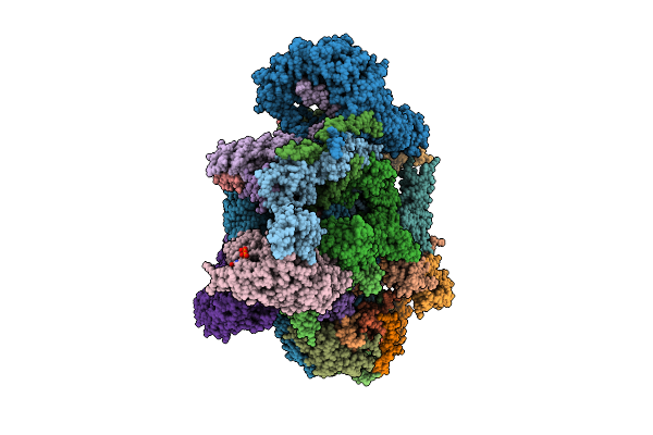 |
Assembly Intermediate Of Human Mitochondrial Ribosome Small Subunit Bound To Mettl15 And Rbfa (Inward Conformation) (State M2)
Organism: Homo sapiens
Method: ELECTRON MICROSCOPY Release Date: 2026-03-11 Classification: RIBOSOME Ligands: MG, FES, ZN, ATP, GDP, NAD, SPM |