
Search Count: 23,639
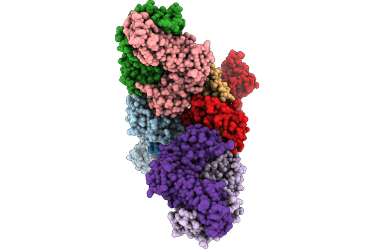 |
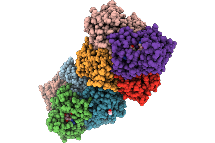
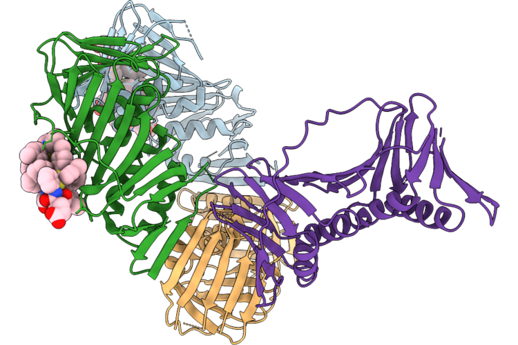
P1-15 T-Cell Receptor Bound To Hla A*2402-Nf9 Pmhc Complex
Organism: Homo sapiens, Severe acute respiratory syndrome coronavirus 2
Method: X-RAY DIFFRACTION Resolution:3.10 Å Release Date: 2026-07-15 Classification: IMMUNE SYSTEM |
Organism: Homo sapiens, Severe acute respiratory syndrome coronavirus 2
Method: X-RAY DIFFRACTION
Release Date: 2026-07-15
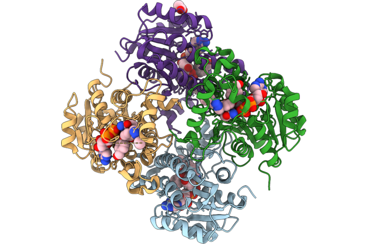 |
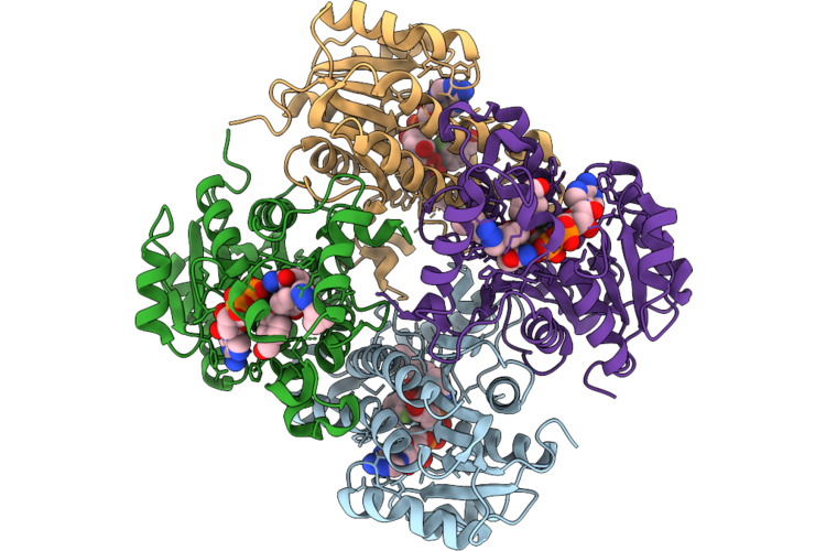
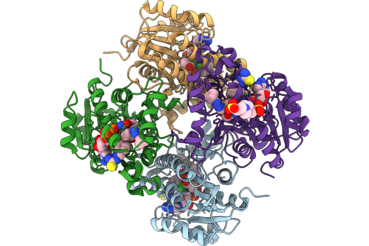
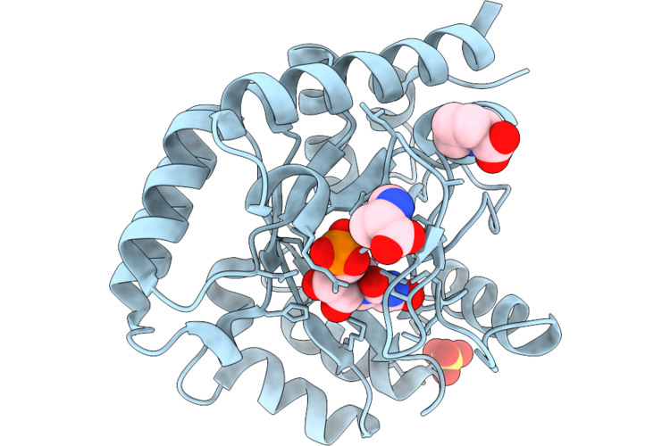
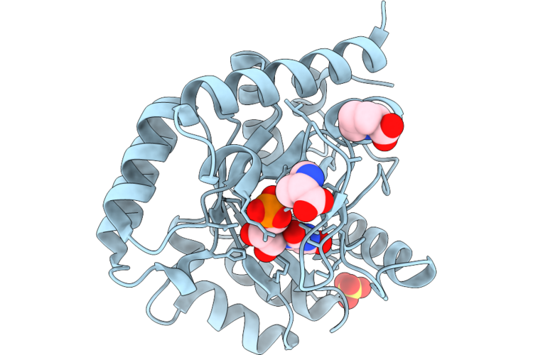
Structure Of Inha From Mycobacterium Tuberculosis In Complex With 4-(4-((4-Cyclopropyl-1H-1,2,3-Triazol-1-Yl)Methyl)-2-Hydroxyphenoxy)Benzaldehyde (Compound 5B)
Organism: Mycobacterium tuberculosis
Method: X-RAY DIFFRACTION Resolution:1.72 Å Release Date: 2026-07-15 Classification: OXIDOREDUCTASE Ligands: NAD, A1JY9, ACT |
Organism: Mycobacterium tuberculosis
Method: X-RAY DIFFRACTION
Release Date: 2026-07-15
Ligands: NAD, A1JY9, ACT
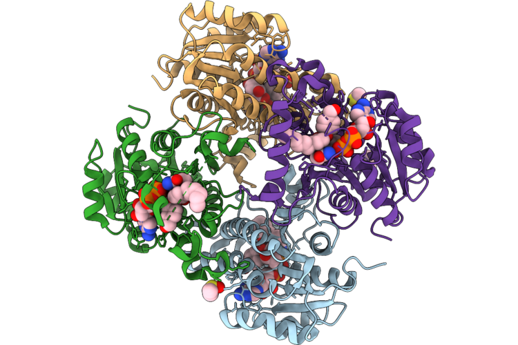 |
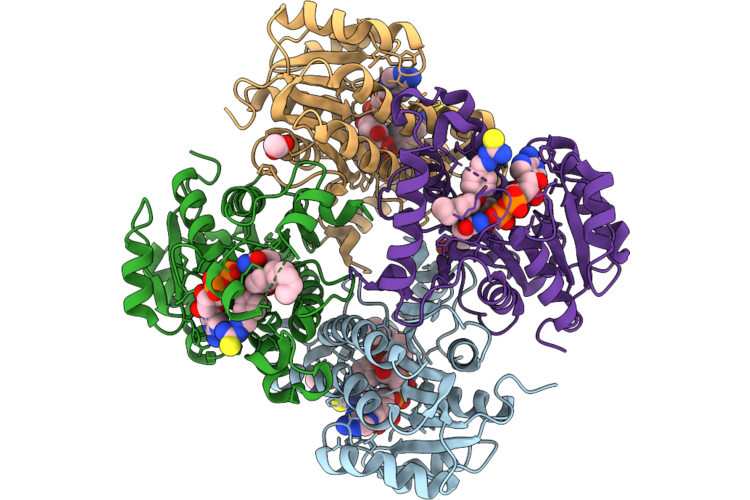
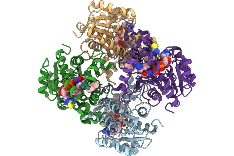
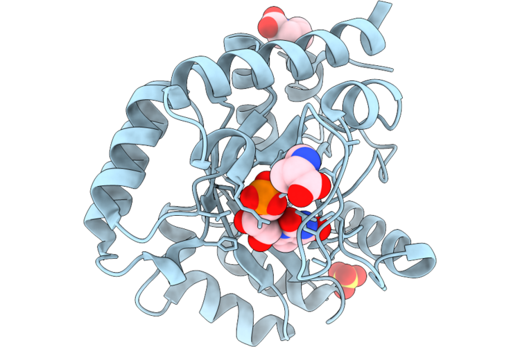
Structure Of Inha From Mycobacterium Tuberculosis In Complex With 4-(4-Hexyl-2-Hydroxyphenoxy)Benzaldehyde (Compound 5A) - Spacegroup C2
Organism: Mycobacterium tuberculosis
Method: X-RAY DIFFRACTION Resolution:1.59 Å Release Date: 2026-07-15 Classification: OXIDOREDUCTASE Ligands: NAD, A1JY8, DMS, GOL |
Organism: Mycobacterium tuberculosis
Method: X-RAY DIFFRACTION
Release Date: 2026-07-15
Ligands: NAD, A1JY8, DMS, GOL
 |
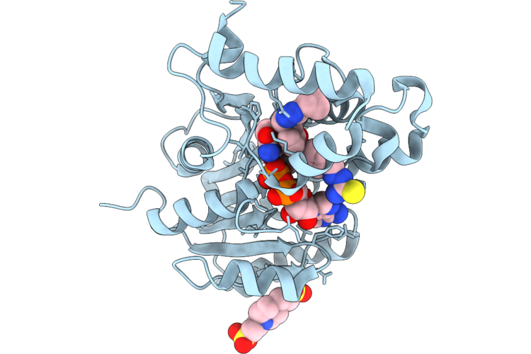
Structure Of Inha From Mycobacterium Tuberculosis In Complex With 4-(4-Hexyl-2-Hydroxyphenoxy)Benzaldehyde (Compound 5A) - Spacegroup P41212
Organism: Mycobacterium tuberculosis
Method: X-RAY DIFFRACTION Resolution:1.51 Å Release Date: 2026-07-15 Classification: OXIDOREDUCTASE Ligands: NAD, DMS, A1JY8 |
Organism: Mycobacterium tuberculosis
Method: X-RAY DIFFRACTION
Release Date: 2026-07-15
Ligands: NAD, DMS, A1JY8
 |
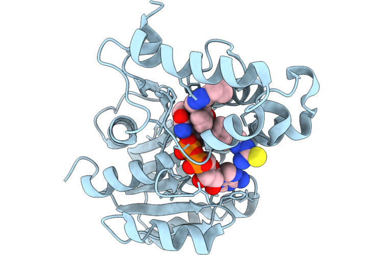
Structure Of Inha From Mycobacterium Tuberculosis In Complex With 4-(4-((4-Cyclopropyl-1H-1,2,3-Triazol-1-Yl)Methyl)-2-Hydroxyphenoxy)-3-Fluorobenzaldehyde (Compound 5C)
Organism: Mycobacterium tuberculosis
Method: X-RAY DIFFRACTION Resolution:1.69 Å Release Date: 2026-07-15 Classification: OXIDOREDUCTASE Ligands: NAD, A1JZB |
Organism: Mycobacterium tuberculosis
Method: X-RAY DIFFRACTION
Release Date: 2026-07-15
Ligands: NAD, A1JZB
 |
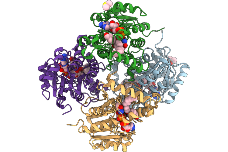
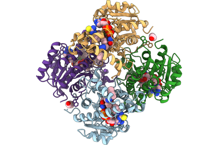
Structure Of Inha From Mycobacterium Tuberculosis In Complex With (E)-2-(4-(4-Hexyl-2-Hydroxyphenoxy)Benzylidene)Hydrazine-1-Carbothioamide (Compound 6A)
Organism: Mycobacterium tuberculosis
Method: X-RAY DIFFRACTION Resolution:2.12 Å Release Date: 2026-07-15 Classification: OXIDOREDUCTASE Ligands: NAD, A1JY7, DMS, ACT |
Organism: Mycobacterium tuberculosis
Method: X-RAY DIFFRACTION
Release Date: 2026-07-15
Ligands: NAD, A1JY7, DMS, ACT
 |
Structure Of Inha From Mycobacterium Tuberculosis In Complex With (E)-2-(4-(4-((4-Cyclopropyl-1H-1,2,3-Triazol-1-Yl)Methyl)-2-Hydroxyphenoxy)Benzylidene)Hydrazine-1-Carbothioamide (Compound 6B)
Organism: Mycobacterium tuberculosis
Method: X-RAY DIFFRACTION Resolution:1.51 Å Release Date: 2026-07-15 Classification: OXIDOREDUCTASE Ligands: NAD, A1JZA, PIN |
Organism: Mycobacterium tuberculosis
Method: X-RAY DIFFRACTION
Release Date: 2026-07-15
Ligands: NAD, A1JZA, PIN
 |
Structure Of Inha From Mycobacterium Tuberculosis In Complex With (E)-2-(4-(4-((4-Cyclopropyl-1H-1,2,3-Triazol-1-Yl)Methyl)-2-Hydroxyphenoxy)-3-Fluorobenzylidene)Hydrazine-1-Carbothioamide (Compound 6C)
Organism: Mycobacterium tuberculosis
Method: X-RAY DIFFRACTION Resolution:2.55 Å Release Date: 2026-07-15 Classification: OXIDOREDUCTASE Ligands: NAD, A1JZC |
Organism: Mycobacterium tuberculosis
Method: X-RAY DIFFRACTION
Release Date: 2026-07-15
Ligands: NAD, A1JZC
 |
Structure Of Inha From Mycobacterium Tuberculosis In Complex With (E)-2-(4-(4-((4-Cyclopropyl-1H-1,2,3-Triazol-1-Yl)Methyl)-2-Hydroxyphenoxy)-3-Chlorobenzylidene)Hydrazine-1-Carbothioamide (Compound 6D)
Organism: Mycobacterium tuberculosis
Method: X-RAY DIFFRACTION Resolution:1.96 Å Release Date: 2026-07-15 Classification: OXIDOREDUCTASE Ligands: NAD, A1JY6, PIN |
Organism: Mycobacterium tuberculosis
Method: X-RAY DIFFRACTION
Release Date: 2026-07-15
Ligands: NAD, A1JY6, PIN
 |
Structure Of Inha From Mycobacterium Tuberculosis In Complex With (E)-2-(4-(4-((4-Butyl-1H-1,2,3-Triazol-1-Yl)Methyl)-2-Hydroxyphenoxy)Benzylidene)Hydrazine-1-Carbothioamide (Compound 6E)
Organism: Mycobacterium tuberculosis
Method: X-RAY DIFFRACTION Resolution:1.96 Å Release Date: 2026-07-15 Classification: OXIDOREDUCTASE Ligands: NAD, A1JY4, CL, NA |
Organism: Mycobacterium tuberculosis
Method: X-RAY DIFFRACTION
Release Date: 2026-07-15
Ligands: NAD, A1JY4, CL, NA
 |
Structure Of Inha From Mycobacterium Tuberculosis In Complex With (E)-2-(4-(4-((4-Butyl-1H-1,2,3-Triazol-1-Yl)Methyl)-2-Hydroxyphenoxy)-3-Fluorobenzylidene)Hydrazine-1-Carbothioamide (Compound 6F)
Organism: Mycobacterium tuberculosis
Method: X-RAY DIFFRACTION Resolution:1.87 Å Release Date: 2026-07-15 Classification: OXIDOREDUCTASE Ligands: NAD, A1JY5, PEG, NA |
Organism: Mycobacterium tuberculosis
Method: X-RAY DIFFRACTION
Release Date: 2026-07-15
Ligands: NAD, A1JY5, PEG, NA
 |
Structure Of Inha From Mycobacterium Tuberculosis In Complex With 4-(4-((4-Butyl-1H-1,2,3-Triazol-1-Yl)Methyl)-2-Hydroxyphenoxy)Benzaldehyde (Compound 5E)
Organism: Mycobacterium tuberculosis
Method: X-RAY DIFFRACTION Resolution:2.10 Å Release Date: 2026-07-15 Classification: OXIDOREDUCTASE Ligands: NAD, A1JZT, NA |
Organism: Mycobacterium tuberculosis
Method: X-RAY DIFFRACTION
Release Date: 2026-07-15
Ligands: NAD, A1JZT, NA
 |
Structure Of Inha From Mycobacterium Tuberculosis In Complex With 4-(4-((4-Butyl-1H-1,2,3-Triazol-1-Yl)Methyl)-2-Hydroxyphenoxy)-3-Fluorobenzaldehyde (Compound 5F)
Organism: Mycobacterium tuberculosis
Method: X-RAY DIFFRACTION Resolution:1.96 Å Release Date: 2026-07-15 Classification: OXIDOREDUCTASE Ligands: NAD, A1JZS, ACT |
Organism: Mycobacterium tuberculosis
Method: X-RAY DIFFRACTION
Release Date: 2026-07-15
Ligands: NAD, A1JZS, ACT
 |
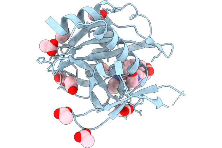
Orotidine 5'-Monophosphate Decarboxylase-Domain Of Human Umps In Complex With 6-Ethyl Ump
Organism: Homo sapiens
Method: X-RAY DIFFRACTION Resolution:1.30 Å Release Date: 2026-07-15 Classification: LYASE Ligands: UEP, SO4, PRO |
Organism: Homo sapiens
Method: X-RAY DIFFRACTION
Release Date: 2026-07-15
Ligands: UEP, SO4, PRO
 |
Orotidine 5'-Monophosphate Decarboxylase-Domain Of Human Umps In Complex With 6-Isopropyl-Ump
Organism: Homo sapiens
Method: X-RAY DIFFRACTION Resolution:1.30 Å Release Date: 2026-07-15 Classification: LYASE Ligands: A1IXW, SO4, PRO |
Organism: Homo sapiens
Method: X-RAY DIFFRACTION
Release Date: 2026-07-15
Ligands: A1IXW, SO4, PRO
 |
Orotidine 5'-Monophosphate Decarboxylase-Domain Of Human Umps In Complex With 6-Isopropenyl Ump
Organism: Homo sapiens
Method: X-RAY DIFFRACTION Resolution:1.15 Å Release Date: 2026-07-15 Classification: LYASE Ligands: A1IXU, SO4, PRO |
Organism: Homo sapiens
Method: X-RAY DIFFRACTION
Release Date: 2026-07-15
Ligands: A1IXU, SO4, PRO
 |
Orotidine 5'-Monophosphate Decarboxylase-Domain Of Human Umps In Complex With 6-Methyl Ump
Organism: Homo sapiens
Method: X-RAY DIFFRACTION Resolution:1.12 Å Release Date: 2026-07-15 Classification: LYASE Ligands: U6M, PRO, SO4 |
Organism: Homo sapiens
Method: X-RAY DIFFRACTION
Release Date: 2026-07-15
Ligands: U6M, PRO, SO4
 |
Co-Crystal Structure Of Capcna Bound To The Aoh1996 Derivative, Aoh2.29-Le
Organism: Homo sapiens
Method: X-RAY DIFFRACTION Resolution:2.87 Å Release Date: 2026-07-15 Classification: DNA BINDING PROTEIN Ligands: XEU, CL |
Organism: Homo sapiens
Method: X-RAY DIFFRACTION
Release Date: 2026-07-15
Ligands: XEU, CL
 |
Crystal Structure Of Nudt7 In Complex With A Novel Inhibitor
Organism: Homo sapiens
Method: X-RAY DIFFRACTION Resolution:1.47 Å Release Date: 2026-07-15 Classification: HYDROLASE Ligands: A1JJG, ACT |
Organism: Homo sapiens
Method: X-RAY DIFFRACTION
Release Date: 2026-07-15
Ligands: A1JJG, ACT
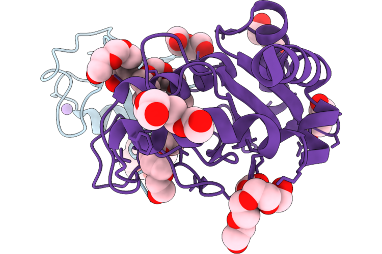 |
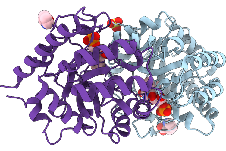
Crystal Structure Of Ube2J2 In Complex With Ubiquitin
Organism: Homo sapiens
Method: X-RAY DIFFRACTION Resolution:2.32 Å Release Date: 2026-07-15 Classification: SIGNALING PROTEIN |
Organism: Homo sapiens
Method: X-RAY DIFFRACTION
Release Date: 2026-07-15