

Search Count: 39
 |
X-Ray Structure Of Homo Sapiens Fumarylacetoacetate Hydrolase Domain Containing Protein 1 (Fahd1) At 1.56A Resolution.
Organism: Homo sapiens
Method: X-RAY DIFFRACTION Resolution:1.56 Å Release Date: 2018-11-07 Classification: HYDROLASE/LYASE Ligands: CL, MG |
Organism: Homo sapiens
Method: X-RAY DIFFRACTION
Release Date: 2018-11-07
Ligands: CL, MG
 |
X-Ray Structure Of Homo Sapiens Fumarylacetoacetate Hydrolase Domain Containing Protein 1 (Fahd1) In Complex With Inhibitor Oxalate At 1.94A Resolution.
Organism: Homo sapiens
Method: X-RAY DIFFRACTION Resolution:1.94 Å Release Date: 2018-11-07 Classification: hydrolase/lyase Ligands: MG, OXL, CL |
Organism: Homo sapiens
Method: X-RAY DIFFRACTION
Release Date: 2018-11-07
Ligands: MG, OXL, CL
 |
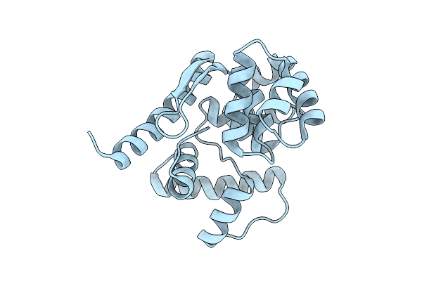
Mvnei1-G86D
Organism: Acanthamoeba polyphaga mimivirus
Method: X-RAY DIFFRACTION Resolution:2.85 Å Release Date: 2014-01-01 Classification: HYDROLASE, LYASE/DNA |
Organism: Acanthamoeba polyphaga mimivirus
Method: X-RAY DIFFRACTION
Release Date: 2014-01-01
 |
Crystal Structure Of Afogg1 In A Dna-Free State
Organism: Archaeoglobus fulgidus
Method: X-RAY DIFFRACTION Resolution:1.93 Å Release Date: 2026-04-15 Classification: HYDROLASE, LYASE Modified Residue(s): MSE |
Organism: Archaeoglobus fulgidus
Method: X-RAY DIFFRACTION
Release Date: 2026-04-15
 |
Catalytic Function And Substrate Recognition Of The Pectate Lyase From Thermotoga Maritima
Organism: Thermotoga maritima
Method: X-RAY DIFFRACTION Resolution:1.94 Å Release Date: 2012-07-11 Classification: LYASE Ligands: GOL, PO4 |
Organism: Thermotoga maritima
Method: X-RAY DIFFRACTION
Release Date: 2012-07-11
Ligands: GOL, PO4
 |
Crystal Structure Of Human Ogg1 In A Dna-Free State
Organism: Homo sapiens
Method: X-RAY DIFFRACTION Resolution:1.85 Å Release Date: 2026-04-15 Classification: HYDROLASE, LYASE/DNA Ligands: SO4 |
Organism: Homo sapiens
Method: X-RAY DIFFRACTION
Release Date: 2026-04-15
Ligands: SO4
 |
Crystal Structure Of S-Hydroxynitrile Lyase From Manihot Esculenta (His103Leu)
Organism: Manihot esculenta
Method: X-RAY DIFFRACTION Resolution:2.60 Å Release Date: 2016-04-13 Classification: LYASE |
Organism: Manihot esculenta
Method: X-RAY DIFFRACTION
Release Date: 2016-04-13
 |
Crystal Structure Of Human Ogg1 (Wt) In A Product Bound State In The Presence Of The Agonist F51
Organism: Homo sapiens, Synthetic construct
Method: X-RAY DIFFRACTION Resolution:2.10 Å Release Date: 2026-07-15 Classification: HYDROLASE,LYASE/DNA Ligands: A1C6R, A1C6S |
Organism: Homo sapiens, Synthetic construct
Method: X-RAY DIFFRACTION
Release Date: 2026-07-15
Ligands: A1C6R, A1C6S
 |
Crystal Structure Of Product-Bound Human Ogg1(Wt)
Organism: Homo sapiens, Synthetic construct
Method: X-RAY DIFFRACTION Resolution:2.00 Å Release Date: 2026-04-15 Classification: HYDROLASE, LYASE/DNA Ligands: EDO |
Organism: Homo sapiens, Synthetic construct
Method: X-RAY DIFFRACTION
Release Date: 2026-04-15
Ligands: EDO
 |
Crystal Structure Of Human Ogg1 (Wt) In A Product Bound State In The Presence Of The Agonist F01
Organism: Homo sapiens, Synthetic construct
Method: X-RAY DIFFRACTION Resolution:2.50 Å Release Date: 2026-04-15 Classification: HYDROLASE, LYASE/DNA Ligands: OXG |
Organism: Homo sapiens, Synthetic construct
Method: X-RAY DIFFRACTION
Release Date: 2026-04-15
Ligands: OXG
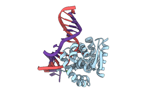 |
Crystal Structure Of Afogg1-D140N Mutant Bound To 8-Og Dna Duplex In An Intermediate State
Organism: Archaeoglobus fulgidus, Synthetic construct
Method: X-RAY DIFFRACTION Resolution:1.95 Å Release Date: 2026-04-15 Classification: HYDROLASE, LYASE/DNA Ligands: MG |
Organism: Archaeoglobus fulgidus, Synthetic construct
Method: X-RAY DIFFRACTION
Release Date: 2026-04-15
Ligands: MG
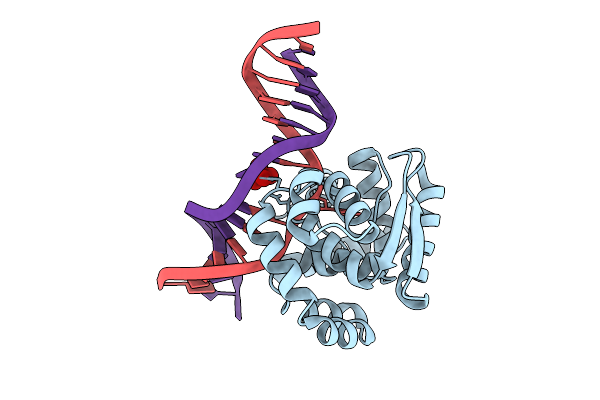 |
Crystal Structure Of Afogg1-K122S Mutant Bound To 8-Og Dna Duplex In An Intermediate State
Organism: Archaeoglobus fulgidus, Synthetic construct
Method: X-RAY DIFFRACTION Resolution:2.50 Å Release Date: 2026-04-15 Classification: HYDROLASE, LYASE/DNA Ligands: MOO |
Organism: Archaeoglobus fulgidus, Synthetic construct
Method: X-RAY DIFFRACTION
Release Date: 2026-04-15
Ligands: MOO
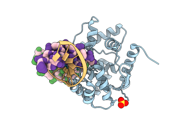 |
Crystal Structure Of Afogg1:8Og-Dna Duplex In A Product-Bound State
Organism: Archaeoglobus fulgidus, Synthetic construct
Method: X-RAY DIFFRACTION Resolution:2.00 Å Release Date: 2026-04-15 Classification: HYDROLASE, LYASE/DNA Ligands: SO4 Modified Residue(s): MSE |
Organism: Archaeoglobus fulgidus, Synthetic construct
Method: X-RAY DIFFRACTION
Release Date: 2026-04-15
Ligands: SO4
 |
Wild Type Poly(Ethylene Terephthalate) Hydrolase
Organism: Ideonella sakaiensis (strain nbrc 110686 / tistr 2288 / 201-f6)
Method: X-RAY DIFFRACTION Resolution:2.02 Å Release Date: 2022-11-30 Classification: HYDROLASE |
 |
Poly(Ethylene Terephthalate) Hydrolase (Ispetase)-Linker
Organism: Ideonella sakaiensis (strain nbrc 110686 / tistr 2288 / 201-f6)
Method: X-RAY DIFFRACTION Resolution:1.50 Å Release Date: 2022-11-30 Classification: HYDROLASE |
 |
Structure Of The N254Y/H258Y Mutant Of The Phosphatidylinositol-Specific Phospholipase C From Staphylococcus Aureus Bound To Hepes
Organism: Staphylococcus aureus subsp. aureus
Method: X-RAY DIFFRACTION Resolution:2.20 Å Release Date: 2013-04-10 Classification: hydrolase, lyase Ligands: EPE, SO4 |
Organism: Staphylococcus aureus subsp. aureus
Method: X-RAY DIFFRACTION
Release Date: 2013-04-10
Ligands: EPE, SO4
 |
Structure Of The H258Y Mutant Of The Phosphatidylinositol-Specific Phospholipase C From Staphylococcus Aureus
Organism: Staphylococcus aureus subsp. aureus
Method: X-RAY DIFFRACTION Resolution:2.00 Å Release Date: 2013-04-10 Classification: hydrolase, lyase Ligands: SO4, INS, PGE |
Organism: Staphylococcus aureus subsp. aureus
Method: X-RAY DIFFRACTION
Release Date: 2013-04-10
Ligands: SO4, INS, PGE
 |
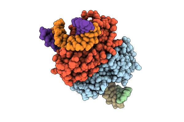
Structure Of The N254Y/H258Y Mutant Of The Phosphatidylinositol-Specific Phospholipase C From S. Aureus Bound To Dic4Pc
Organism: Staphylococcus aureus subsp. aureus
Method: X-RAY DIFFRACTION Resolution:1.85 Å Release Date: 2013-04-10 Classification: hydrolase, lyase Ligands: ACT, XP5 |
Organism: Staphylococcus aureus subsp. aureus
Method: X-RAY DIFFRACTION
Release Date: 2013-04-10
Ligands: ACT, XP5
 |
Crystal Structure Of Afnth1-K122A Mutant Bound To Tg-Dna Duplex Complex In An Intermediate State
Organism: Archaeoglobus fulgidus, Synthetic construct
Method: X-RAY DIFFRACTION Resolution:3.10 Å Release Date: 2026-04-15 Classification: LYASE/DNA Ligands: SF4 |
Organism: Archaeoglobus fulgidus, Synthetic construct
Method: X-RAY DIFFRACTION
Release Date: 2026-04-15
Ligands: SF4
 |
Fructose-1,6-Bisphosphate Aldolase/Phosphatase From Thermoproteus Neutrophilus, Dhap-Bound Form
Organism: Thermoproteus neutrophilus
Method: X-RAY DIFFRACTION Resolution:1.30 Å Release Date: 2011-10-26 Classification: LYASE, HYDROLASE Ligands: MG, 13P |
Organism: Thermoproteus neutrophilus
Method: X-RAY DIFFRACTION
Release Date: 2011-10-26
Ligands: MG, 13P