

Search Count: 1,662
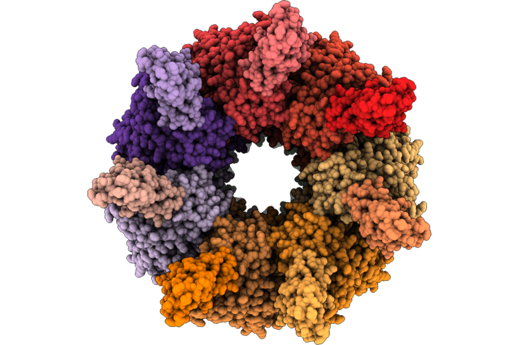 |
Amp-Pnp Bound E.Coli Cnox-Groel Complex, State I
Organism: Escherichia coli k-12
Method: ELECTRON MICROSCOPY Release Date: 2026-08-05 Classification: CHAPERONE Ligands: ANP, MG, K |
Organism: Escherichia coli k-12
Method: ELECTRON MICROSCOPY
Release Date: 2026-08-05
Ligands: ANP, MG, K
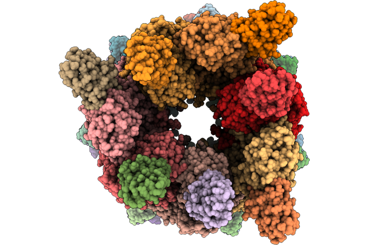 |
Amp-Pnp Bound E.Coli Cnox-Groel Complex, State Ii
Organism: Escherichia coli k-12
Method: ELECTRON MICROSCOPY Release Date: 2026-08-05 Classification: CHAPERONE Ligands: MG, K, ANP |
Organism: Escherichia coli k-12
Method: ELECTRON MICROSCOPY
Release Date: 2026-08-05
Ligands: MG, K, ANP
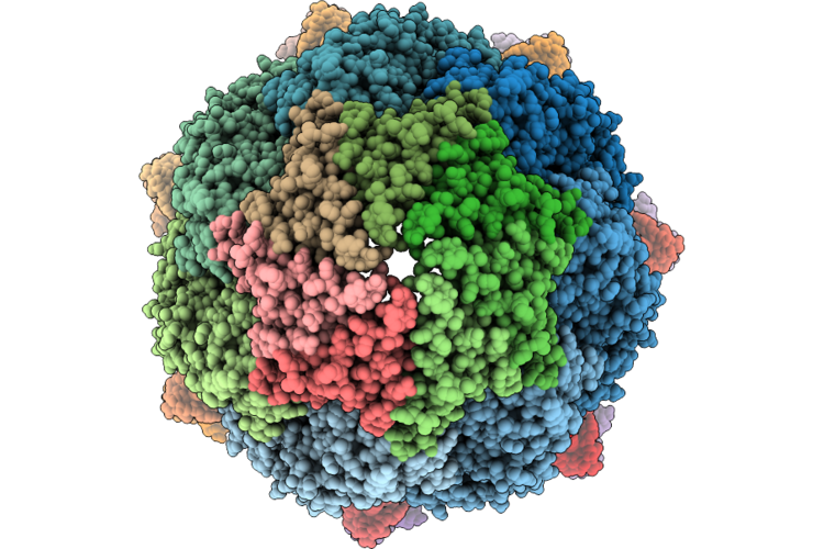 |
Amp-Pnp Bound E.Coli Cnox-Groel/Es Complex, State Iv
Organism: Escherichia coli k-12
Method: ELECTRON MICROSCOPY Release Date: 2026-08-05 Classification: CHAPERONE Ligands: MG, ANP, K |
Organism: Escherichia coli k-12
Method: ELECTRON MICROSCOPY
Release Date: 2026-08-05
Ligands: MG, ANP, K
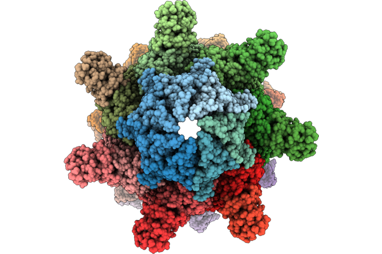 |
Amp-Pnp Bound E.Coli Cnox-Groel/Es Complex, State Iii
Organism: Escherichia coli k-12
Method: ELECTRON MICROSCOPY Release Date: 2026-08-05 Classification: CHAPERONE Ligands: MG, ANP, K |
Organism: Escherichia coli k-12
Method: ELECTRON MICROSCOPY
Release Date: 2026-08-05
Ligands: MG, ANP, K
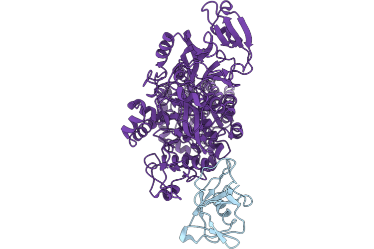 |
Cryo-Em Structure Of Lars1:Iars1 Complex
Organism: Homo sapiens
Method: ELECTRON MICROSCOPY Release Date: 2026-07-29 Classification: TRANSLATION |
Organism: Homo sapiens
Method: ELECTRON MICROSCOPY
Release Date: 2026-07-29
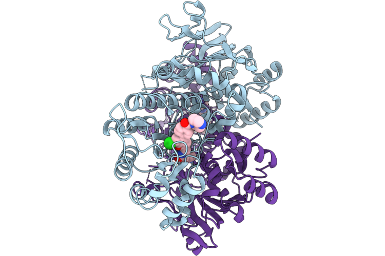 |
Crystal Structure Of Nampt-Kpt-7523 Complex
Organism: Homo sapiens
Method: X-RAY DIFFRACTION Resolution:1.45 Å Release Date: 2026-07-29 Classification: TRANSFERASE Ligands: A1MBG |
Organism: Homo sapiens
Method: X-RAY DIFFRACTION
Release Date: 2026-07-29
Ligands: A1MBG
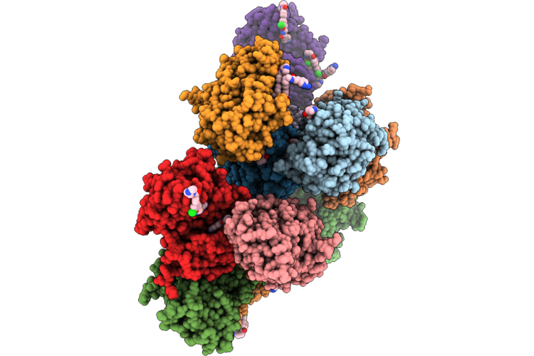 |
Crystal Structure Of Pak4-Kpt-7523 Complex
Organism: Homo sapiens
Method: X-RAY DIFFRACTION Resolution:2.20 Å Release Date: 2026-07-29 Classification: STRUCTURAL PROTEIN Ligands: A1MBG |
Organism: Homo sapiens
Method: X-RAY DIFFRACTION
Release Date: 2026-07-29
Ligands: A1MBG
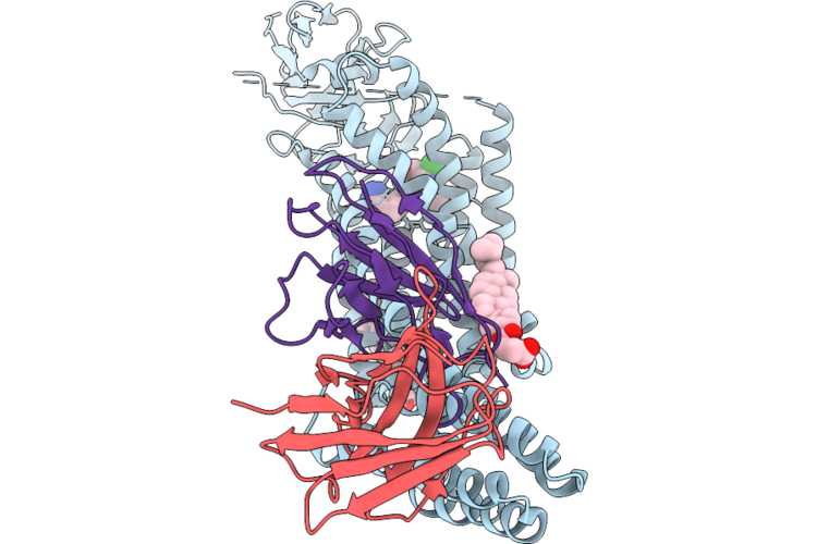 |
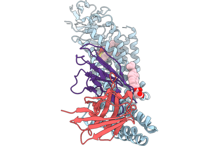
Structure Of At118-R Nanobody In Complex With The Angiotensin Ii Type I Receptor Bound To Losartan
Organism: Synthetic construct, Homo sapiens, Escherichia coli
Method: ELECTRON MICROSCOPY Resolution:3.40 Å Release Date: 2026-07-29 Classification: MEMBRANE PROTEIN Ligands: Y01, LSN |
Organism: Synthetic construct, Homo sapiens, Escherichia coli
Method: ELECTRON MICROSCOPY
Release Date: 2026-07-29
Ligands: Y01, LSN
 |
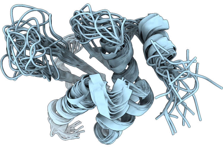
Structure Of At118-R Nanobody In Complex With The Angiotensin Ii Type I Receptor Bound To L-162,313
Organism: Synthetic construct, Homo sapiens, Escherichia coli
Method: ELECTRON MICROSCOPY Resolution:3.20 Å Release Date: 2026-07-29 Classification: MEMBRANE PROTEIN Ligands: Y01, A1C42 |
Organism: Synthetic construct, Homo sapiens, Escherichia coli
Method: ELECTRON MICROSCOPY
Release Date: 2026-07-29
Ligands: Y01, A1C42
 |
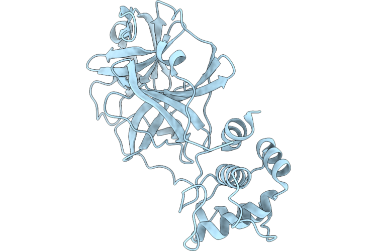
The Solution Structure Of Escherichia Coli Iscu(G64V)
Organism: Escherichia coli k-12
Method: SOLUTION NMR Release Date: 2026-07-22 Classification: METAL TRANSPORT |
Organism: Escherichia coli k-12
Method: SOLUTION NMR
Release Date: 2026-07-22
 |
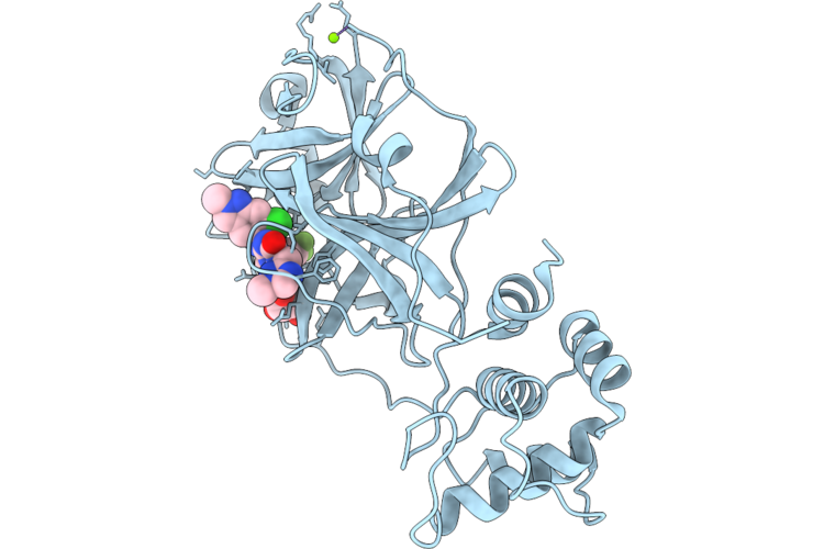
Crystal Structure Of The Apo Form Of Main Protease (Mpro) From Hku5-Cov-1
Organism: Pipistrellus bat coronavirus hku5
Method: X-RAY DIFFRACTION Resolution:1.75 Å Release Date: 2026-07-15 Classification: VIRAL PROTEIN |
Organism: Pipistrellus bat coronavirus hku5
Method: X-RAY DIFFRACTION
Release Date: 2026-07-15
 |
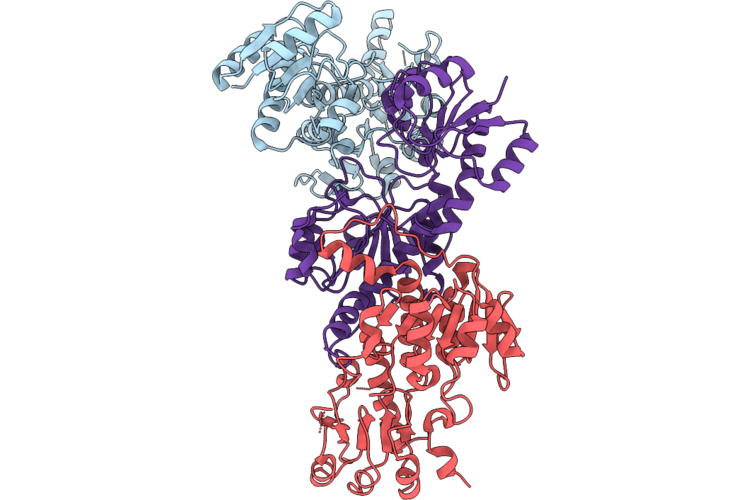
Crystal Structure Of Hku5-Cov-1 Main Protease(Mpro) In Complex With Ensitrelvir
Organism: Pipistrellus bat coronavirus hku5
Method: X-RAY DIFFRACTION Resolution:1.55 Å Release Date: 2026-07-15 Classification: VIRAL PROTEIN Ligands: 7YY, GOL, MG |
Organism: Pipistrellus bat coronavirus hku5
Method: X-RAY DIFFRACTION
Release Date: 2026-07-15
Ligands: 7YY, GOL, MG
 |
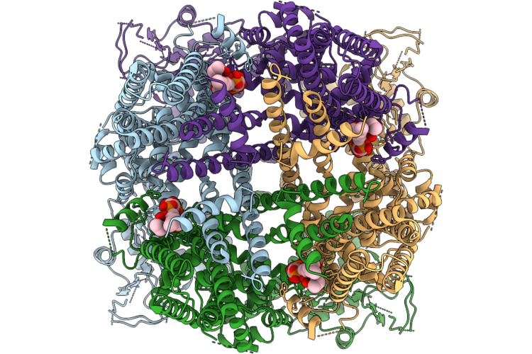
Crystal Structure Of Vanh From Acinetobacter Baumannii
Organism: Acinetobacter baumannii
Method: X-RAY DIFFRACTION Resolution:2.39 Å Release Date: 2026-07-08 Classification: ANTIBIOTIC |
Organism: Acinetobacter baumannii
Method: X-RAY DIFFRACTION
Release Date: 2026-07-08
 |
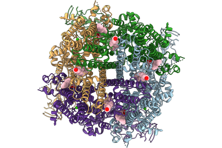
Cryo-Em Structure Of Trp Melastatin Channel In The Intermediate 3 State
Organism: Mus musculus
Method: ELECTRON MICROSCOPY Resolution:3.48 Å Release Date: 2026-07-01 Classification: MEMBRANE PROTEIN Ligands: PIO |
Organism: Mus musculus
Method: ELECTRON MICROSCOPY
Release Date: 2026-07-01
Ligands: PIO
 |
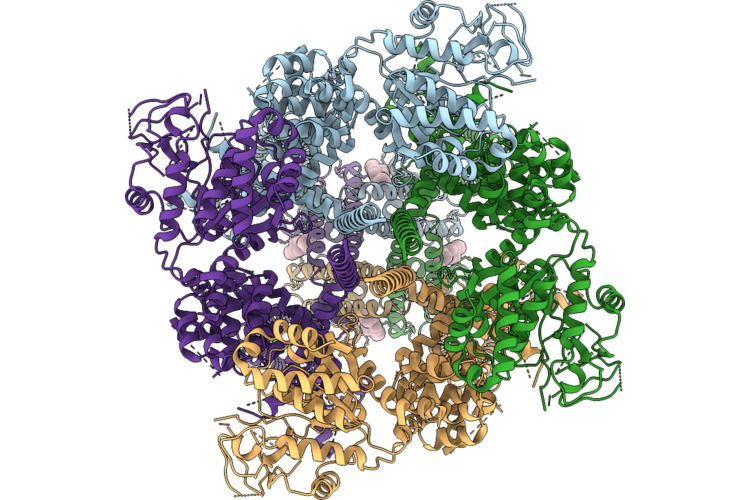
Cryo-Em Structure Of Trp Melastatin Channel In The Desensitized State, With Chs
Organism: Mus musculus
Method: ELECTRON MICROSCOPY Resolution:3.25 Å Release Date: 2026-07-01 Classification: MEMBRANE PROTEIN Ligands: Y01, CA, CLR |
Organism: Mus musculus
Method: ELECTRON MICROSCOPY
Release Date: 2026-07-01
Ligands: Y01, CA, CLR
 |
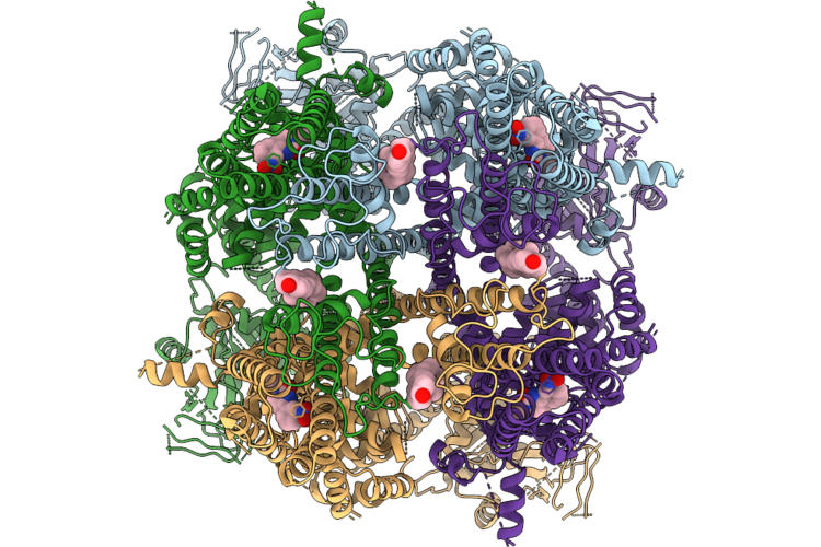
Cryo-Em Structure Of Trp Melastatin Channel In The Desensitized State, Without Chs
Organism: Mus musculus
Method: ELECTRON MICROSCOPY Resolution:2.81 Å Release Date: 2026-07-01 Classification: MEMBRANE PROTEIN Ligands: CA, CLR |
Organism: Mus musculus
Method: ELECTRON MICROSCOPY
Release Date: 2026-07-01
Ligands: CA, CLR
 |
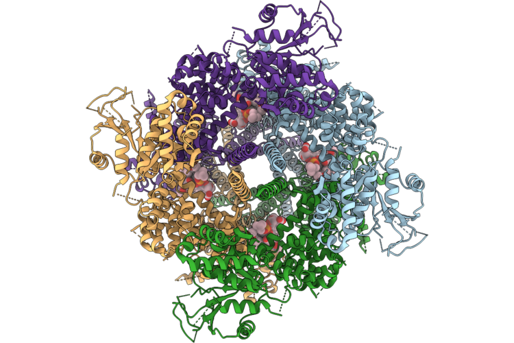
Cryo-Em Structure Of Trp Melastatin Channel In The Desensitized State, With Icilin
Organism: Mus musculus
Method: ELECTRON MICROSCOPY Resolution:3.45 Å Release Date: 2026-07-01 Classification: MEMBRANE PROTEIN Ligands: CA, KX7, CLR |
Organism: Mus musculus
Method: ELECTRON MICROSCOPY
Release Date: 2026-07-01
Ligands: CA, KX7, CLR
 |
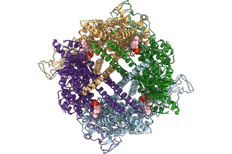
Cryo-Em Structure Of Trp Melastatin Channel In The Closed State
Organism: Mus musculus
Method: ELECTRON MICROSCOPY Resolution:3.43 Å Release Date: 2026-06-24 Classification: MEMBRANE PROTEIN Ligands: Y01, POV |
Organism: Mus musculus
Method: ELECTRON MICROSCOPY
Release Date: 2026-06-24
Ligands: Y01, POV
 |
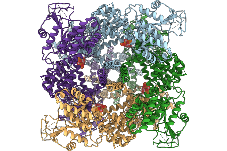
Cryo-Em Structure Of Trp Melastatin Channel In The Intermediate 1 State
Organism: Mus musculus
Method: ELECTRON MICROSCOPY Resolution:2.82 Å Release Date: 2026-06-24 Classification: MEMBRANE PROTEIN Ligands: PIO, CA |
Organism: Mus musculus
Method: ELECTRON MICROSCOPY
Release Date: 2026-06-24
Ligands: PIO, CA
 |
Cryo-Em Structure Of Trp Melastatin Channel In The Intermediate 2 State
Organism: Mus musculus
Method: ELECTRON MICROSCOPY Resolution:3.29 Å Release Date: 2026-06-24 Classification: MEMBRANE PROTEIN Ligands: PIO, CA |
Organism: Mus musculus
Method: ELECTRON MICROSCOPY
Release Date: 2026-06-24
Ligands: PIO, CA