

Search Count: 271
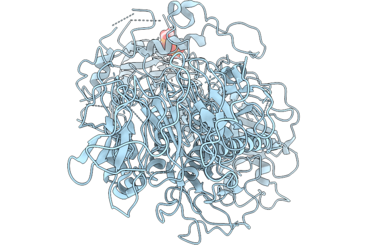 |
Leucine Rich Repeat Kinase 2 Bound To Gmp-Pnp (Inactive)
Organism: Homo sapiens
Method: ELECTRON MICROSCOPY Resolution:4.39 Å Release Date: 2026-08-19 Classification: HYDROLASE Ligands: GNP |
Organism: Homo sapiens
Method: ELECTRON MICROSCOPY
Release Date: 2026-08-19
Ligands: GNP
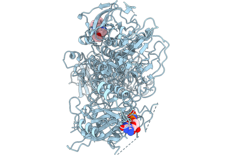 |
Leucine Rich Repeat Kinase 2 Bound To Gmp-Pnp (Active)
Organism: Homo sapiens
Method: ELECTRON MICROSCOPY Resolution:3.73 Å Release Date: 2026-08-19 Classification: HYDROLASE Ligands: GNP, A1N |
Organism: Homo sapiens
Method: ELECTRON MICROSCOPY
Release Date: 2026-08-19
Ligands: GNP, A1N
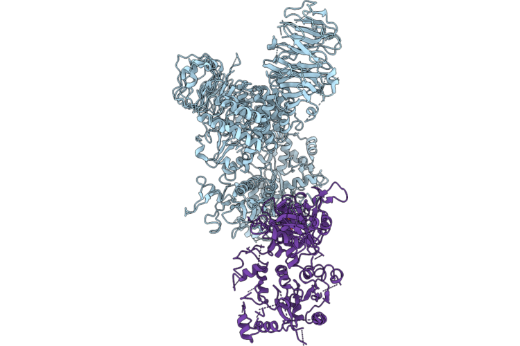 |
Lrrk2 Asymmetric Dimer
Organism: Homo sapiens
Method: ELECTRON MICROSCOPY Resolution:3.44 Å Release Date: 2026-08-19 Classification: HYDROLASE Ligands: GDP |
Organism: Homo sapiens
Method: ELECTRON MICROSCOPY
Release Date: 2026-08-19
Ligands: GDP
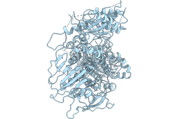 |
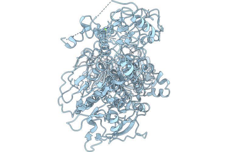
Leucine Rich Repeat Kinase 2 Activate State (Apo Roc State 2)
Organism: Homo sapiens
Method: ELECTRON MICROSCOPY Resolution:4.00 Å Release Date: 2026-08-19 Classification: HYDROLASE |
Organism: Homo sapiens
Method: ELECTRON MICROSCOPY
Release Date: 2026-08-19
 |
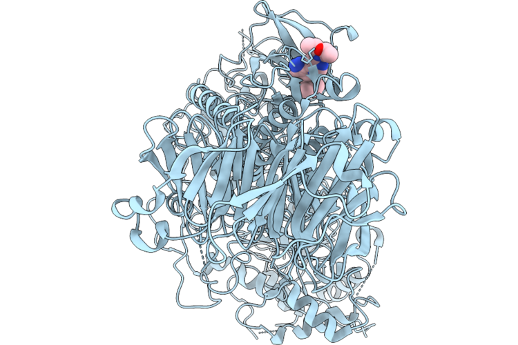
Leucine Rich Repeat Kinase 2 Activate State (Apo Roc State 1)
Organism: Homo sapiens
Method: ELECTRON MICROSCOPY Resolution:4.02 Å Release Date: 2026-08-19 Classification: HYDROLASE Ligands: MG |
Organism: Homo sapiens
Method: ELECTRON MICROSCOPY
Release Date: 2026-08-19
Ligands: MG
 |
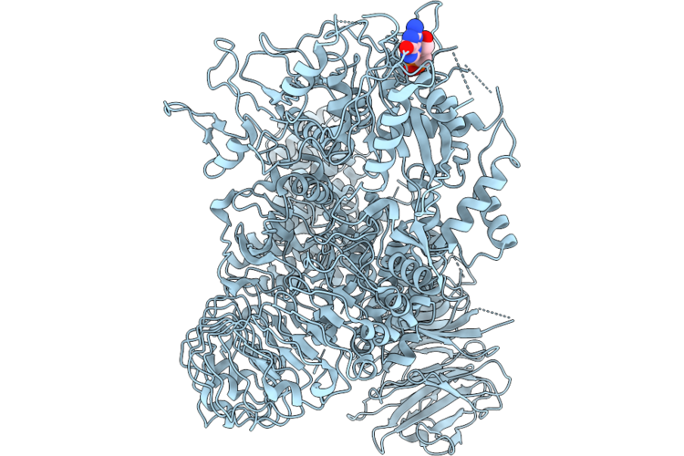
Leucine Rich Repeat Kinase 2 R1441C Mutant (Active)
Organism: Homo sapiens
Method: ELECTRON MICROSCOPY Resolution:3.63 Å Release Date: 2026-08-19 Classification: HYDROLASE Ligands: A1N |
Organism: Homo sapiens
Method: ELECTRON MICROSCOPY
Release Date: 2026-08-19
Ligands: A1N
 |
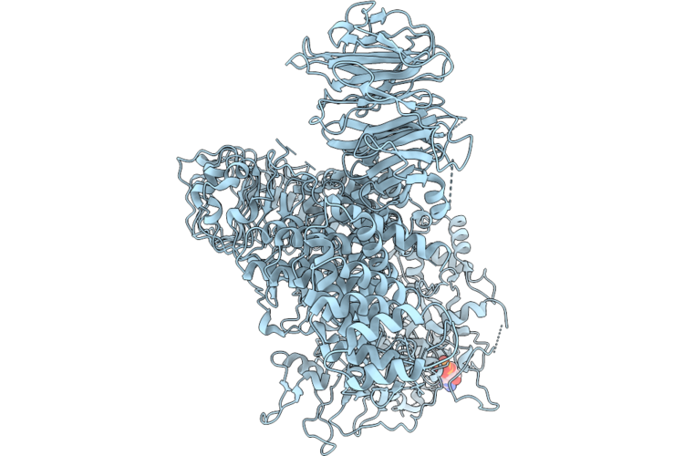
Leucine Rich Repeat Kinase 2 Bound To Gdp (From Gmppnp Data)
Organism: Homo sapiens
Method: ELECTRON MICROSCOPY Resolution:3.42 Å Release Date: 2026-08-19 Classification: HYDROLASE Ligands: GDP |
Organism: Homo sapiens
Method: ELECTRON MICROSCOPY
Release Date: 2026-08-19
Ligands: GDP
 |
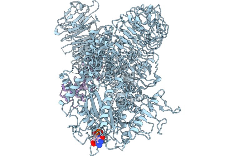
Full-Length Lrrk2 Autoinhibited
Organism: Homo sapiens
Method: ELECTRON MICROSCOPY Resolution:3.31 Å Release Date: 2026-08-12 Classification: TRANSFERASE Ligands: GDP |
Organism: Homo sapiens
Method: ELECTRON MICROSCOPY
Release Date: 2026-08-12
Ligands: GDP
 |
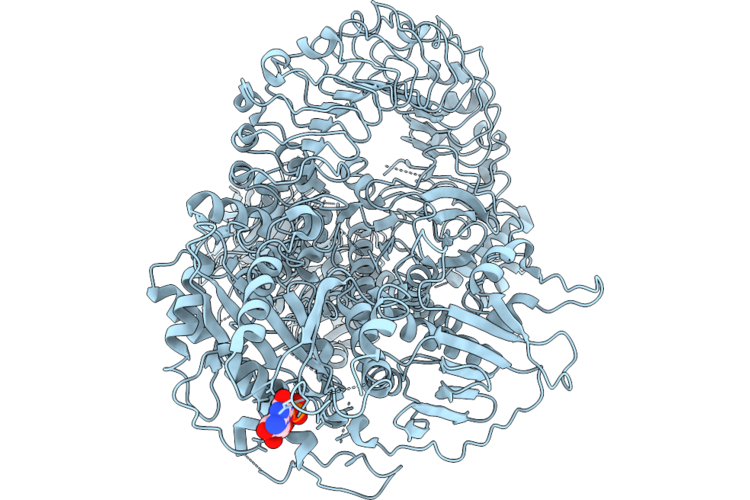
Full Length Lrrk2 Bound To G10 Darpin
Organism: Homo sapiens
Method: ELECTRON MICROSCOPY Resolution:3.15 Å Release Date: 2026-08-12 Classification: STRUCTURAL PROTEIN Ligands: MG, GDP |
Organism: Homo sapiens
Method: ELECTRON MICROSCOPY
Release Date: 2026-08-12
Ligands: MG, GDP
 |
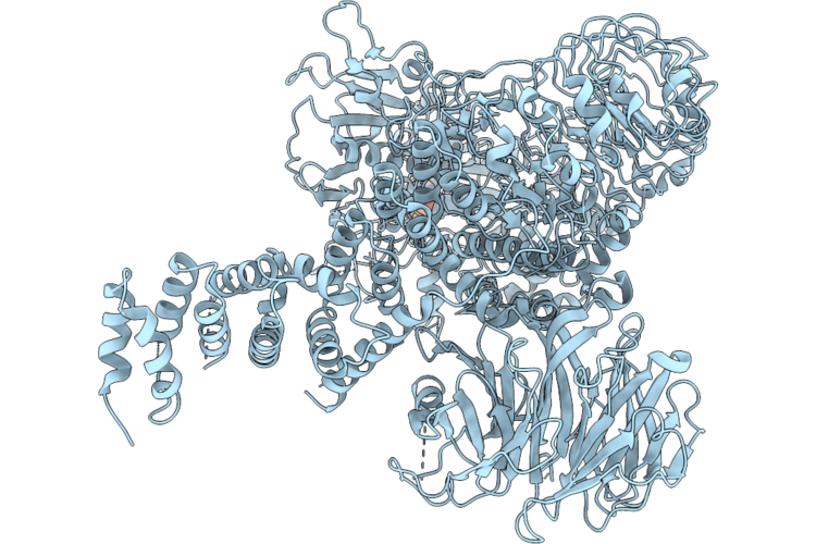
Full-Length Lrrk2 Autonhibited (Flexible)
Organism: Homo sapiens
Method: ELECTRON MICROSCOPY Release Date: 2026-08-12 Classification: HYDROLASE Ligands: GDP |
Organism: Homo sapiens
Method: ELECTRON MICROSCOPY
Release Date: 2026-08-12
Ligands: GDP
 |
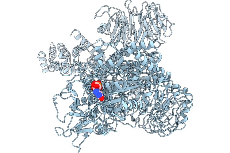
Full Lrrk2 (R1441C Mutant) After Symmetry Expansion
Organism: Homo sapiens
Method: ELECTRON MICROSCOPY Release Date: 2026-08-12 Classification: HYDROLASE Ligands: GDP |
Organism: Homo sapiens
Method: ELECTRON MICROSCOPY
Release Date: 2026-08-12
Ligands: GDP
 |
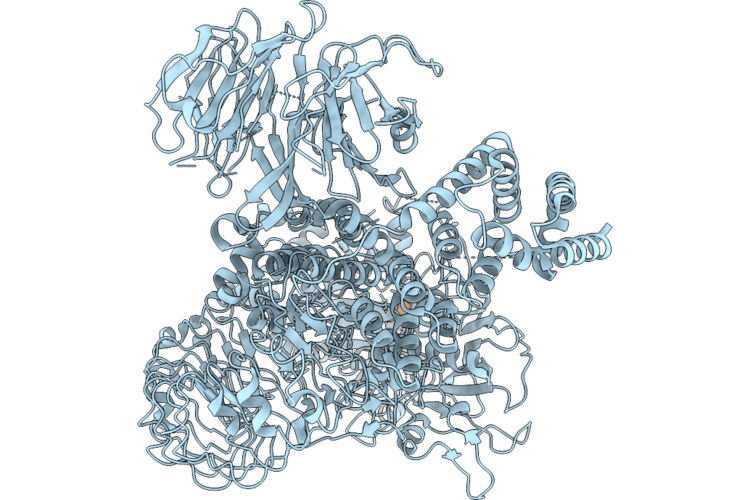
Full Length Lrrk2 After Symmetry Expansion
Organism: Homo sapiens
Method: ELECTRON MICROSCOPY Resolution:3.72 Å Release Date: 2026-08-12 Classification: HYDROLASE Ligands: GDP |
Organism: Homo sapiens
Method: ELECTRON MICROSCOPY
Release Date: 2026-08-12
Ligands: GDP
 |
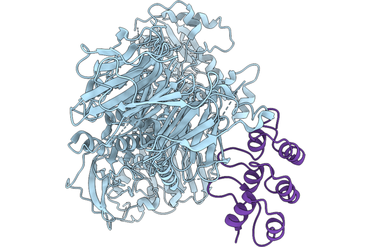
Full Length Lrrk2 (G2019S) After Symmetry Expansion
Organism: Homo sapiens
Method: ELECTRON MICROSCOPY Resolution:3.25 Å Release Date: 2026-08-12 Classification: HYDROLASE Ligands: GDP |
Organism: Homo sapiens
Method: ELECTRON MICROSCOPY
Release Date: 2026-08-12
Ligands: GDP
 |
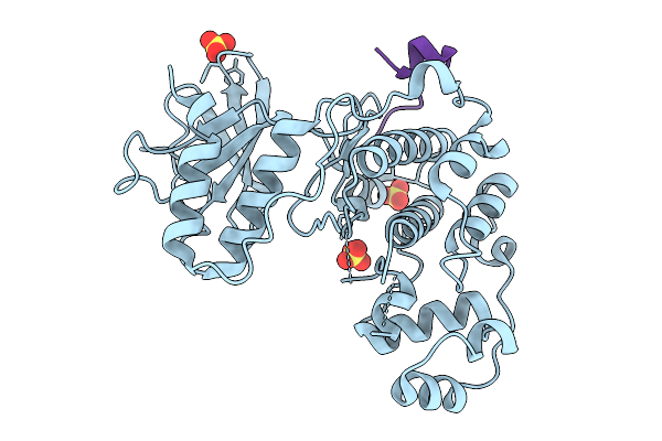
C-Terminal Lrrk2 Bound To C12 Darpin
Organism: Homo sapiens, Synthetic construct
Method: ELECTRON MICROSCOPY Resolution:3.60 Å Release Date: 2026-08-12 Classification: PROTEIN BINDING |
Organism: Homo sapiens, Synthetic construct
Method: ELECTRON MICROSCOPY
Release Date: 2026-08-12
 |
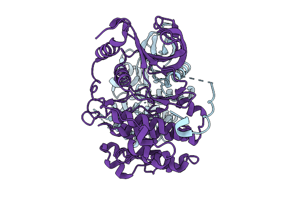
Crystal Structure Of Human Erk1 In Complex With The Kim1 Motif Of The T. Gondii Protein Gra24
Organism: Homo sapiens, Toxoplasma gondii
Method: X-RAY DIFFRACTION Resolution:2.17 Å Release Date: 2026-02-25 Classification: TRANSFERASE Ligands: SO4 |
Organism: Homo sapiens, Toxoplasma gondii
Method: X-RAY DIFFRACTION
Release Date: 2026-02-25
Ligands: SO4
 |
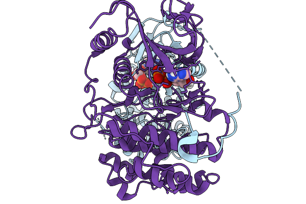
Structure Of The Map2K Mek1 Without Bound Nucleotide In Complex With Its Substrate Mapk Erk2
Organism: Homo sapiens, Toxoplasma gondii me49
Method: ELECTRON MICROSCOPY Resolution:3.60 Å Release Date: 2026-02-25 Classification: SIGNALING PROTEIN |
Organism: Homo sapiens, Toxoplasma gondii me49
Method: ELECTRON MICROSCOPY
Release Date: 2026-02-25
 |
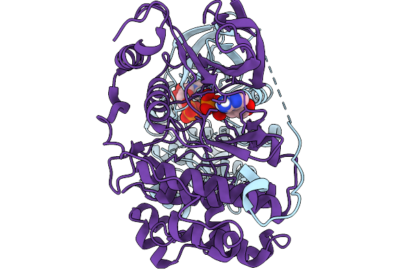
Structure Of The Map2K Mek1 In An Inactive Conformation In Complex With Its Substrate Mapk Erk2
Organism: Homo sapiens, Toxoplasma gondii me49
Method: ELECTRON MICROSCOPY Release Date: 2026-02-11 Classification: SIGNALING PROTEIN Ligands: ADP, MG |
Organism: Homo sapiens, Toxoplasma gondii me49
Method: ELECTRON MICROSCOPY
Release Date: 2026-02-11
Ligands: ADP, MG
 |
Structure Of The Map2K Mek1 In An Active Conformation In Complex With Its Substrate Mapk Erk2
Organism: Homo sapiens, Toxoplasma gondii me49
Method: ELECTRON MICROSCOPY Release Date: 2026-02-11 Classification: SIGNALING PROTEIN Ligands: ADP |
Organism: Homo sapiens, Toxoplasma gondii me49
Method: ELECTRON MICROSCOPY
Release Date: 2026-02-11
Ligands: ADP
 |
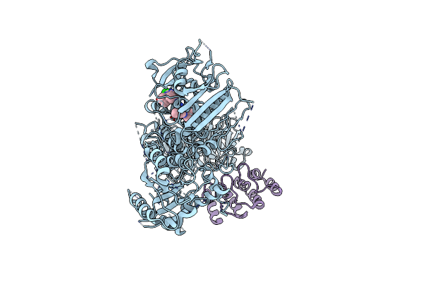
Solution Structure Of Homodimeric Tmem106B
Organism: Homo sapiens
Method: SOLUTION NMR Release Date: 2025-11-26 Classification: MEMBRANE PROTEIN Ligands: ZN |
Organism: Homo sapiens
Method: SOLUTION NMR
Release Date: 2025-11-26
Ligands: ZN
 |
Structure Of The C-Terminal Half Of Lrrk2 Bound To Rn277 (Type-Ii Inhibitor)
Organism: Homo sapiens, Synthetic construct
Method: ELECTRON MICROSCOPY Resolution:3.35 Å Release Date: 2025-06-18 Classification: PROTEIN BINDING Ligands: A1A7Q |
Organism: Homo sapiens, Synthetic construct
Method: ELECTRON MICROSCOPY
Release Date: 2025-06-18
Ligands: A1A7Q