
Search Count: 769
All
Selected
 |
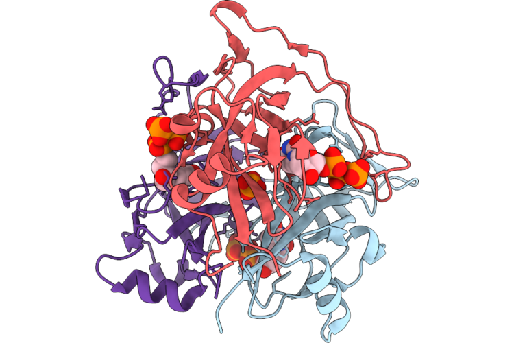
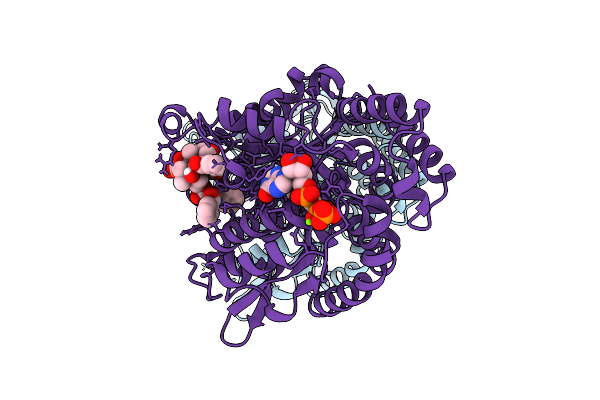
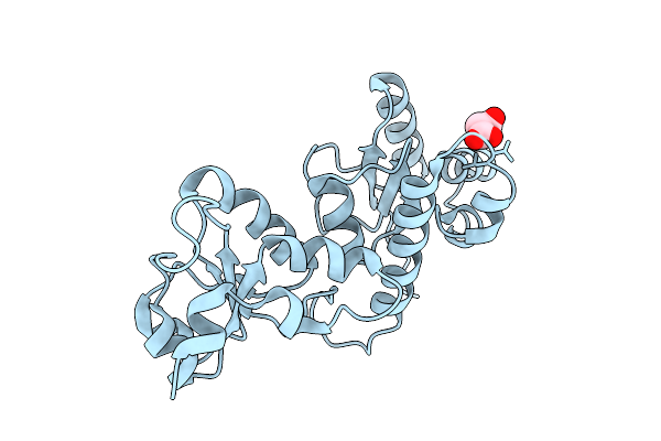
Organism: Methanosarcina mazei
Method: X-RAY DIFFRACTION Resolution:1.45 Å Release Date: 2026-04-29 Classification: HYDROLASE Ligands: PO4 |
 |
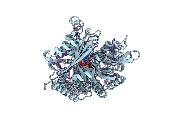
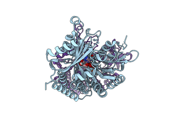
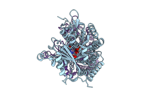
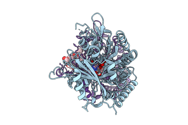
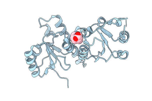
Organism: Methanosarcina mazei
Method: X-RAY DIFFRACTION Resolution:1.53 Å Release Date: 2026-04-29 Classification: HYDROLASE Ligands: DUT, PO4 |
 |
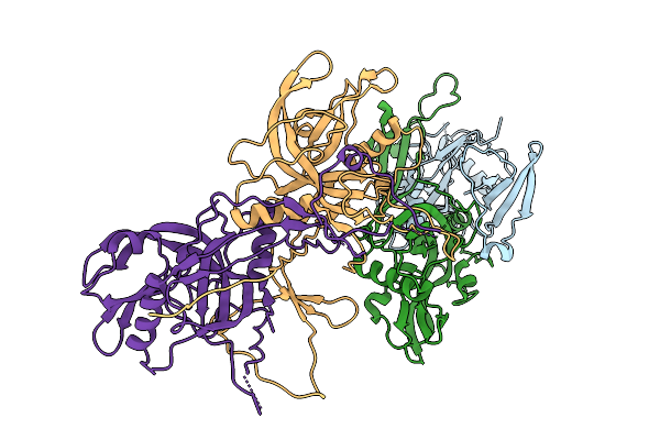
Crystal Structure Of A Zikv E Glycoprotein Di-Diii Vaccine Candidate In Complex With Human Neutralizing Antibody Mz4
Organism: Homo sapiens, Zika virus zikv/h. sapiens/frenchpolynesia/10087pf/2013
Method: X-RAY DIFFRACTION Resolution:2.85 Å Release Date: 2026-04-15 Classification: VIRAL PROTEIN Ligands: NAG |
 |
Organism: Homo sapiens
Method: ELECTRON MICROSCOPY Resolution:2.48 Å Release Date: 2026-04-01 Classification: STRUCTURAL PROTEIN Ligands: MG, GDP, GTP |
 |
Organism: Homo sapiens
Method: ELECTRON MICROSCOPY Resolution:2.41 Å Release Date: 2026-04-01 Classification: STRUCTURAL PROTEIN Ligands: GDP, MG, GTP |
 |
Organism: Homo sapiens
Method: ELECTRON MICROSCOPY Release Date: 2026-04-01 Classification: STRUCTURAL PROTEIN Ligands: GTP, MG, G2P |
 |
Organism: Homo sapiens
Method: ELECTRON MICROSCOPY Release Date: 2026-04-01 Classification: STRUCTURAL PROTEIN Ligands: GTP, MG, G2P |
 |
Organism: Homo sapiens
Method: ELECTRON MICROSCOPY Release Date: 2026-04-01 Classification: STRUCTURAL PROTEIN Ligands: MG, GTP, TA1, G2P |
 |
Organism: Homo sapiens
Method: ELECTRON MICROSCOPY Release Date: 2026-04-01 Classification: STRUCTURAL PROTEIN Ligands: GTP, MG, TA1, G2P |
 |
Crystal Structure Of Human Pkmyt1 Protein Kinase Domain With Naphthyridinone Inhibitor Compound 11
Organism: Homo sapiens
Method: X-RAY DIFFRACTION Resolution:1.74 Å Release Date: 2026-03-25 Classification: TRANSFERASE Ligands: A1E5A, SO4 |
 |
Crystal Structure Of Human Pkmyt1 Protein Kinase Domain With Naphthyridinone Inhibitor Compound 16
Organism: Homo sapiens
Method: X-RAY DIFFRACTION Resolution:1.59 Å Release Date: 2026-03-25 Classification: TRANSFERASE Ligands: A1E5J, DMS, EDO, SO4 |
 |
Organism: Homo sapiens
Method: X-RAY DIFFRACTION Resolution:1.65 Å Release Date: 2026-03-18 Classification: ISOMERASE Ligands: MLA |
 |
Crystal Structure Of Topbp1Brct7-8 Domain In Complex With 1-Adamantaneacetic Acid
Organism: Homo sapiens
Method: X-RAY DIFFRACTION Resolution:2.45 Å Release Date: 2026-03-18 Classification: ISOMERASE Ligands: A1ENP |
 |
Organism: Homo sapiens
Method: ELECTRON MICROSCOPY Release Date: 2026-03-04 Classification: HYDROLASE Ligands: MG, ATP, ADP |
 |
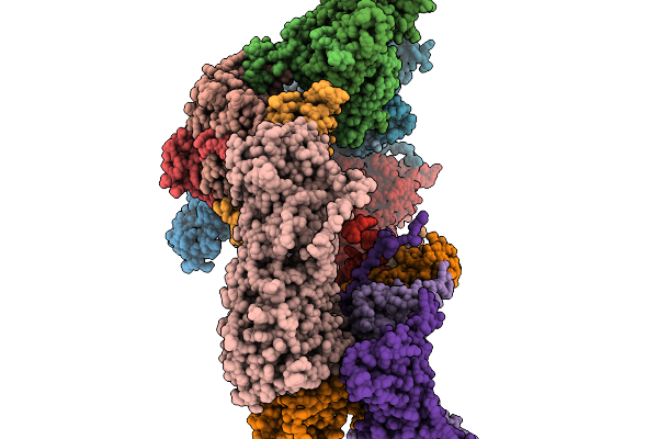
Organism: Escherichia coli nctc 86
Method: ELECTRON MICROSCOPY Resolution:2.90 Å Release Date: 2026-02-18 Classification: ANTIVIRAL PROTEIN Ligands: MG |
 |
Organism: Pseudomonas phage phikz
Method: ELECTRON MICROSCOPY Resolution:3.37 Å Release Date: 2026-02-11 Classification: VIRAL PROTEIN |
 |
Organism: Pseudomonas phage phikz
Method: ELECTRON MICROSCOPY Resolution:3.15 Å Release Date: 2026-02-11 Classification: VIRAL PROTEIN |
 |
Organism: Pseudomonas phage phikz
Method: ELECTRON MICROSCOPY Resolution:4.14 Å Release Date: 2026-02-11 Classification: VIRAL PROTEIN |
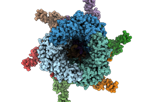 |
Organism: Pseudomonas phage phikz
Method: ELECTRON MICROSCOPY Resolution:4.02 Å Release Date: 2026-02-11 Classification: VIRAL PROTEIN |
 |
Organism: Pseudomonas phage phikz
Method: ELECTRON MICROSCOPY Resolution:3.52 Å Release Date: 2026-02-11 Classification: VIRAL PROTEIN |