
Search Count: 42
All
Selected
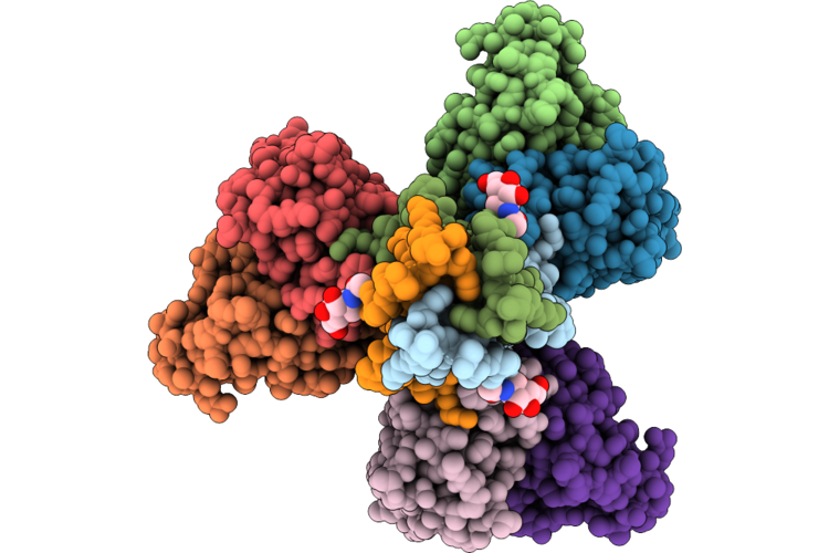 |
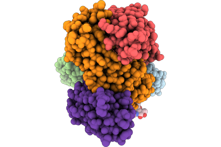
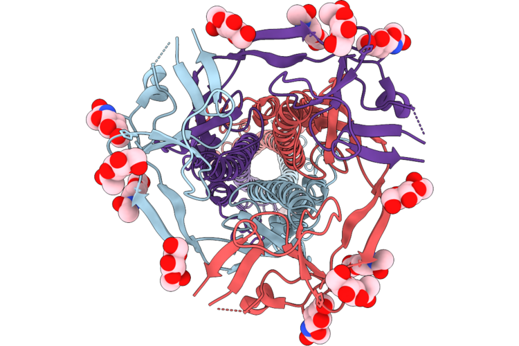
Sars-Cov-2 Spike S2 Trimer Stabilized In The Early Fusion Intermediate Conformation (E-Fics-V3) Bound To The Vn01H1 Fab (Fab Local Refinement)
Organism: Saccharomyces cerevisiae s288c, Severe acute respiratory syndrome coronavirus 2, Homo sapiens
Method: ELECTRON MICROSCOPY Release Date: 2026-05-13 Classification: VIRAL PROTEIN Ligands: NAG |
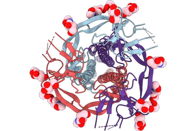 |
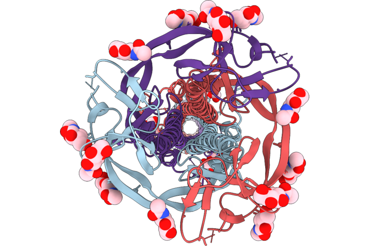
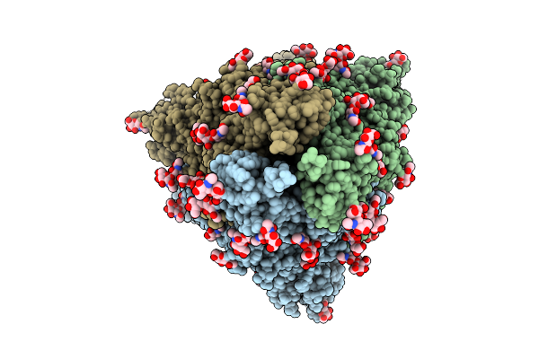
Sars-Cov-2 Spike S2 Trimer Stabilized In The Early Fusion Intermediate Conformation (E-Fics-V3) Bound To The Vn01H1 Fab (S2 Local Refinement)
Organism: Saccharomyces cerevisiae s288c, Severe acute respiratory syndrome coronavirus 2
Method: ELECTRON MICROSCOPY Release Date: 2026-05-13 Classification: VIRAL PROTEIN Ligands: NAG |
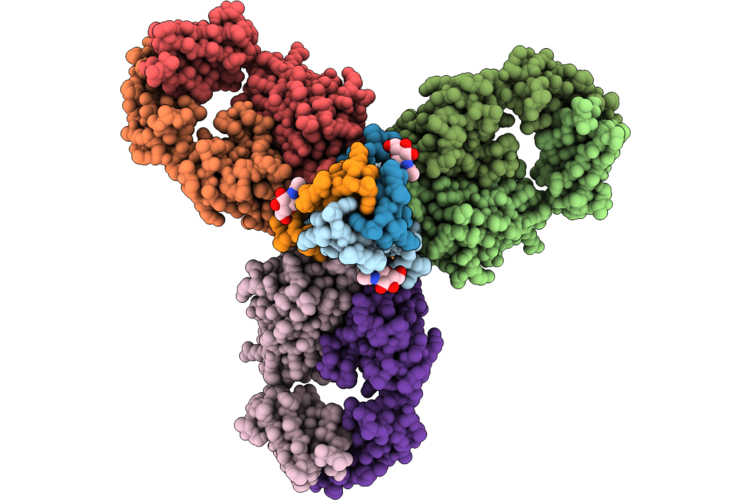 |
Sars-Cov-2 Spike S2 Trimer Stabilized In The Early Fusion Intermediate Conformation (E-Fics-V3) Bound To C77G12 (Fab Local Refinement)
Organism: Saccharomyces cerevisiae s288c, Severe acute respiratory syndrome coronavirus 2, Homo sapiens
Method: ELECTRON MICROSCOPY Release Date: 2026-05-13 Classification: VIRAL PROTEIN Ligands: NAG |
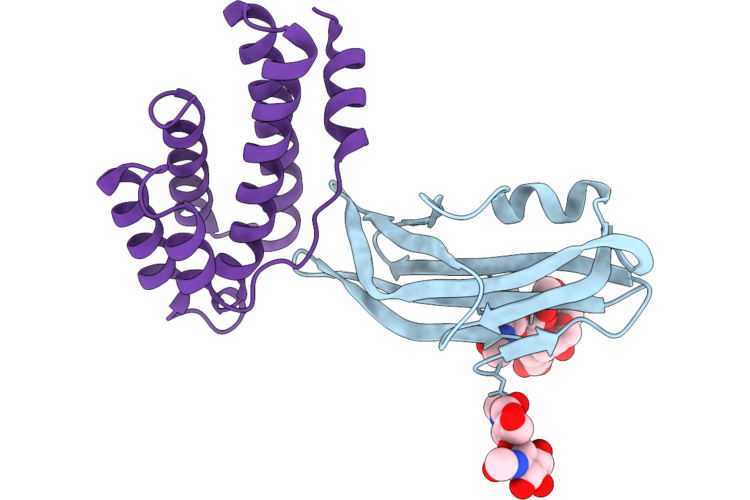 |
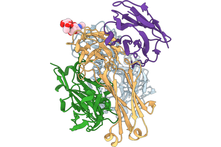
Structure Of The Porcine Deltacoronavirus (Pdcov) Receptor-Binding Domain Bound To The Rbd Minibinder 11, The Pd3 Fab, And The Kappa Light Chain Nanobody (Local Refinement)
Organism: Porcine deltacoronavirus, Synthetic construct
Method: ELECTRON MICROSCOPY Resolution:2.84 Å Release Date: 2026-05-13 Classification: VIRAL PROTEIN |
 |
Structure Of The Porcine Deltacoronavirus (Pdcov) Receptor-Binding Domain Bound To The Rbd Minibinder 11, The Pd3 Fab, And The Kappa Light Chain Nanobody
Organism: Porcine deltacoronavirus, Synthetic construct, Mus musculus, Lama glama
Method: ELECTRON MICROSCOPY Resolution:2.80 Å Release Date: 2026-05-13 Classification: VIRAL PROTEIN |
 |
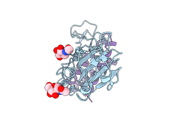
Tmprss2 (S441A) Bound To The Hcov-Nl63 S2'Region Genetically Fused To The Hcov-Hku1 Rbd
Organism: Human coronavirus hku1, Homo sapiens
Method: ELECTRON MICROSCOPY Resolution:3.30 Å Release Date: 2026-05-13 Classification: VIRAL PROTEIN/HYDROLASE Ligands: NAG |
 |
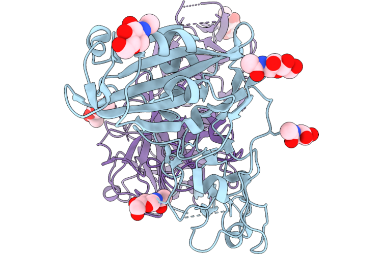
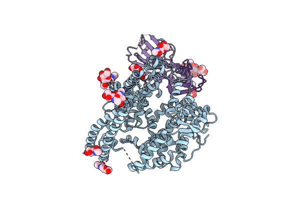
Tmprss2 S441A In Complex With The H1H7 Fab And Anti-Kappa Light Chain Nanobody
Organism: Homo sapiens, Lama glama
Method: ELECTRON MICROSCOPY Resolution:3.20 Å Release Date: 2026-05-13 Classification: HYDROLASE/IMMUNE SYSTEM |
 |
Sars-Cov-2 Spike Trimer In The Early Fusion Intermediate Conformation Bound To The Vn01H1 Fab (Fab Local Refinement)
Organism: Severe acute respiratory syndrome coronavirus 2, Homo sapiens
Method: ELECTRON MICROSCOPY Release Date: 2026-05-13 Classification: VIRAL PROTEIN/IMMUNE SYSTEM Ligands: NAG |
 |
Sars-Cov-2 Spike Trimer In The Early Fusion Intermediate Conformation Bound To The Vn01H1 Fab (S2 Local Refinement)
Organism: Severe acute respiratory syndrome coronavirus 2
Method: ELECTRON MICROSCOPY Resolution:3.30 Å Release Date: 2026-05-13 Classification: VIRAL PROTEIN Ligands: NAG |
 |
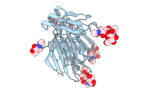
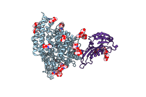
Hcov-Nl63 S2' Peptide Bound To Tmprss2 S441A (Complexed With The H1H7 Fab And An Anti-Kappa-Nanobody)
Organism: Human coronavirus nl63, Homo sapiens, Lama glama
Method: ELECTRON MICROSCOPY Resolution:2.80 Å Release Date: 2026-05-13 Classification: VIRAL PROTEIN/Immune System |
 |
Sars-Cov-2 S2 Trimer Stabilized In The Early Fusion Intermediate Conformation By Circular Permutation And Clamping By Gp41 (E-Fics-V1)
Organism: Severe acute respiratory syndrome coronavirus 2
Method: ELECTRON MICROSCOPY Release Date: 2026-05-13 Classification: VIRAL PROTEIN Ligands: NAG |
 |
Sars-Cov-2 Spike S2 Trimer Stabilized In The Early Fusion Intermediate Conformation (E-Fics-V3) Bound To C77G12 (S2 Local Refinement)
Organism: Saccharomyces cerevisiae s288c, Severe acute respiratory syndrome coronavirus 2
Method: ELECTRON MICROSCOPY Release Date: 2026-03-18 Classification: VIRAL PROTEIN Ligands: NAG |
 |
Local Refinement Of The Sars-Cov-2 Ba.2.86 Rbd In Complex With Tri2-2 Minibinder
Organism: Severe acute respiratory syndrome coronavirus 2, Synthetic construct
Method: ELECTRON MICROSCOPY Release Date: 2026-01-21 Classification: VIRAL PROTEIN Ligands: NAG |
 |
Organism: Severe acute respiratory syndrome coronavirus 2
Method: ELECTRON MICROSCOPY Release Date: 2026-01-21 Classification: VIRAL PROTEIN Ligands: NAG |
 |
Organism: Severe acute respiratory syndrome coronavirus 2, Synthetic construct
Method: ELECTRON MICROSCOPY Resolution:2.40 Å Release Date: 2026-01-21 Classification: VIRAL PROTEIN Ligands: NAG |
 |
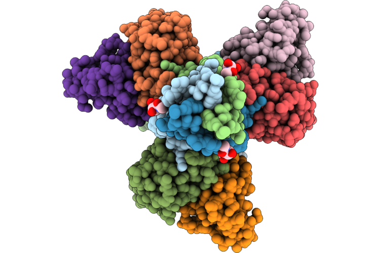
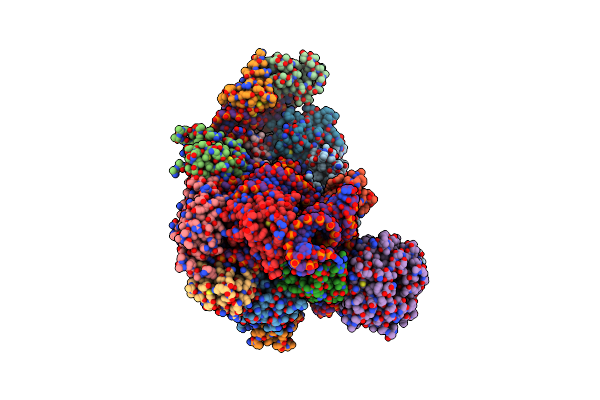
Sars-Cov-2 Nsp1 Bound To The Rhinolophus Lepidus 40S Ribosomal Subunit (Local Refinement Of The 40S Body)
Organism: Severe acute respiratory syndrome coronavirus 2, Homo sapiens, Rhinolophus lepidus
Method: ELECTRON MICROSCOPY Resolution:2.10 Å Release Date: 2025-06-11 Classification: RIBOSOME Ligands: MG, K, ZN |
 |
Sars-Cov-2 Nsp1 Bound To The Rhinolophus Lepidus 40S Ribosome (Local Refinement Of The 40S Head)
Organism: Rhinolophus lepidus
Method: ELECTRON MICROSCOPY Resolution:2.10 Å Release Date: 2025-06-11 Classification: RIBOSOME Ligands: MG, ZN, K |
 |
Organism: Merbecovirus, Pteronotus davyi
Method: ELECTRON MICROSCOPY Release Date: 2025-03-05 Classification: VIRAL PROTEIN Ligands: NAG |
 |
Merbecovirus Pnnl2018B Spike Glycoprotein Rbd Bound To The P. Nathusii Ace2
Organism: Pipistrellus nathusii, Merbecovirus
Method: ELECTRON MICROSCOPY Release Date: 2025-02-19 Classification: VIRAL PROTEIN Ligands: NAG, ZN |
 |
Organism: Pipistrellus nathusii, Middle east respiratory syndrome-related coronavirus
Method: ELECTRON MICROSCOPY Release Date: 2025-02-12 Classification: VIRAL PROTEIN/HYDROLASE Ligands: NAG, ZN |