
Search Count: 4,552
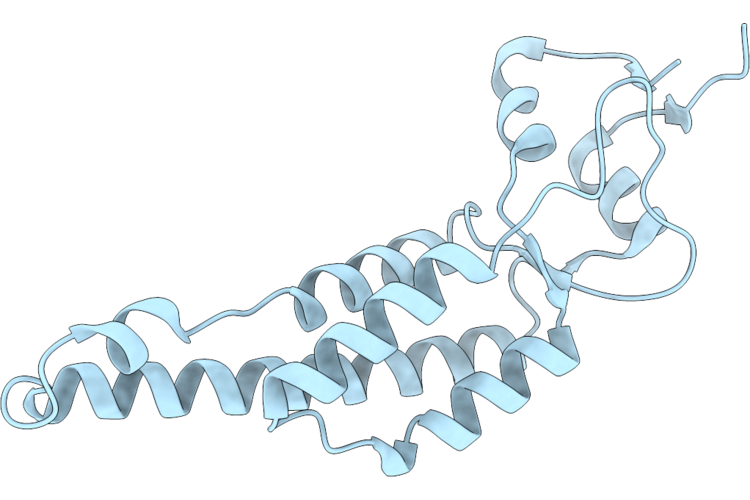 |
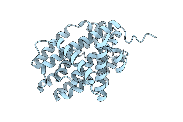
Cucumber Green Mottle Mosaic Virus (Cgmmv) Coat Protein
Organism: Cucumber green mottle mosaic virus (strain watermelon sh)
Method: ELECTRON MICROSCOPY Resolution:2.86 Å Release Date: 2026-07-15 Classification: VIRAL PROTEIN |
Organism: Cucumber green mottle mosaic virus (strain watermelon sh)
Method: ELECTRON MICROSCOPY
Release Date: 2026-07-15
 |
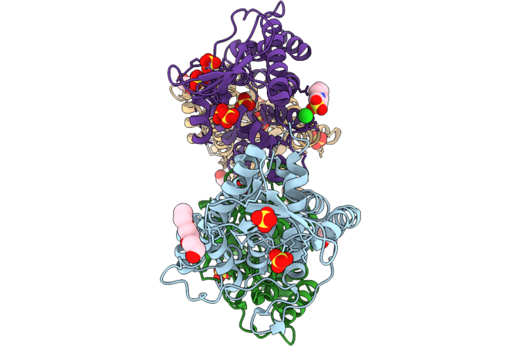
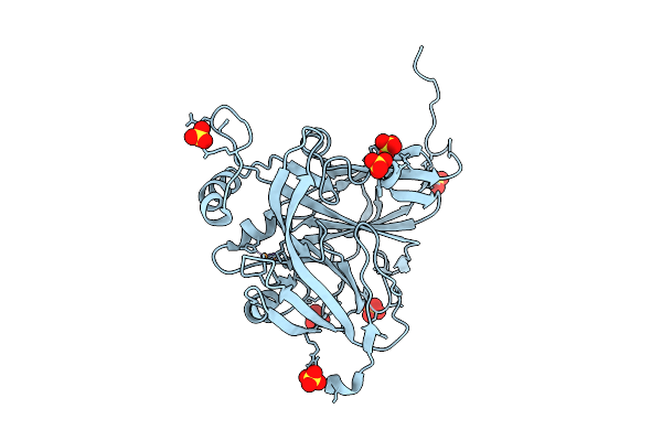
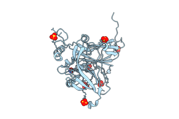
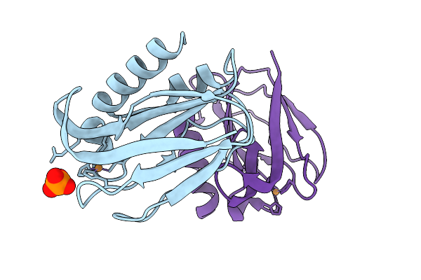
Cryo-Em Structure Of A Soluble Hcv E1E2 Antigen In Complex With At1211 Fab
Organism: Hepacivirus hominis, Homo sapiens
Method: ELECTRON MICROSCOPY Resolution:3.06 Å Release Date: 2026-07-01 Classification: VIRAL PROTEIN Ligands: NAG |
Organism: Hepacivirus hominis, Homo sapiens
Method: ELECTRON MICROSCOPY
Release Date: 2026-07-01
Ligands: NAG
 |
Crystal Structure Of Conserved Hypothetical Protein From Stenotrophomonas Maltophilia (Strain K279A)
Organism: Stenotrophomonas maltophilia k279a
Method: X-RAY DIFFRACTION Resolution:1.85 Å Release Date: 2026-06-17 Classification: UNKNOWN FUNCTION Ligands: SO4, PGE, CL, EDO, EPE |
Organism: Stenotrophomonas maltophilia k279a
Method: X-RAY DIFFRACTION
Release Date: 2026-06-17
Ligands: SO4, PGE, CL, EDO, EPE
 |
Crystal Structure Of Alcohol Oxidase Pcaox(M59V/Q60P/R61N)(Phanerochaete Chrysosporium)
Organism: Phanerodontia chrysosporium
Method: X-RAY DIFFRACTION Resolution:2.90 Å Release Date: 2026-06-03 Classification: OXIDOREDUCTASE Ligands: FAD, SO4 |
Organism: Phanerodontia chrysosporium
Method: X-RAY DIFFRACTION
Release Date: 2026-06-03
Ligands: FAD, SO4
 |
Cryo-Electron Microscopic Structure Of A Novel Amidohydrolase With Three Mutations
Organism: Novilysobacter luteus
Method: ELECTRON MICROSCOPY Resolution:2.69 Å Release Date: 2026-05-06 Classification: HYDROLASE Ligands: ZN, 97U |
Organism: Novilysobacter luteus
Method: ELECTRON MICROSCOPY
Release Date: 2026-05-06
Ligands: ZN, 97U
 |
Cryo-Electron Microscopic Structure Of A Highly Efficient Ochratoxin Detoxification Enzyme Lladh
Organism: Novilysobacter luteus
Method: ELECTRON MICROSCOPY Resolution:2.90 Å Release Date: 2026-05-06 Classification: HYDROLASE Ligands: ZN |
Organism: Novilysobacter luteus
Method: ELECTRON MICROSCOPY
Release Date: 2026-05-06
Ligands: ZN
 |
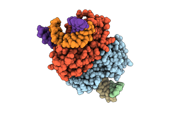
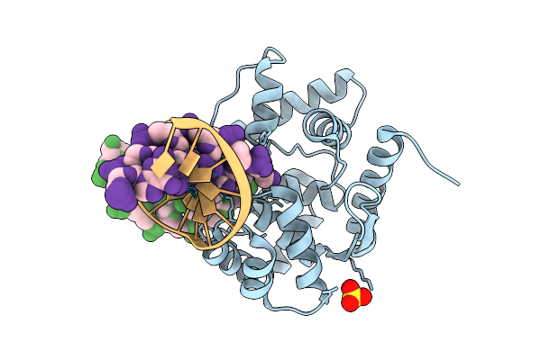
Crystal Structure Of Afnth1:Tg-Dna Duplex Complex In A Pre-Intermediate State
Organism: Archaeoglobus fulgidus, Synthetic construct
Method: X-RAY DIFFRACTION Resolution:3.50 Å Release Date: 2026-04-15 Classification: LYASE/DNA Ligands: SF4 |
Organism: Archaeoglobus fulgidus, Synthetic construct
Method: X-RAY DIFFRACTION
Release Date: 2026-04-15
Ligands: SF4
 |
Crystal Structure Of Afnth1-K122A Mutant Bound To Tg-Dna Duplex Complex In An Intermediate State
Organism: Archaeoglobus fulgidus, Synthetic construct
Method: X-RAY DIFFRACTION Resolution:3.10 Å Release Date: 2026-04-15 Classification: LYASE/DNA Ligands: SF4 |
Organism: Archaeoglobus fulgidus, Synthetic construct
Method: X-RAY DIFFRACTION
Release Date: 2026-04-15
Ligands: SF4
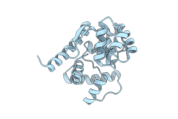 |
Crystal Structure Of Afogg1 In A Dna-Free State
Organism: Archaeoglobus fulgidus
Method: X-RAY DIFFRACTION Resolution:1.93 Å Release Date: 2026-04-15 Classification: HYDROLASE, LYASE |
Organism: Archaeoglobus fulgidus
Method: X-RAY DIFFRACTION
Release Date: 2026-04-15
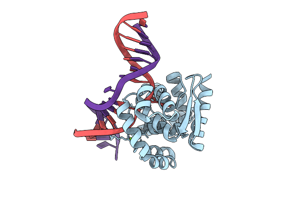 |
Crystal Structure Of Afogg1-D140N Mutant Bound To 8-Og Dna Duplex In An Intermediate State
Organism: Archaeoglobus fulgidus, Synthetic construct
Method: X-RAY DIFFRACTION Resolution:1.95 Å Release Date: 2026-04-15 Classification: HYDROLASE, LYASE/DNA Ligands: MG |
Organism: Archaeoglobus fulgidus, Synthetic construct
Method: X-RAY DIFFRACTION
Release Date: 2026-04-15
Ligands: MG
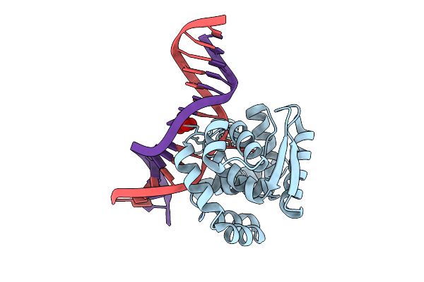 |
Crystal Structure Of Afogg1-K122S Mutant Bound To 8-Og Dna Duplex In An Intermediate State
Organism: Archaeoglobus fulgidus, Synthetic construct
Method: X-RAY DIFFRACTION Resolution:2.50 Å Release Date: 2026-04-15 Classification: HYDROLASE, LYASE/DNA Ligands: MOO |
Organism: Archaeoglobus fulgidus, Synthetic construct
Method: X-RAY DIFFRACTION
Release Date: 2026-04-15
Ligands: MOO
 |
Crystal Structure Of Afogg1:8Og-Dna Duplex In A Product-Bound State
Organism: Archaeoglobus fulgidus, Synthetic construct
Method: X-RAY DIFFRACTION Resolution:2.00 Å Release Date: 2026-04-15 Classification: HYDROLASE, LYASE/DNA Ligands: SO4 |
Organism: Archaeoglobus fulgidus, Synthetic construct
Method: X-RAY DIFFRACTION
Release Date: 2026-04-15
Ligands: SO4
 |
Crystal Structure Of Isoprimeverose-Producing Enzyme From Phaeoacremonium Minimum
Organism: Rattus rattus, Phaeoacremonium minimum
Method: X-RAY DIFFRACTION Resolution:2.80 Å Release Date: 2026-04-08 Classification: HYDROLASE Ligands: NAG, ACT, PG4, CA, PGE, P6G |
Organism: Rattus rattus, Phaeoacremonium minimum
Method: X-RAY DIFFRACTION
Release Date: 2026-04-08
Ligands: NAG, ACT, PG4, CA, PGE, P6G
 |
Structure Of The Cyanobacteria Specific Prk From Gloeocapsa Pcc 73106
Organism: Gloeocapsa sp. pcc 73106
Method: X-RAY DIFFRACTION Resolution:3.20 Å Release Date: 2026-04-08 Classification: PHOTOSYNTHESIS |
Organism: Gloeocapsa sp. pcc 73106
Method: X-RAY DIFFRACTION
Release Date: 2026-04-08
 |
O-Acetylserine Sulfhydrylase A (Cysk) From Neisseria Gonorrhoeae
Organism: Neisseria gonorrhoeae fa 1090
Method: X-RAY DIFFRACTION Resolution:2.50 Å Release Date: 2026-04-01 Classification: BIOSYNTHETIC PROTEIN Ligands: SO4, PEG, GOL |
Organism: Neisseria gonorrhoeae fa 1090
Method: X-RAY DIFFRACTION
Release Date: 2026-04-01
Ligands: SO4, PEG, GOL
 |
Crystal Structure Of Selenomethionine-Substituted N-Oxygenase Rohs From Pseudomonas Syringae Pv. Tomato Str. Dc3000 (Pstorohs)
Organism: Pseudomonas syringae pv. tomato str. dc3000
Method: X-RAY DIFFRACTION Resolution:2.40 Å Release Date: 2026-04-01 Classification: BIOSYNTHETIC PROTEIN |
Organism: Pseudomonas syringae pv. tomato str. dc3000
Method: X-RAY DIFFRACTION
Release Date: 2026-04-01
 |
Sub-Atomic Resolution (0.95 A) Xfel Structure Of As-Isolated Copper Nitrite Reductase From Achromobacter Cycloclastes Determined By Serial Femtosecond Rotation Crystallography (Sf-Rox)
Organism: Achromobacter cycloclastes
Method: X-RAY DIFFRACTION Resolution:0.95 Å Release Date: 2026-03-18 Classification: OXIDOREDUCTASE Ligands: CU, SO4 |
Organism: Achromobacter cycloclastes
Method: X-RAY DIFFRACTION
Release Date: 2026-03-18
Ligands: CU, SO4
 |
Sub-Atomic Resolution (0.95 A) Xfel Structure Of Nitrite-Bound Copper Nitrite Reductase From Achromobacter Cycloclastes Determined By Serial Femtosecond Rotation Crystallography (Sf-Rox) At 100 K
Organism: Achromobacter cycloclastes
Method: X-RAY DIFFRACTION Resolution:0.95 Å Release Date: 2026-03-18 Classification: OXIDOREDUCTASE Ligands: CU, SO4, NO2 |
Organism: Achromobacter cycloclastes
Method: X-RAY DIFFRACTION
Release Date: 2026-03-18
Ligands: CU, SO4, NO2
 |
X-Ray Crystal Structure Of Pseudoazurin Met16Arg Variant, Oxidized Form At Ph 7.0
Organism: Achromobacter cycloclastes
Method: X-RAY DIFFRACTION Resolution:1.22 Å Release Date: 2026-03-11 Classification: ELECTRON TRANSPORT Ligands: CU, PO4 |
Organism: Achromobacter cycloclastes
Method: X-RAY DIFFRACTION
Release Date: 2026-03-11
Ligands: CU, PO4
 |
Low-Dose (14.9 Kgy) Structure Of Copper-Containing Nitrite Reductase From Achromobacter Cycloclastes At Room Temperature
Organism: Achromobacter cycloclastes
Method: X-RAY DIFFRACTION Resolution:1.60 Å Release Date: 2026-03-04 Classification: OXIDOREDUCTASE Ligands: CU, SO4 |
Organism: Achromobacter cycloclastes
Method: X-RAY DIFFRACTION
Release Date: 2026-03-04
Ligands: CU, SO4