

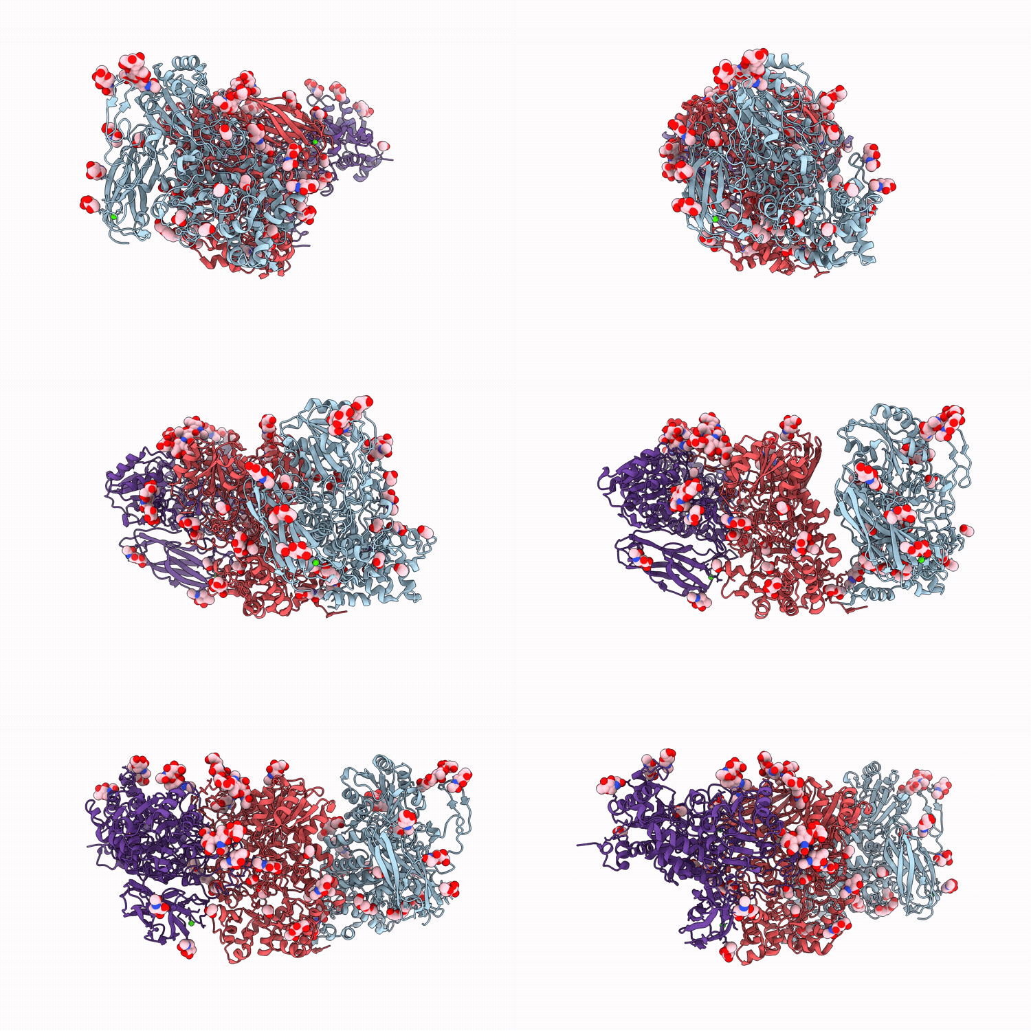
21JI image
Deposition Date 2025-12-15
Release Date 2026-04-08
Last Version Date 2026-04-15
Entry Detail
PDB ID:
21JI
Keywords:
Title:
Crystal structure of isoprimeverose-producing enzyme from Phaeoacremonium minimum
Biological Source:
Source Organism(s):
Expression System(s):
Method Details:
Experimental Method:
Resolution:
2.80 Å
R-Value Free:
0.26
R-Value Work:
0.21
R-Value Observed:
0.21
Space Group:
C 1 2 1

Macromolecular Entities
Protein Blast
Polymer Type:polypeptide(L)
Molecule:Isoprimeverose-producing olig
Gene (Uniprot):RAG, UCRPA7_4649
Chain IDs:A, B, C
Chain Length:871
Number of Molecules:3
Biological Source:Rattus rattus, Phaeoacremonium minimum
Primary Citation
Structural basis for galactose side-chain recognition in isoprimeverose-producing oligoxyloglucan hydrolase from Phaeoacremonium minimum.
Int. J. Biol. Macromol. 357 151665 151665 (2026)
PMID: 41903633 DOI: 10.1016/j.ijbiomac.2026.151665

Abstact

Isoprimeverose-producing oligoxyloglucan hydrolases (IPases), which belong to glycoside hydrolase family 3 (GH3), release isoprimeverose (alpha-d-xylopyranosyl-(1-->6)-d-glucopyranose) from the non-reducing end of xyloglucan oligosaccharides. The IPase from Phaeoacremonium minimum (PmIPase) exhibits distinctive features compared to its homologous IPase in Aspergillus oryzae, IpeA. The hydrolytic activity of A. oryzae IpeA was inhibited by galactosylation of the second xylopyranosyl residue at the nonreducing end of the substrate, whereas PmIPase was unaffected. To elucidate the structural basis of the unique features of PmIPase, its crystal structure was determined. PmIPase forms a homo-dimer, and its active site is formed by two monomers. Structural comparison with IpeA revealed that the active-site structures are highly conserved between both enzymes, but with some differences, including Phe172 at subsite -1 and Trp624 at a positive subsite in PmIPase (Met173 and Ile625 in IpeA, respectively). Trp624 is from the adjacent monomer and seems to be located at the putative binding position of the galactose residue at subsite +1. When the substrate was a mixture of XXXG, XXLG/XLXG, and XLLG, the W624I mutant showed lower specificity for XLLG than XXXG and XXLG/XLXG, although the wild-type enzyme degraded all three substrates with equal efficiency. This result indicates that Trp624 in PmIPase is involved in the recognition of the galactose residue at subsite +1. These findings provide structural insights into the characteristics and diversity of IPases that adapt to the side chain structures of xyloglucan.

Legend

Protein

Chemical

Disease

Primary Citation of related structures
Feedback Form
Name
Email
Institute
Feedback