
Search Count: 351
All
Selected
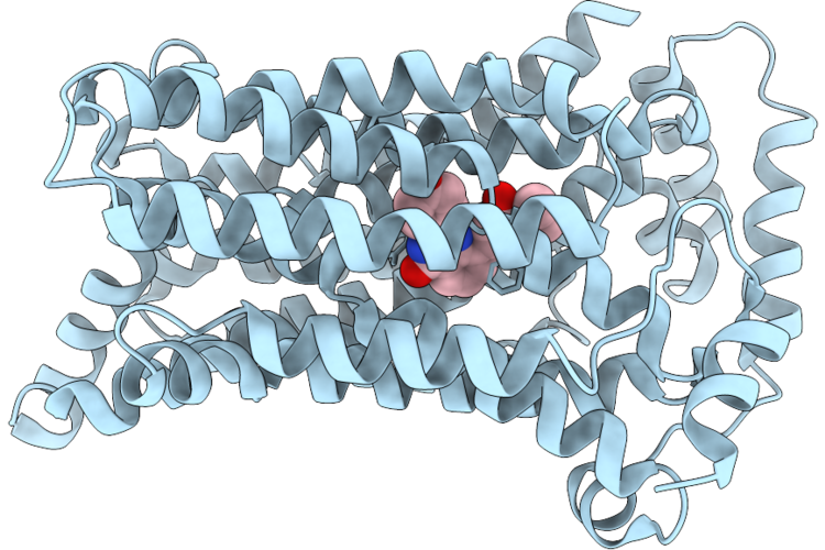 |
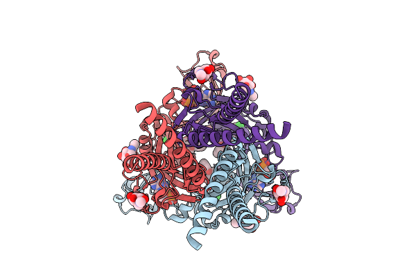
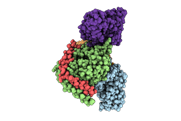
Cryo-Em Structure Of Human Urate Transporter Glut9 Bound To A Selective Inhibitor Sg4
Organism: Homo sapiens
Method: ELECTRON MICROSCOPY Release Date: 2026-05-13 Classification: TRANSPORT PROTEIN Ligands: A1ETZ |
 |
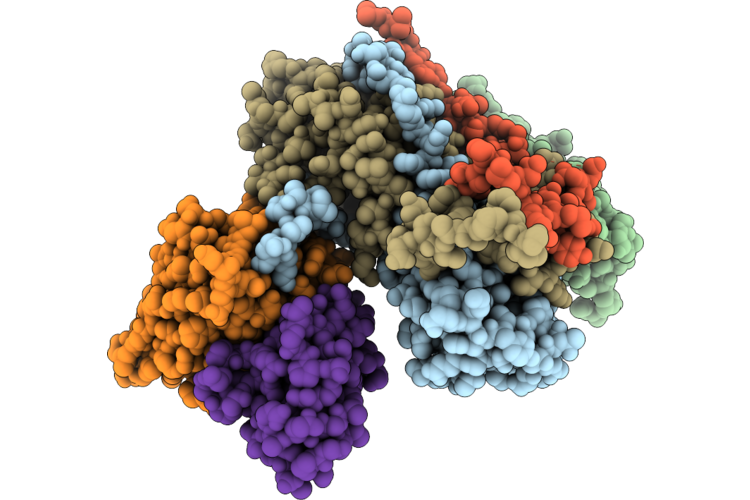
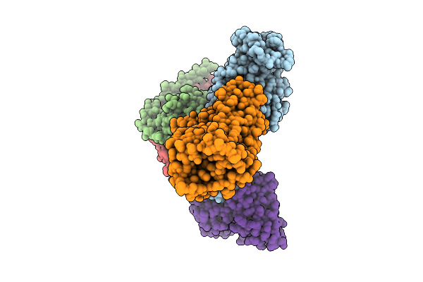
Organism: Listeria monocytogenes egd-e
Method: ELECTRON MICROSCOPY Release Date: 2026-05-06 Classification: TRANSCRIPTION |
 |
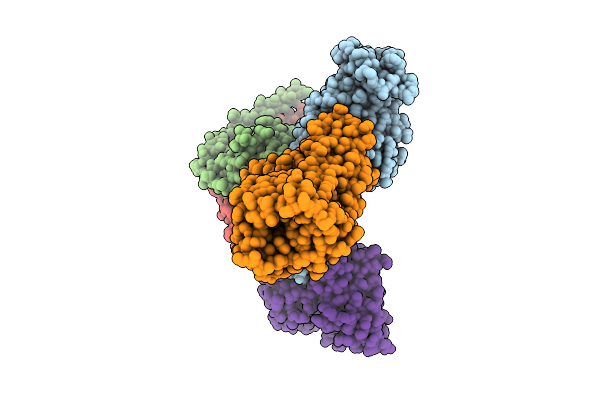
Organism: Listeria monocytogenes
Method: ELECTRON MICROSCOPY Release Date: 2026-05-06 Classification: TRANSCRIPTION |
 |
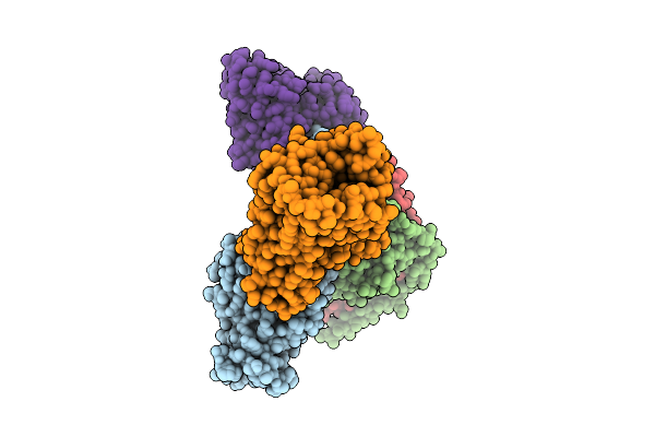
Organism: Listeria monocytogenes
Method: ELECTRON MICROSCOPY Release Date: 2026-05-06 Classification: TRANSCRIPTION |
 |
Organism: Listeria monocytogenes
Method: ELECTRON MICROSCOPY Release Date: 2026-05-06 Classification: TRANSCRIPTION |
 |
Organism: Listeria monocytogenes egd-e
Method: ELECTRON MICROSCOPY Release Date: 2026-05-06 Classification: TRANSCRIPTION |
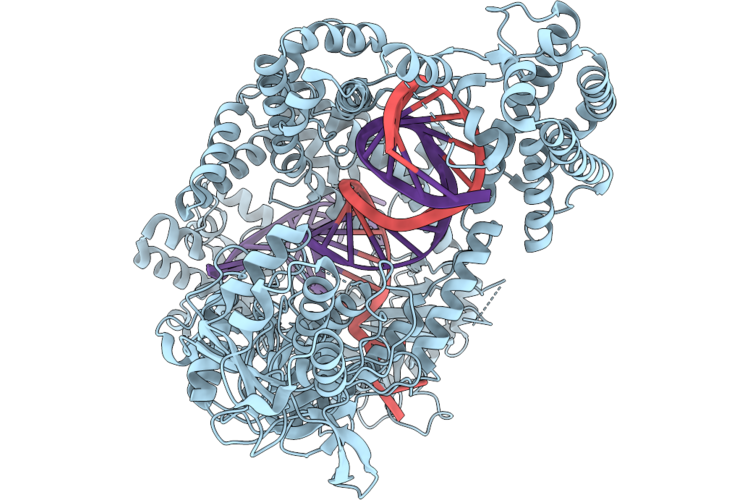 |
Organism: Acidaminococcus sp. bv3l6, Synthetic construct
Method: ELECTRON MICROSCOPY Resolution:3.17 Å Release Date: 2026-04-29 Classification: DNA BINDING PROTEIN |
 |
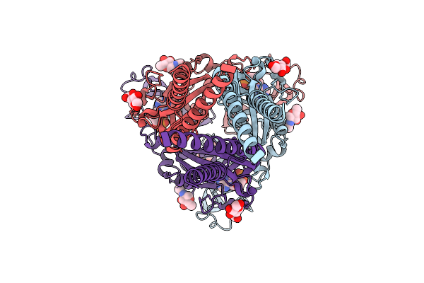
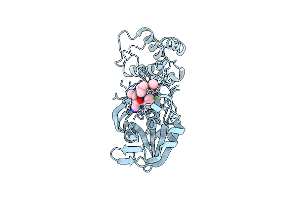
Crystal Structure Of The V165A/S219E/A225P Mutant Of Alanine Dehyrogenase From Geobacillus Stearothermophilus In Complex With Nicotinamide Cytosine Dinucleotide
Organism: Geobacillus stearothermophilus
Method: X-RAY DIFFRACTION Resolution:2.56 Å Release Date: 2026-04-29 Classification: OXIDOREDUCTASE Ligands: A1EP0 |
 |
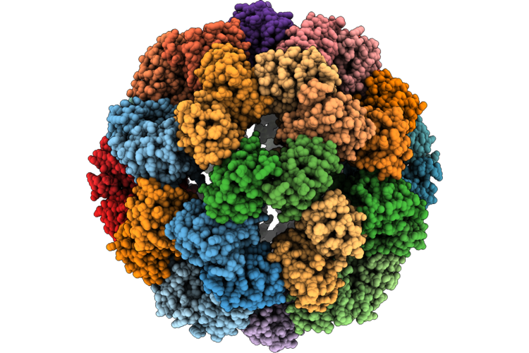
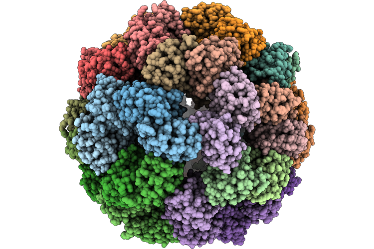
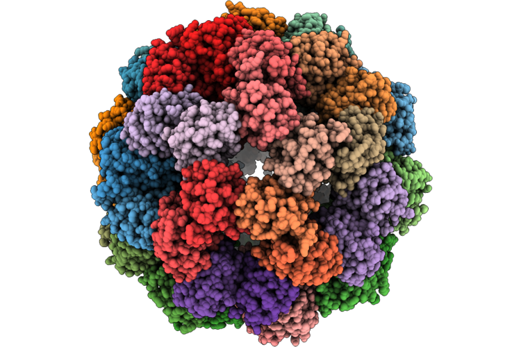
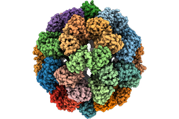
Complex Of Fmdv O/18074 And Porcine-Derived Neutralizing Monoclonal Antibody Po18-10
Organism: Sus scrofa, Foot-and-mouth disease virus o
Method: ELECTRON MICROSCOPY Resolution:2.27 Å Release Date: 2026-04-22 Classification: VIRUS |
 |
Cryo-Em Structure Of The Human P2X3 Receptor In The Atp-Bound, Desensitized State
Organism: Homo sapiens
Method: ELECTRON MICROSCOPY Release Date: 2026-04-01 Classification: TRANSPORT PROTEIN Ligands: ATP, NAG |
 |
Cryo-Em Structure Of The Human P2X3 Receptor In The Atp- And Sivopixant-Bound Closed State
Organism: Homo sapiens
Method: ELECTRON MICROSCOPY Release Date: 2026-04-01 Classification: TRANSPORT PROTEIN Ligands: A1E2N, NAG, ATP |
 |
Organism: Homo sapiens, Synthetic construct
Method: ELECTRON MICROSCOPY Release Date: 2026-03-18 Classification: MEMBRANE PROTEIN/IMMUNE SYSTEM Ligands: AKG |
 |
Organism: Homo sapiens, Synthetic construct
Method: ELECTRON MICROSCOPY Release Date: 2026-03-18 Classification: MEMBRANE PROTEIN/IMMUNE SYSTEM Ligands: ITN |
 |
Organism: Homo sapiens, Synthetic construct
Method: ELECTRON MICROSCOPY Release Date: 2026-03-18 Classification: MEMBRANE PROTEIN/IMMUNE SYSTEM Ligands: A1E2R |
 |
Organism: Homo sapiens, Synthetic construct
Method: ELECTRON MICROSCOPY Release Date: 2026-03-18 Classification: MEMBRANE PROTEIN/IMMUNE SYSTEM |
 |
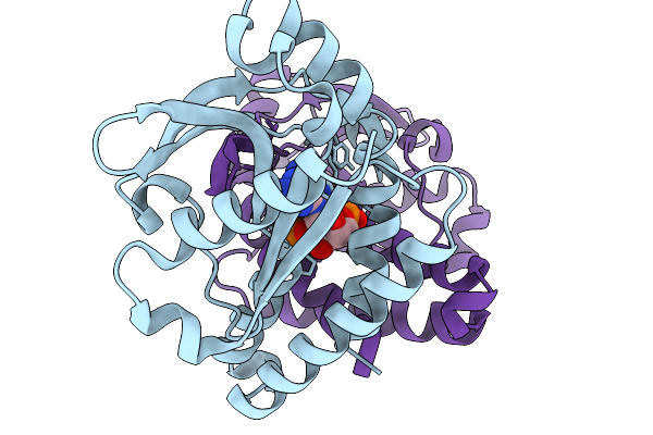
Crystal Structure Of Sars-Cov-2 Main Protease E166N Mutant In Complex With Ibuzatrelvir
Organism: Severe acute respiratory syndrome coronavirus 2
Method: X-RAY DIFFRACTION Resolution:2.07 Å Release Date: 2026-03-11 Classification: VIRAL PROTEIN Ligands: YDL |
 |
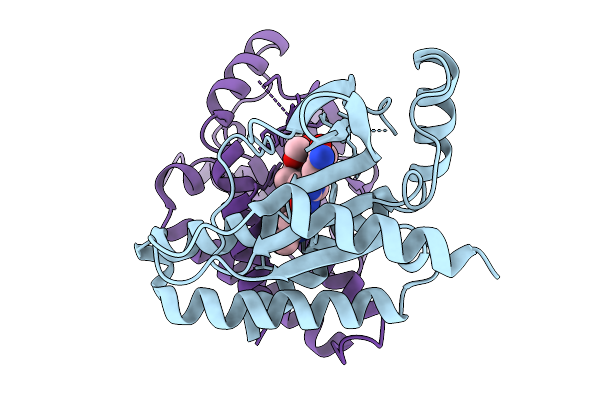
Crystal Structure Of Sars-Cov-2 Main Protease E166R Mutant In Complex With Ibuzatrelvir
Organism: Severe acute respiratory syndrome coronavirus 2
Method: X-RAY DIFFRACTION Resolution:1.92 Å Release Date: 2026-03-11 Classification: VIRAL PROTEIN Ligands: YDL |
 |
Organism: Anas platyrhynchos
Method: X-RAY DIFFRACTION Resolution:2.49 Å Release Date: 2026-03-04 Classification: SIGNALING PROTEIN Ligands: 1SY |
 |
Organism: Homo sapiens
Method: X-RAY DIFFRACTION Resolution:1.90 Å Release Date: 2026-03-04 Classification: SIGNALING PROTEIN Ligands: A1AZ0 |
 |
Organism: Bos taurus
Method: X-RAY DIFFRACTION Resolution:1.81 Å Release Date: 2026-03-04 Classification: SIGNALING PROTEIN Ligands: A1AZ0 |