

Search Count: 2,075
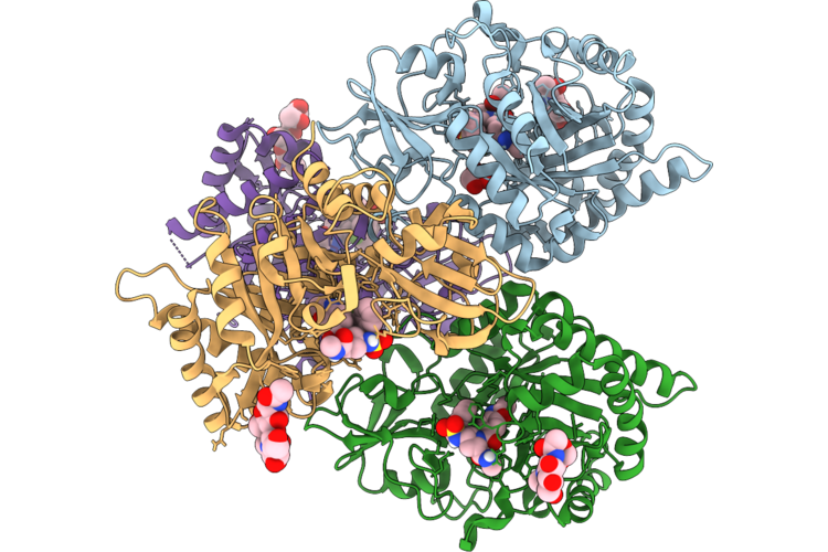 |
Crystal Structure Of Human Chi3L1 In Complex With The Chi-Del Small Molecule
Organism: Homo sapiens
Method: X-RAY DIFFRACTION Resolution:2.81 Å Release Date: 2026-08-05 Classification: HYDROLASE Ligands: GOL, A1C9M |
Organism: Homo sapiens
Method: X-RAY DIFFRACTION
Release Date: 2026-08-05
Ligands: GOL, A1C9M
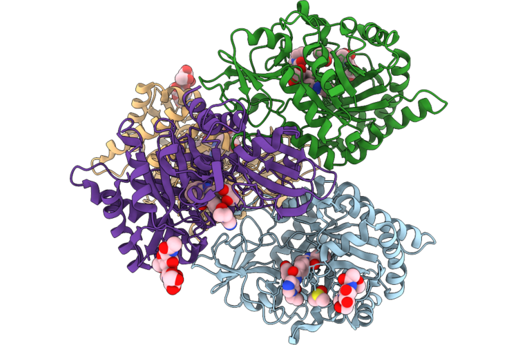 |
Structure Of The Human Chi3L1-Lz6 Complex
Organism: Homo sapiens
Method: X-RAY DIFFRACTION Resolution:2.75 Å Release Date: 2026-08-05 Classification: HYDROLASE Ligands: DMS, GOL, A1DAW |
Organism: Homo sapiens
Method: X-RAY DIFFRACTION
Release Date: 2026-08-05
Ligands: DMS, GOL, A1DAW
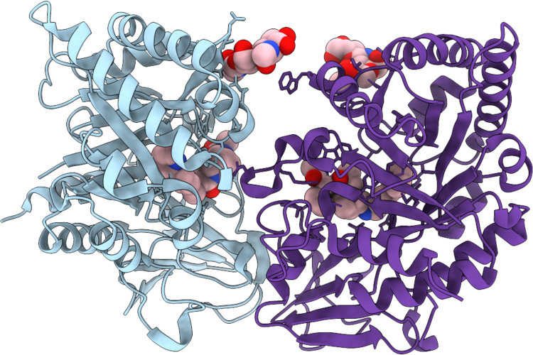 |
Co-Crystal Structure Of Chi3L1 Bound To Delc2 Small Molecule Ligand
Organism: Homo sapiens
Method: X-RAY DIFFRACTION Resolution:2.47 Å Release Date: 2026-08-05 Classification: HYDROLASE Ligands: A1C9L |
Organism: Homo sapiens
Method: X-RAY DIFFRACTION
Release Date: 2026-08-05
Ligands: A1C9L
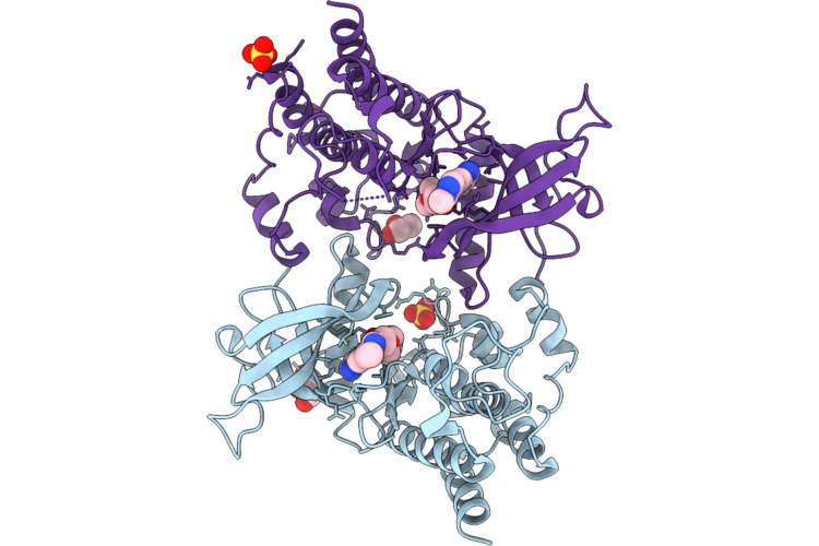 |
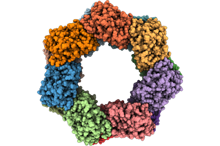
Crystal Structure Of Fgfr2 With A Macrocyclic Compound Xs9001
Organism: Homo sapiens
Method: X-RAY DIFFRACTION Resolution:2.10 Å Release Date: 2026-08-05 Classification: TRANSFERASE Ligands: A1EVI, GOL, SO4 |
Organism: Homo sapiens
Method: X-RAY DIFFRACTION
Release Date: 2026-08-05
Ligands: A1EVI, GOL, SO4
 |
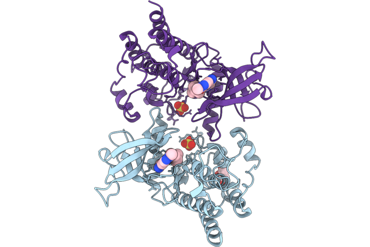
Ycfa From Erwinia Amylovora
Organism: Erwinia amylovora atcc 49946
Method: ELECTRON MICROSCOPY Release Date: 2026-08-05 Classification: BIOSYNTHETIC PROTEIN |
Organism: Erwinia amylovora atcc 49946
Method: ELECTRON MICROSCOPY
Release Date: 2026-08-05
 |
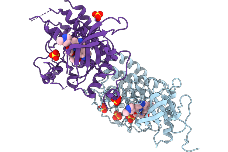
Crystal Structure Of Fgfr2 With A Macrocyclic Compound Xs8148
Organism: Homo sapiens
Method: X-RAY DIFFRACTION Resolution:2.63 Å Release Date: 2026-07-29 Classification: TRANSFERASE Ligands: A1EUM, SO4, GOL |
Organism: Homo sapiens
Method: X-RAY DIFFRACTION
Release Date: 2026-07-29
Ligands: A1EUM, SO4, GOL
 |
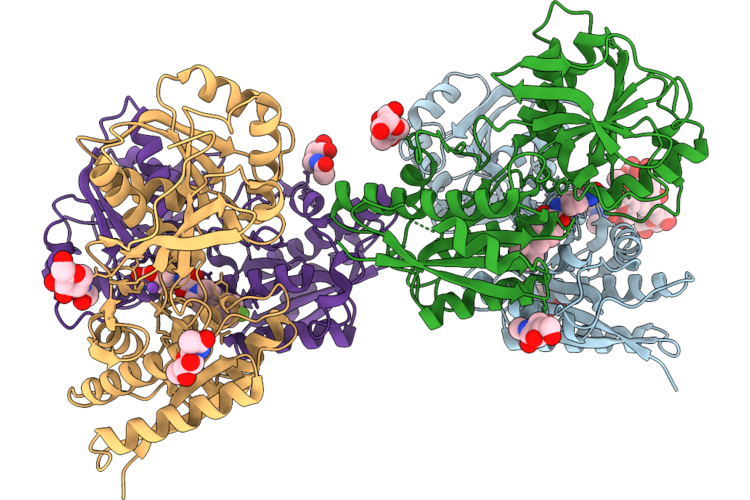
Crystal Structure Of Fgfr1 With A Macrocyclic Compound Xs8148
Organism: Homo sapiens
Method: X-RAY DIFFRACTION Resolution:1.96 Å Release Date: 2026-07-29 Classification: TRANSFERASE Ligands: A1EUM, SO4 |
Organism: Homo sapiens
Method: X-RAY DIFFRACTION
Release Date: 2026-07-29
Ligands: A1EUM, SO4
 |
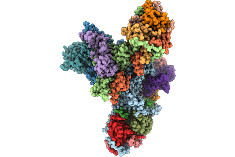
Glun1B-Glun2B Atd In Complex With Radiprodil
Organism: Xenopus laevis, Rattus norvegicus
Method: X-RAY DIFFRACTION Resolution:2.74 Å Release Date: 2026-07-22 Classification: MEMBRANE PROTEIN Ligands: NAG, A1CNW, NA, CL |
Organism: Xenopus laevis, Rattus norvegicus
Method: X-RAY DIFFRACTION
Release Date: 2026-07-22
Ligands: NAG, A1CNW, NA, CL
 |
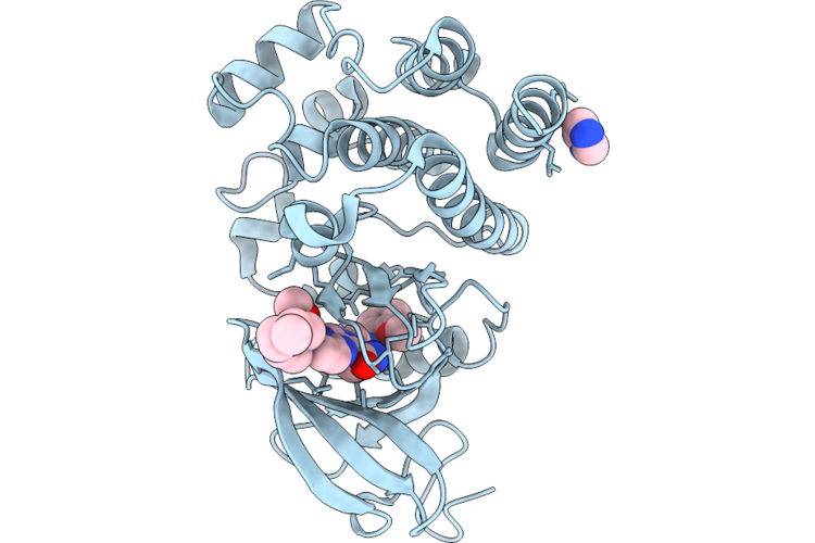
Trypanosoma Brucei Mitochondrial Rna-Editing Catalytic Complex 2, U-Insertion (Recc2)
Organism: Trypanosoma brucei
Method: ELECTRON MICROSCOPY Release Date: 2026-07-22 Classification: RNA BINDING PROTEIN Ligands: ZN, MG |
Organism: Trypanosoma brucei
Method: ELECTRON MICROSCOPY
Release Date: 2026-07-22
Ligands: ZN, MG
 |
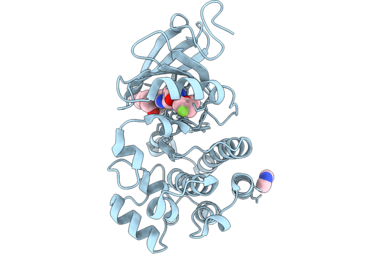
Co-Crystal Structure Of Btk Kinase Domain With Non-Covalent Inhibitor
Organism: Homo sapiens
Method: X-RAY DIFFRACTION Resolution:1.60 Å Release Date: 2026-07-15 Classification: TRANSFERASE Ligands: IMD, A1CSI |
Organism: Homo sapiens
Method: X-RAY DIFFRACTION
Release Date: 2026-07-15
Ligands: IMD, A1CSI
 |
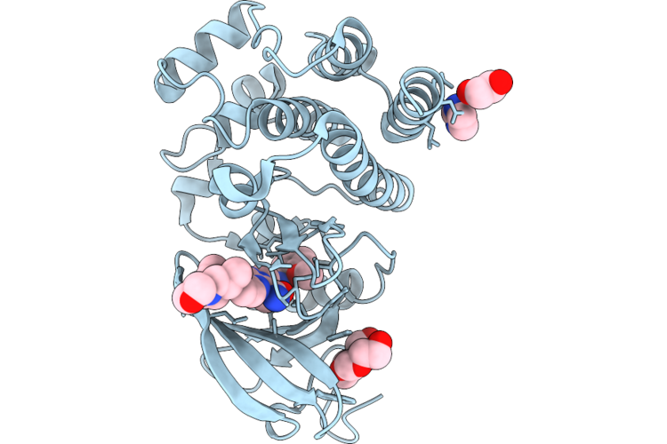
Co-Crystal Structure Of Btk Kinase Domain With Non-Covalent Inhibitor
Organism: Homo sapiens
Method: X-RAY DIFFRACTION Resolution:1.40 Å Release Date: 2026-07-15 Classification: TRANSFERASE Ligands: IMD, A1CSJ |
Organism: Homo sapiens
Method: X-RAY DIFFRACTION
Release Date: 2026-07-15
Ligands: IMD, A1CSJ
 |
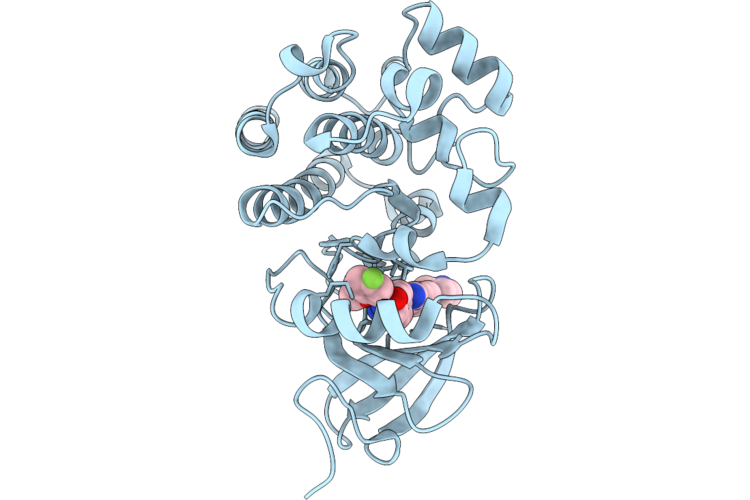
Co-Crystal Structure Of Btk Kinase Domain With Non-Covalent Inhibitor
Organism: Homo sapiens
Method: X-RAY DIFFRACTION Resolution:1.70 Å Release Date: 2026-07-15 Classification: TRANSFERASE Ligands: IMD, PG4, A1CSK, EDO |
Organism: Homo sapiens
Method: X-RAY DIFFRACTION
Release Date: 2026-07-15
Ligands: IMD, PG4, A1CSK, EDO
 |
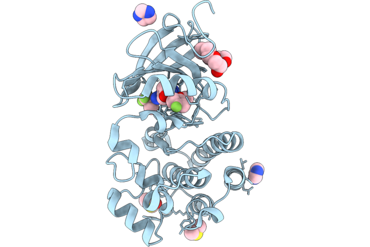
Co-Crystal Structure Of Btk Kinase Domain With Non-Covalent Inhibitor
Organism: Homo sapiens
Method: X-RAY DIFFRACTION Resolution:1.90 Å Release Date: 2026-07-15 Classification: TRANSFERASE Ligands: A1CSL |
Organism: Homo sapiens
Method: X-RAY DIFFRACTION
Release Date: 2026-07-15
Ligands: A1CSL
 |
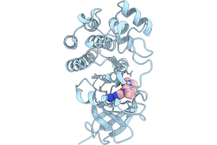
Co-Crystal Structure Of Btk Kinase Domain With Non-Covalent Inhibitor
Organism: Homo sapiens
Method: X-RAY DIFFRACTION Resolution:1.66 Å Release Date: 2026-07-15 Classification: TRANSFERASE Ligands: IMD, 1PE, DMS, A1CSM |
Organism: Homo sapiens
Method: X-RAY DIFFRACTION
Release Date: 2026-07-15
Ligands: IMD, 1PE, DMS, A1CSM
 |
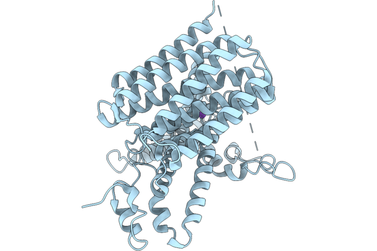
Co-Crystal Structure Of Btk Kinase Domain With Non-Covalent Inhibitor
Organism: Homo sapiens
Method: X-RAY DIFFRACTION Resolution:2.51 Å Release Date: 2026-07-15 Classification: TRANSFERASE Ligands: A1CSG |
Organism: Homo sapiens
Method: X-RAY DIFFRACTION
Release Date: 2026-07-15
Ligands: A1CSG
 |
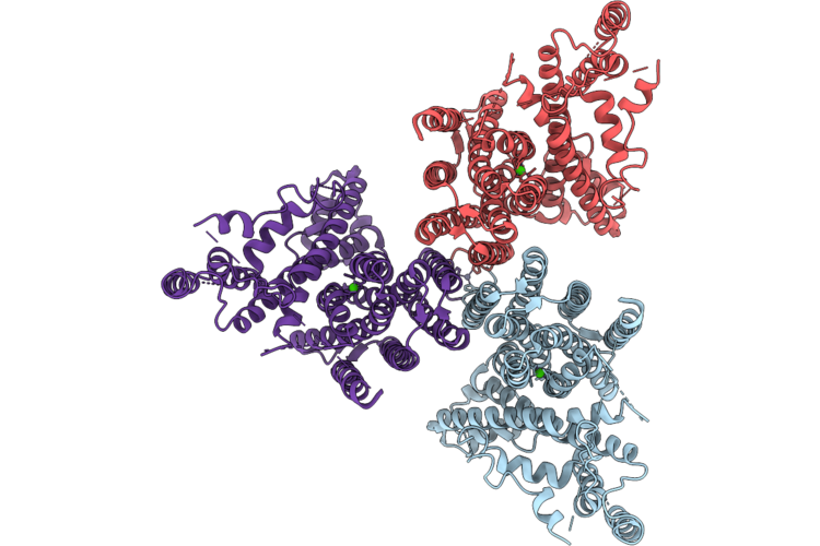
Cryo-Em Structure Of The Mitochondrial Nclx In Its Monomeric State
Organism: Rattus norvegicus
Method: ELECTRON MICROSCOPY Resolution:3.62 Å Release Date: 2026-07-15 Classification: MEMBRANE PROTEIN |
Organism: Rattus norvegicus
Method: ELECTRON MICROSCOPY
Release Date: 2026-07-15
 |
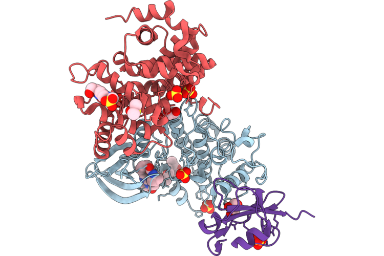
Cryo-Em Structure Of The Mitochondrial Nclx In Its Trimeric State
Organism: Rattus norvegicus
Method: ELECTRON MICROSCOPY Resolution:3.15 Å Release Date: 2026-07-15 Classification: MEMBRANE PROTEIN Ligands: CA, ZN |
Organism: Rattus norvegicus
Method: ELECTRON MICROSCOPY
Release Date: 2026-07-15
Ligands: CA, ZN
 |
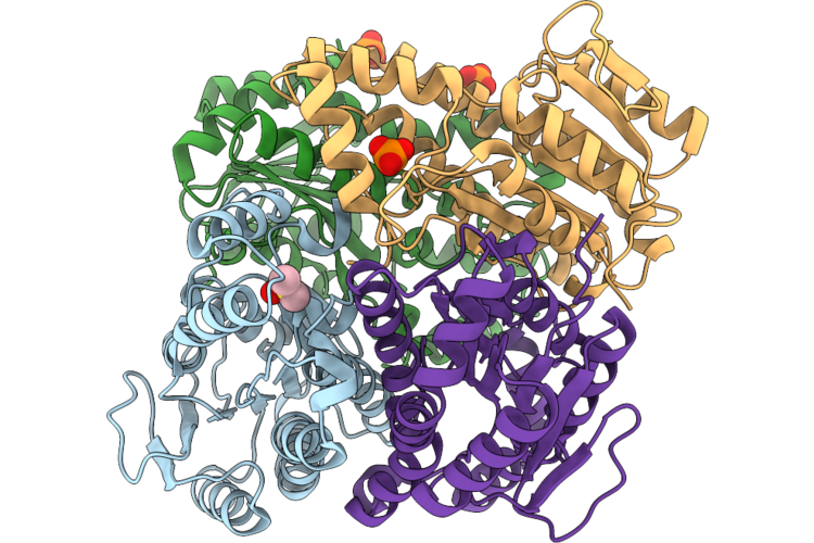
Crystal Structure Of Selective Inhibitor 16 Bound At The Active Site Of Cdk1
Organism: Homo sapiens
Method: X-RAY DIFFRACTION Resolution:2.40 Å Release Date: 2026-07-08 Classification: SIGNALING PROTEIN Ligands: A1DEQ, A1DJL, GOL, SO4, DMS, EPE |
Organism: Homo sapiens
Method: X-RAY DIFFRACTION
Release Date: 2026-07-08
Ligands: A1DEQ, A1DJL, GOL, SO4, DMS, EPE
 |
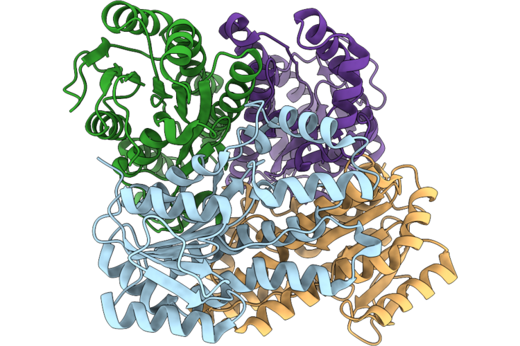
Crystal Structure Of Kred Mutant-Y199A/N149L
Organism: Novosphingobium aromaticivorans dsm 12444
Method: X-RAY DIFFRACTION Resolution:2.94 Å Release Date: 2026-07-08 Classification: OXIDOREDUCTASE Ligands: DMS, PO4 |
Organism: Novosphingobium aromaticivorans dsm 12444
Method: X-RAY DIFFRACTION
Release Date: 2026-07-08
Ligands: DMS, PO4
 |
Crystal Structure Of Kred
Organism: Novosphingobium aromaticivorans dsm 12444
Method: X-RAY DIFFRACTION Resolution:2.53 Å Release Date: 2026-07-08 Classification: OXIDOREDUCTASE |
Organism: Novosphingobium aromaticivorans dsm 12444
Method: X-RAY DIFFRACTION
Release Date: 2026-07-08