
Search Count: 3,353
 |
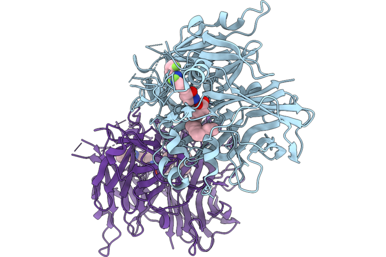
Organism: Homo sapiens
Method: X-RAY DIFFRACTION Resolution:1.70 Å Release Date: 2026-06-03 Classification: ONCOPROTEIN Ligands: A1C3J, GDP, MG, PO4 |
 |
Organism: Homo sapiens
Method: X-RAY DIFFRACTION Resolution:1.68 Å Release Date: 2026-06-03 Classification: ONCOPROTEIN Ligands: MG, A1C3N, GDP, SO4 |
 |
Organism: Homo sapiens
Method: X-RAY DIFFRACTION Resolution:1.91 Å Release Date: 2026-06-03 Classification: ONCOPROTEIN Ligands: MG, A1C3O, GDP |
 |
Organism: Bos taurus
Method: X-RAY DIFFRACTION Resolution:1.75 Å Release Date: 2026-05-27 Classification: HYDROLASE Ligands: FE2, PLM, A1B98 |
 |
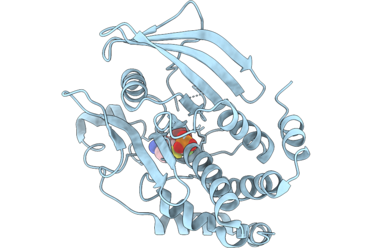
Structure Of Ptp1B Complexed With Difluoromethylphosphonate Inhibitor Compound 2
Organism: Homo sapiens
Method: X-RAY DIFFRACTION Resolution:1.73 Å Release Date: 2026-05-27 Classification: HYDROLASE/INHIBITOR Ligands: A1C3A |
 |
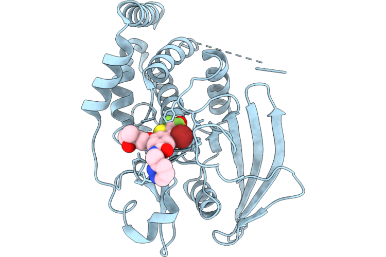
Structure Of Ptp1B Complexed With Difluoromethylphosphonate Inhibitor Compound 10
Organism: Homo sapiens
Method: X-RAY DIFFRACTION Resolution:2.51 Å Release Date: 2026-05-27 Classification: HYDROLASE/INHIBITOR Ligands: A1C3B |
 |
Structure Of Ptp1B Complexed With Difluoromethylphosphonate Inhibitor Compound 15
Organism: Homo sapiens
Method: X-RAY DIFFRACTION Resolution:2.15 Å Release Date: 2026-05-27 Classification: HYDROLASE/INHIBITOR Ligands: A1C3C, DMS |
 |
Structure Of Ptp1B Complexed With Difluoromethylphosphonate Inhibitor Compound 30
Organism: Homo sapiens
Method: X-RAY DIFFRACTION Resolution:1.94 Å Release Date: 2026-05-27 Classification: HYDROLASE/INHIBITOR Ligands: A1C3D |
 |
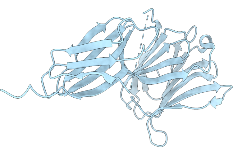
Crystal Structure Of Galectin-3 Bound To Fn3Con-9 And Fn3Con-41, A Cooperative Binder That Recognises The Galectin-3-Fn3Con-9 Interface
Organism: Synthetic construct, Homo sapiens
Method: X-RAY DIFFRACTION Resolution:1.96 Å Release Date: 2026-05-20 Classification: CARBOHYDRATE |
 |
Crystal Structure Of The Galectin-3 Complex With Fn3Con-7, An Mrna Display-Derived Binding Protein
Organism: Homo sapiens, Synthetic construct
Method: X-RAY DIFFRACTION Resolution:1.92 Å Release Date: 2026-05-20 Classification: CARBOHYDRATE |
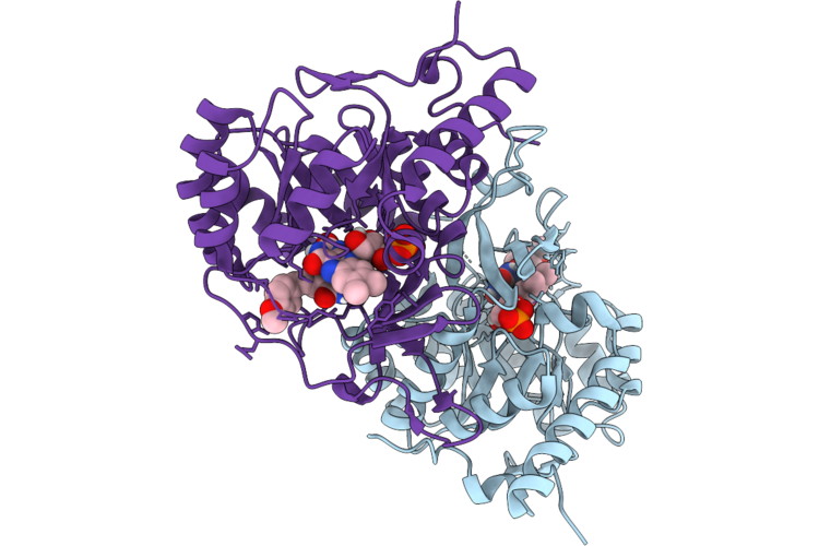 |
Crystal Structure Of Dihydroorotate Dehydrogenase From Leishmania Brasiliensis In Complex With 5-[(E)-3-(P-Methoxyphenyl)-2-Propenylidene]-2,4,6(1H,3H,5H)-Pyrimidinetrione
Organism: Leishmania braziliensis
Method: X-RAY DIFFRACTION Resolution:2.32 Å Release Date: 2026-05-06 Classification: OXIDOREDUCTASE Ligands: FMN, 5TI |
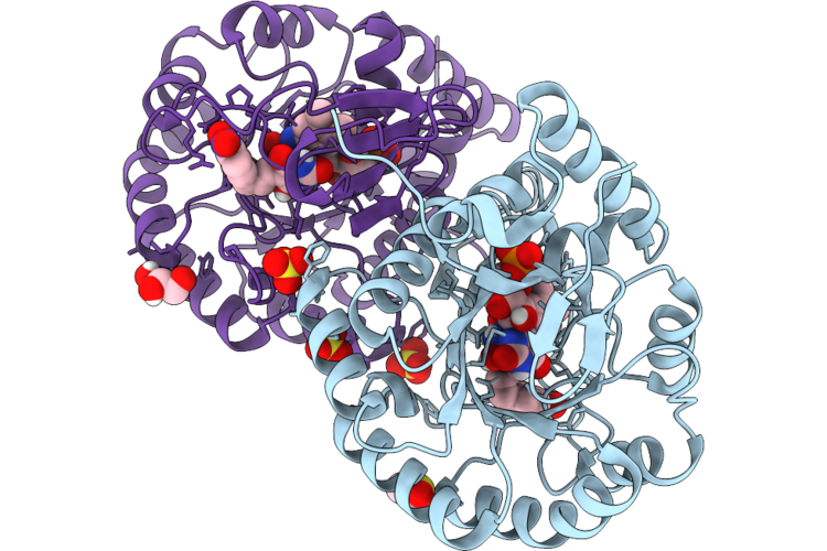 |
Crystal Structure Of Dihydroorotate Dehydrogenase From Leishmania Brasiliensis In Complex With (E)-5-(3-(4-Nitrophenyl)Allylidene)Pyrimidine-2,4,6(1H,3H,5H)-Trione
Organism: Leishmania braziliensis
Method: X-RAY DIFFRACTION Resolution:1.97 Å Release Date: 2026-05-06 Classification: OXIDOREDUCTASE Ligands: FMN, DMS, A1CAU, SO4, GOL |
 |
Organism: Bacillus pumilus atcc 7061
Method: X-RAY DIFFRACTION Resolution:2.16 Å Release Date: 2026-04-29 Classification: HYDROLASE Ligands: GOL, CIT |
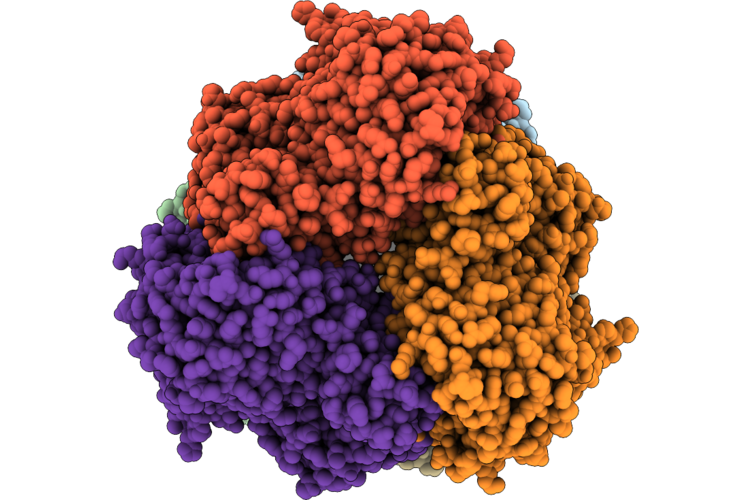 |
Organism: Heyndrickxia coagulans
Method: ELECTRON MICROSCOPY Release Date: 2026-04-29 Classification: HYDROLASE Ligands: ZN, CL |
 |
Organism: Klebsiella oxytoca
Method: X-RAY DIFFRACTION Resolution:1.97 Å Release Date: 2026-04-29 Classification: OXIDOREDUCTASE Ligands: EDO |
 |
Crystal Structure Of Klebsiella Oxytoca Ribitol Dehydrogenase In Complex With D-Allose
Organism: Klebsiella oxytoca
Method: X-RAY DIFFRACTION Resolution:1.73 Å Release Date: 2026-04-29 Classification: OXIDOREDUCTASE Ligands: ALL |
 |
Crystal Structure Of Klebsiella Oxytoca Ribitol Dehydrogenase In Complex With D-Allulose
Organism: Klebsiella oxytoca
Method: X-RAY DIFFRACTION Resolution:1.56 Å Release Date: 2026-04-29 Classification: OXIDOREDUCTASE Ligands: IPA, WEB, PSJ, PSV |
 |
Crystal Structure Of Klebsiella Oxytoca Ribitol Dehydrogenase In Complex With Nad+
Organism: Klebsiella oxytoca
Method: X-RAY DIFFRACTION Resolution:1.89 Å Release Date: 2026-04-29 Classification: OXIDOREDUCTASE Ligands: NAD |
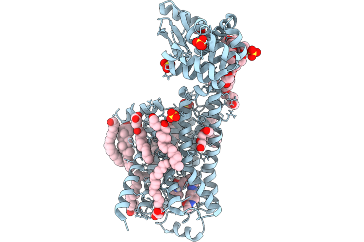 |
Dark Structure Of Beta-2 Adrenergic Receptor With Photoazolol In Dark State Recorded At Swissfel
Organism: Homo sapiens, Enterobacteria phage t4
Method: X-RAY DIFFRACTION Resolution:2.45 Å Release Date: 2026-04-22 Classification: MEMBRANE PROTEIN Ligands: SO4, CLR, 12P, OLC, 1PE, EDO, GOL, A1JHU, PLM |
 |
Mixed Model Refinement Of Beta-2 Adrenergic Receptor With Photoazolol In Dark State And Light State, 10 Seconds After Light Activation, Recorded At Swissfel
Organism: Homo sapiens, Tequatrovirus t4
Method: X-RAY DIFFRACTION Resolution:2.45 Å Release Date: 2026-04-22 Classification: MEMBRANE PROTEIN Ligands: SO4, ACM, CLR, PLM, 12P, OLC, 1PE, EDO, GOL, A1JHU |