
Search Count: 1,09,266
All
Selected
 |
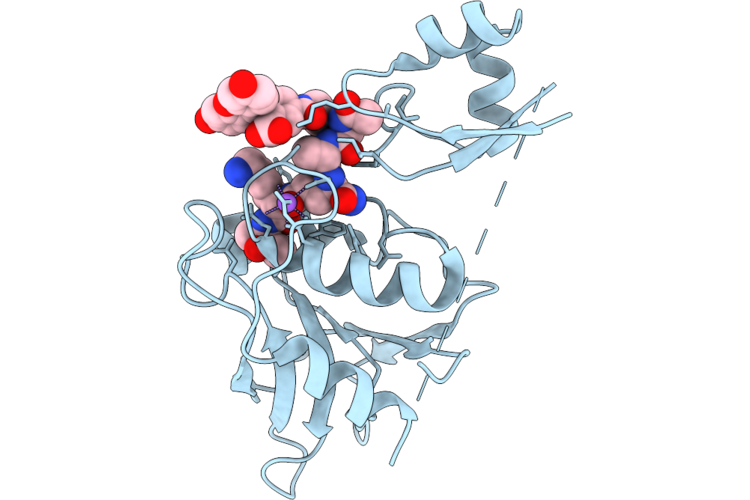
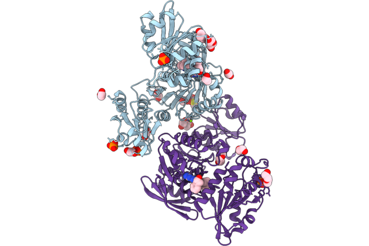
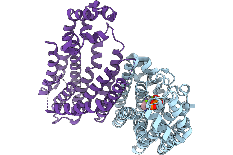
Room Temperature X-Ray Structure Of Sars Cov-2 Main Protease Intermediate Precursor With Ensitrelvir (Esv)
Organism: Homo sapiens
Method: X-RAY DIFFRACTION Resolution:2.05 Å Release Date: 2026-05-06 Classification: HYDROLASE/HYDROLASE INHIBITOR Ligands: 7YY |
 |
Organism: Bacillus thuringiensis
Method: X-RAY DIFFRACTION Resolution:2.00 Å Release Date: 2026-05-06 Classification: TRANSFERASE Ligands: UDP, NOV |
 |
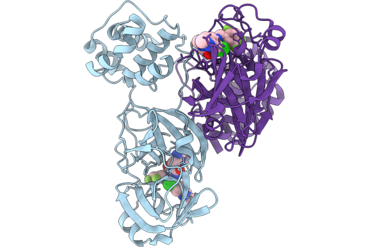
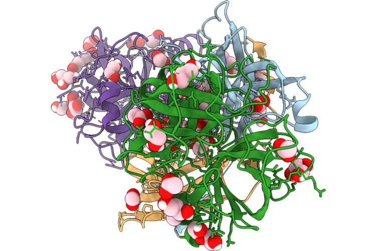
Chap Domain Of Staphylococcus Aureus-Specific Lysin L1 Covalently Complexed To Pep1A-Cmk Substrate Mimic
Organism: Staphylococcus aureus
Method: X-RAY DIFFRACTION Resolution:1.09 Å Release Date: 2026-05-06 Classification: HYDROLASE Ligands: A1DEZ, NA, SCN |
 |
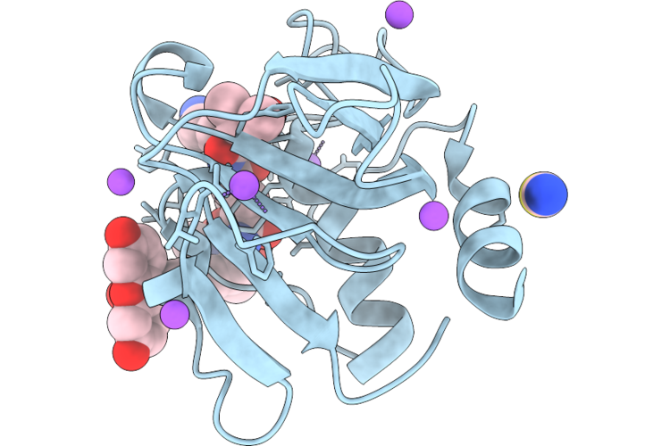
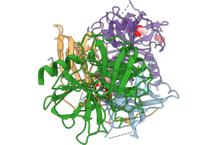
Staphylococcus Aureus-Specific Lysin L1-3 (Lysm-Chap) Covalently Complexed To Pep1A-Cmk Substrate Mimic
Organism: Staphylococcus aureus
Method: X-RAY DIFFRACTION Resolution:2.83 Å Release Date: 2026-05-06 Classification: HYDROLASE Ligands: A1DEZ, NA |
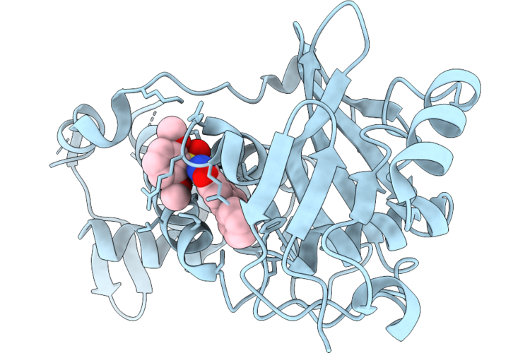 |
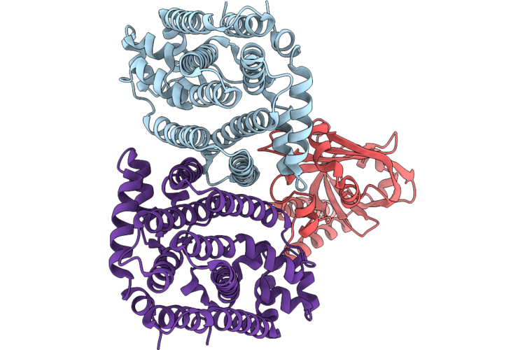
Crystal Structure Of Myst Histone Acetyltransferase Kat6A In Complex With Inhibitor Compound 9
Organism: Homo sapiens
Method: X-RAY DIFFRACTION Resolution:2.29 Å Release Date: 2026-05-06 Classification: TRANSCRIPTION Ligands: A1MGC |
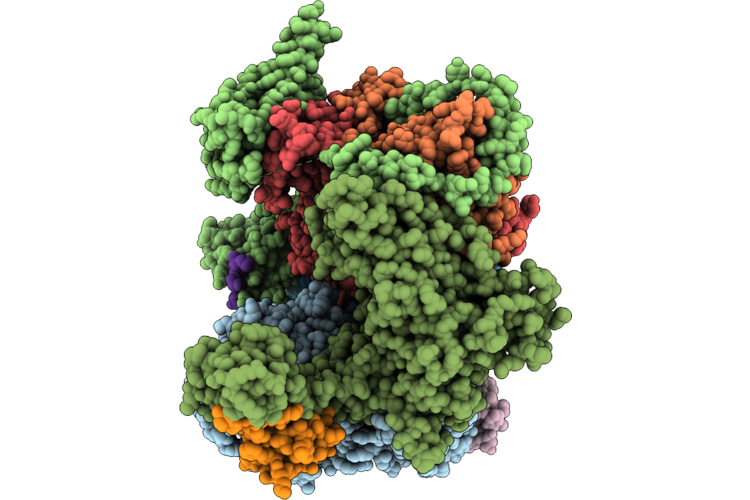 |
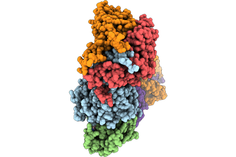
Cryo-Em Structure Of The Human Holo-Tfiih And Xpc Initial Encounter Complex
Organism: Homo sapiens
Method: ELECTRON MICROSCOPY Resolution:3.29 Å Release Date: 2026-05-06 Classification: DNA BINDING PROTEIN Ligands: SF4, ZN |
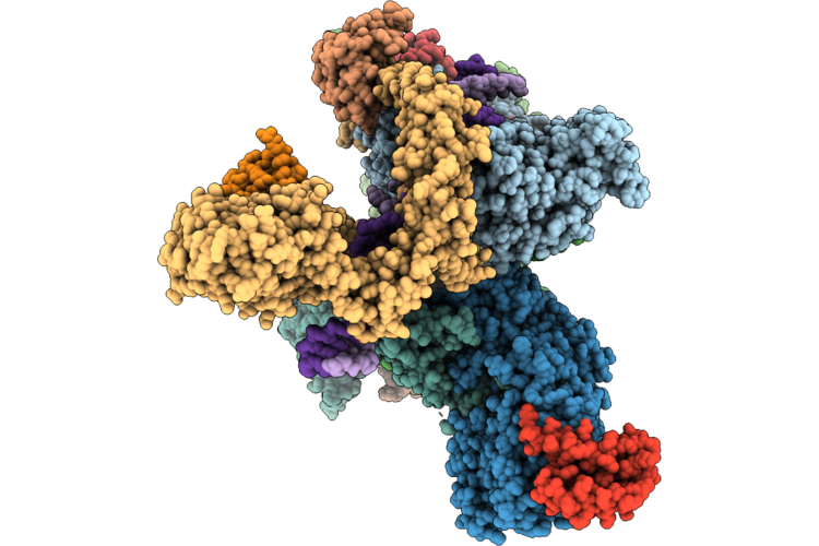 |
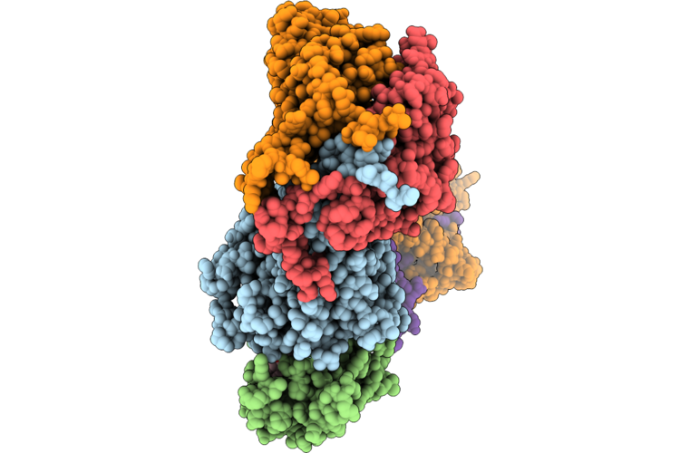
Cryo-Em Structure Of The Human Holo-Tfiih-Xpc Complex Bound To Bulky Lesion-Mimic Dna (Composite Map)
Organism: Homo sapiens, Synthetic construct
Method: ELECTRON MICROSCOPY Resolution:3.32 Å Release Date: 2026-05-06 Classification: DNA BINDING PROTEIN Ligands: SF4, ZN, CA |
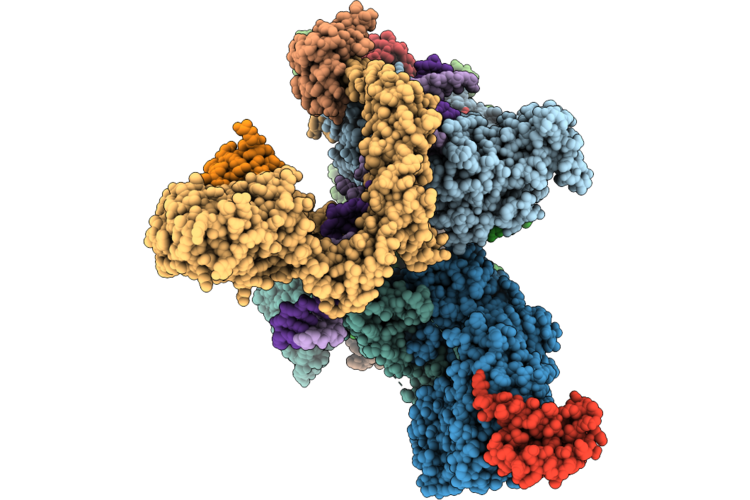 |
Cryo-Em Structure Of The Human Holo-Tfiih-Xpc Complex Bound To Bulky Lesion-Mimic Dna (Consensus Map)
Organism: Homo sapiens, Synthetic construct
Method: ELECTRON MICROSCOPY Resolution:3.91 Å Release Date: 2026-05-06 Classification: DNA BINDING PROTEIN Ligands: SF4, ZN, CA |
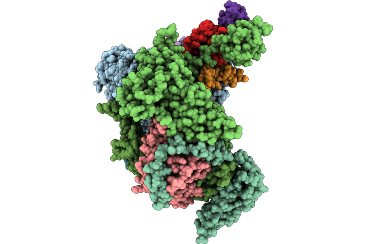 |
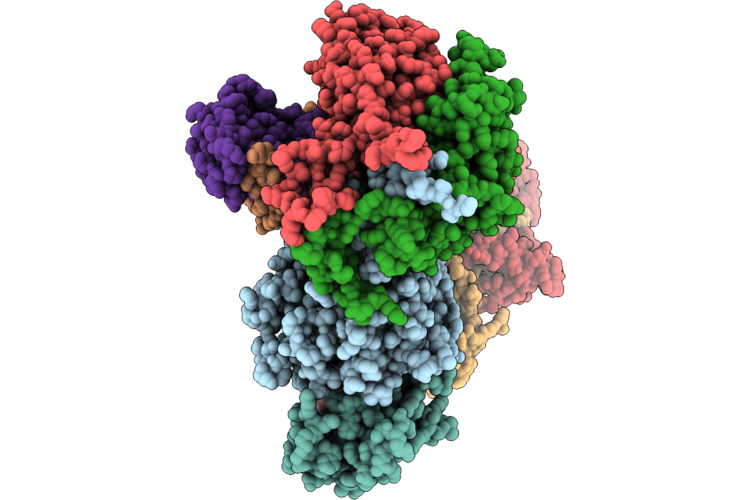
Cryo-Em Structure Of The Human Holo-Tfiih-Xpc-Xpa Complex Bound To Bulky Lesion-Mimic Dna
Organism: Homo sapiens, Synthetic construct
Method: ELECTRON MICROSCOPY Resolution:3.60 Å Release Date: 2026-05-06 Classification: DNA BINDING PROTEIN Ligands: SF4, ZN |
 |
Cryo-Em Structure Of The Cul1-Rbx1-Skp1-Fbxo22 Scf Ubiquition Ligase In Complex With Nsd2 Via Unc10088
Organism: Homo sapiens
Method: ELECTRON MICROSCOPY Release Date: 2026-05-06 Classification: LIGASE Ligands: A1J20 |
 |
Cryo-Em Structure Of The Cul1-Rbx1-Skp1-Fbxo22 Scf Ubiquition Ligase In Complex With Nsd2, Unc10088 And Bach1
Organism: Homo sapiens
Method: ELECTRON MICROSCOPY Release Date: 2026-05-06 Classification: LIGASE Ligands: A1J20 |
 |
Cryo-Em Structure Of The Cul1-Rbx1-Skp1-Fbxo22 Scf Ubiquition Ligase In Complex With Nsd2 Via Unc10415667
Organism: Homo sapiens
Method: ELECTRON MICROSCOPY Release Date: 2026-05-06 Classification: LIGASE Ligands: A1J21 |
 |
Crystal Structure Of Udp-N-Acetylmuramate-L-Alanine Ligase (Murc) From Pseudomonas Aeruginosa In Complex With Compound Osa_001175 (Wyh77)
Organism: Pseudomonas aeruginosa
Method: X-RAY DIFFRACTION Resolution:2.40 Å Release Date: 2026-05-06 Classification: LIGASE Ligands: PO4, MG, A1J52, EDO, DMS, PEG |
 |
Organism: Leishmania braziliensis, Bos taurus
Method: X-RAY DIFFRACTION Resolution:1.90 Å Release Date: 2026-05-06 Classification: HYDROLASE Ligands: PEG, ACT, GOL, EDO, CL |
 |
Organism: Bos taurus, Leishmania major
Method: X-RAY DIFFRACTION Resolution:1.80 Å Release Date: 2026-05-06 Classification: HYDROLASE Ligands: ACT, GOL, PG4 |
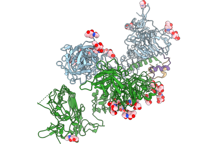 |
Drosophila Melanogaster Insulin Receptor Ectodomain In Complex With One Dilp2 Molecule
Organism: Drosophila melanogaster
Method: ELECTRON MICROSCOPY Resolution:3.70 Å Release Date: 2026-05-06 Classification: SIGNALING PROTEIN Ligands: NAG |
 |
Drosophila Melanogaster Insulin Receptor Ectodomain In Complex With Two Dilp2 Molecules
Organism: Drosophila melanogaster
Method: ELECTRON MICROSCOPY Release Date: 2026-05-06 Classification: SIGNALING PROTEIN Ligands: NAG |
 |
Organism: Oryza sativa subsp. japonica
Method: ELECTRON MICROSCOPY Release Date: 2026-05-06 Classification: TRANSFERASE |
 |
Organism: Oryza sativa japonica group
Method: ELECTRON MICROSCOPY Release Date: 2026-05-06 Classification: TRANSFERASE |
 |
Organism: Sorghum bicolor
Method: X-RAY DIFFRACTION Resolution:2.43 Å Release Date: 2026-05-06 Classification: TRANSFERASE Ligands: A1EE9, MG |