

Search Count: 38
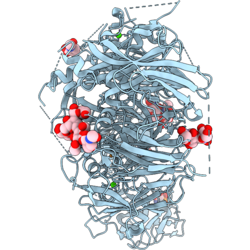 |
Coagulation Factor V Dtqq, Membrane-Bound, On Ptd-Choline : Ptd-Serine 75:25 Vesicles
Organism: Homo sapiens
Method: ELECTRON MICROSCOPY Resolution:2.90 Å Release Date: 2026-09-16 Classification: MEMBRANE PROTEIN Ligands: NAG, CA, CU1, SEP |
Organism: Homo sapiens
Method: ELECTRON MICROSCOPY
Release Date: 2026-09-16
Ligands: NAG, CA, CU1, SEP
 |
Coagulation Factor Viii, Full-Length, Membrane-Bound, On Ptd-Choline : Ptd-Serine 75:25 Vesicles
Organism: Homo sapiens
Method: ELECTRON MICROSCOPY Resolution:3.15 Å Release Date: 2026-09-16 Classification: MEMBRANE PROTEIN Ligands: NAG, CA, CU1, SEP, CPS |
Organism: Homo sapiens
Method: ELECTRON MICROSCOPY
Release Date: 2026-09-16
Ligands: NAG, CA, CU1, SEP, CPS
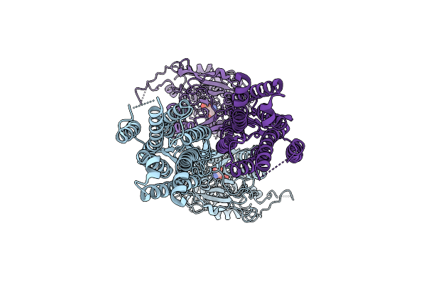 |
Cryo-Em Structure Of A Class C Gpcr (Global, Without Symmetry)
Organism: Homo sapiens
Method: ELECTRON MICROSCOPY Resolution:3.20 Å Release Date: 2026-03-18 Classification: MEMBRANE PROTEIN Ligands: SEP |
Organism: Homo sapiens
Method: ELECTRON MICROSCOPY
Release Date: 2026-03-18
Ligands: SEP
 |
Cryo-Em Structure Of A Class C Gpcr (Class 1)
Organism: Homo sapiens
Method: ELECTRON MICROSCOPY Resolution:3.39 Å Release Date: 2026-03-18 Classification: MEMBRANE PROTEIN Ligands: SEP |
Organism: Homo sapiens
Method: ELECTRON MICROSCOPY
Release Date: 2026-03-18
Ligands: SEP
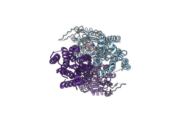 |
Cryo-Em Structure Of A Class C Gpcr (Class 3)
Organism: Homo sapiens
Method: ELECTRON MICROSCOPY Resolution:3.51 Å Release Date: 2026-03-18 Classification: MEMBRANE PROTEIN Ligands: SEP |
Organism: Homo sapiens
Method: ELECTRON MICROSCOPY
Release Date: 2026-03-18
Ligands: SEP
 |
Cryo-Em Structure Of A Class C Gpcr (Class 2)
Organism: Homo sapiens
Method: ELECTRON MICROSCOPY Resolution:3.27 Å Release Date: 2026-03-18 Classification: MEMBRANE PROTEIN Ligands: SEP |
Organism: Homo sapiens
Method: ELECTRON MICROSCOPY
Release Date: 2026-03-18
Ligands: SEP
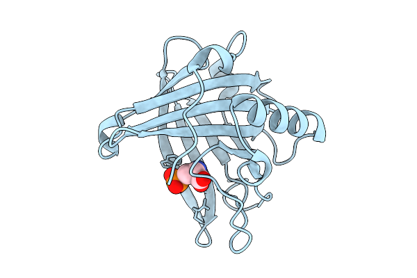 |
Lcn2 In Complex With Phosphoserine
Organism: Homo sapiens
Method: X-RAY DIFFRACTION Resolution:2.85 Å Release Date: 2025-09-03 Classification: LIPID BINDING PROTEIN Ligands: SEP |
Organism: Homo sapiens
Method: X-RAY DIFFRACTION
Release Date: 2025-09-03
Ligands: SEP
 |
Crystal Structure Of Phosphoserine Phosphatase (Serb) From Brucella Melitensis In Copmplex With O-Phosphoserine
Organism: Brucella melitensis
Method: X-RAY DIFFRACTION Resolution:2.35 Å Release Date: 2025-06-25 Classification: HYDROLASE Ligands: SEP, GOL, CA, CL |
Organism: Brucella melitensis
Method: X-RAY DIFFRACTION
Release Date: 2025-06-25
Ligands: SEP, GOL, CA, CL
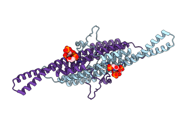 |
Compact State - Pil1 Dimer With Lipid Headgroups Fitted In Native Eisosome Lattice Bound To Plasma Membrane Microdomain
Organism: Saccharomyces cerevisiae
Method: ELECTRON MICROSCOPY Release Date: 2024-07-24 Classification: LIPID BINDING PROTEIN Ligands: I3P, SEP |
Organism: Saccharomyces cerevisiae
Method: ELECTRON MICROSCOPY
Release Date: 2024-07-24
Ligands: I3P, SEP
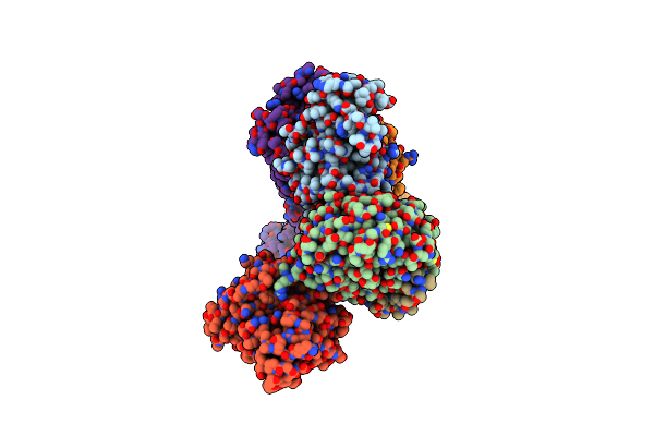 |
Cryo-Em Structure Of Gi1-Bound Metabotropic Glutamate Receptor Mglu4
Organism: Homo sapiens, Escherichia coli
Method: ELECTRON MICROSCOPY Release Date: 2023-06-21 Classification: MEMBRANE PROTEIN Ligands: BK0, SEP |
Organism: Homo sapiens, Escherichia coli
Method: ELECTRON MICROSCOPY
Release Date: 2023-06-21
Ligands: BK0, SEP
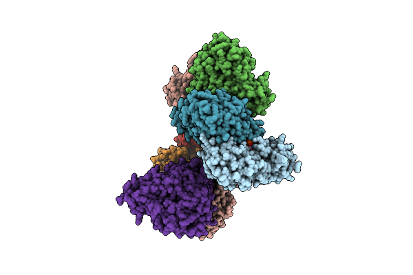 |
Crystal Structure Of The Human Phosphoserine Aminotransferase (Psat) In Complex With O-Phosphoserine
Organism: Homo sapiens
Method: X-RAY DIFFRACTION Resolution:2.78 Å Release Date: 2023-03-29 Classification: TRANSFERASE Ligands: E1U, SEP, PMP, SO4 |
Organism: Homo sapiens
Method: X-RAY DIFFRACTION
Release Date: 2023-03-29
Ligands: E1U, SEP, PMP, SO4
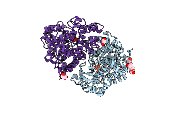 |
Structure Of Human Enolase 2 In Complex With Phosphoserine
Organism: Homo sapiens
Method: X-RAY DIFFRACTION Resolution:2.10 Å Release Date: 2022-11-09 Classification: LYASE Ligands: PO4, MG, ACT, EDO, PGE, SEP |
Organism: Homo sapiens
Method: X-RAY DIFFRACTION
Release Date: 2022-11-09
Ligands: PO4, MG, ACT, EDO, PGE, SEP
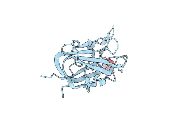 |
Crystal Structure Of Porcine Factor Viii C2 Domain Bound To Phosphatidylserine
Organism: Sus scrofa
Method: X-RAY DIFFRACTION Resolution:1.30 Å Release Date: 2021-10-27 Classification: LIPID BINDING PROTEIN Ligands: SEP |
Organism: Sus scrofa
Method: X-RAY DIFFRACTION
Release Date: 2021-10-27
Ligands: SEP
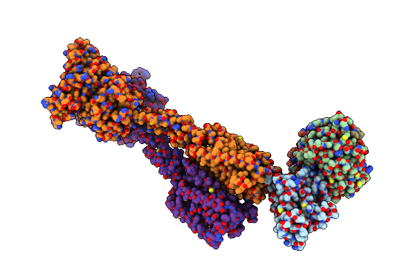 |
Cryo-Em Structure Of Gi-Bound Metabotropic Glutamate Receptor Mglu4
Organism: Homo sapiens
Method: ELECTRON MICROSCOPY Release Date: 2021-06-23 Classification: MEMBRANE PROTEIN Ligands: SEP |
Organism: Homo sapiens
Method: ELECTRON MICROSCOPY
Release Date: 2021-06-23
Ligands: SEP
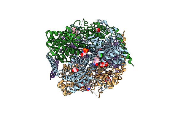 |
Crystal Structure Of Phosphofructokinase From Trypanosoma Brucei In Complex With An Allosteric Inhibitor Ctcb360
Organism: Trypanosoma brucei brucei
Method: X-RAY DIFFRACTION Resolution:2.35 Å Release Date: 2020-03-18 Classification: TRANSFERASE Ligands: JJ5, CIT, GOL, SEP |
Organism: Trypanosoma brucei brucei
Method: X-RAY DIFFRACTION
Release Date: 2020-03-18
Ligands: JJ5, CIT, GOL, SEP
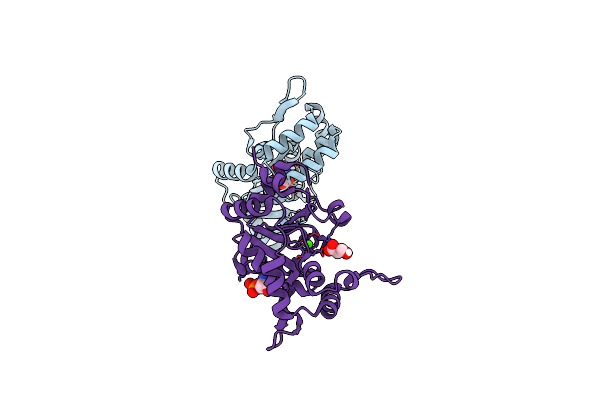 |
Psph Human Phosphoserine Phosphatase
Organism: Homo sapiens
Method: X-RAY DIFFRACTION Resolution:1.93 Å Release Date: 2019-06-12 Classification: HYDROLASE Ligands: SEP, CA, SER |
Organism: Homo sapiens
Method: X-RAY DIFFRACTION
Release Date: 2019-06-12
Ligands: SEP, CA, SER
 |
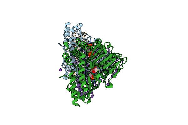
Crystal Structure Of Phosphoserine Phosphatase Mutant (H9A) From Entamoeba Histolytica In Complex With Phosphoserine
Organism: Entamoeba histolytica hm-1:imss-a
Method: X-RAY DIFFRACTION Resolution:2.95 Å Release Date: 2019-04-24 Classification: HYDROLASE Ligands: SEP, SO4, PG4, PEG |
Organism: Entamoeba histolytica hm-1:imss-a
Method: X-RAY DIFFRACTION
Release Date: 2019-04-24
Ligands: SEP, SO4, PG4, PEG
 |
Crystal Structure Of Arabidopsis Thaliana Phosphoserine Aminotransferase Isoform 1 (Atpsat1) In Complex With Plp-Phosphoserine Geminal Diamine Intermediate
Organism: Arabidopsis thaliana
Method: X-RAY DIFFRACTION Resolution:1.70 Å Release Date: 2018-05-23 Classification: TRANSFERASE Ligands: PLP, SEP, NA |
Organism: Arabidopsis thaliana
Method: X-RAY DIFFRACTION
Release Date: 2018-05-23
Ligands: PLP, SEP, NA
 |
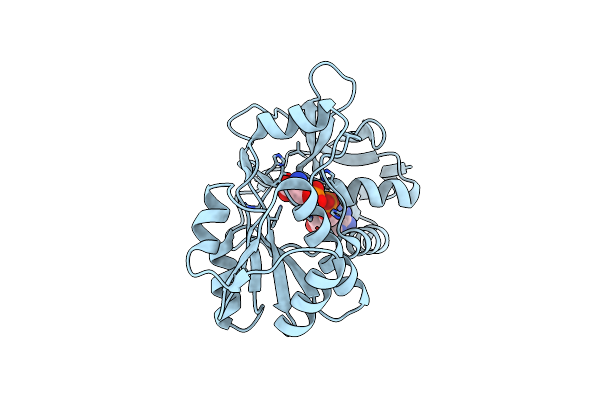
14-3-3C In Complex With Cpp And Fusicoccin
Organism: Nicotiana tabacum, Arabidopsis thaliana
Method: X-RAY DIFFRACTION Resolution:3.30 Å Release Date: 2017-11-22 Classification: SIGNALING PROTEIN Ligands: FSC, SEP |
Organism: Nicotiana tabacum, Arabidopsis thaliana
Method: X-RAY DIFFRACTION
Release Date: 2017-11-22
Ligands: FSC, SEP
 |
Free Serine Kinase (E30A Mutant) In Complex With Phosphoserine And Amp
Organism: Thermococcus kodakarensis kod1
Method: X-RAY DIFFRACTION Resolution:2.00 Å Release Date: 2017-04-12 Classification: TRANSFERASE Ligands: AMP, SEP, MG |
Organism: Thermococcus kodakarensis kod1
Method: X-RAY DIFFRACTION
Release Date: 2017-04-12
Ligands: AMP, SEP, MG