

Search Count: 466
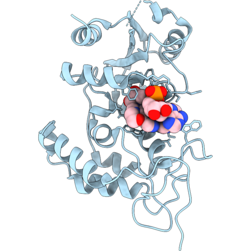 |
Crystal Structure Of Mouse Cd38 With Compound 9
Organism: Mus musculus
Method: X-RAY DIFFRACTION Resolution:1.96 Å Release Date: 2026-10-07 Classification: HYDROLASE Ligands: A1C6U |
Organism: Mus musculus
Method: X-RAY DIFFRACTION
Release Date: 2026-10-07
Ligands: A1C6U
 |
Crystal Structure Of Mouse Cd38 In Complex With Compound 6
Organism: Mus musculus
Method: X-RAY DIFFRACTION Resolution:2.15 Å Release Date: 2026-10-07 Classification: HYDROLASE Ligands: A1C64, ACT |
Organism: Mus musculus
Method: X-RAY DIFFRACTION
Release Date: 2026-10-07
Ligands: A1C64, ACT
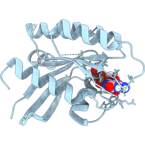 |
Structure Of Gdp-Bound Kras-G12C/H95Q, A Switch-Ii Pocket Inhibitor Resistance Mutant
Organism: Homo sapiens
Method: X-RAY DIFFRACTION Resolution:1.35 Å Release Date: 2026-10-07 Classification: SIGNALING PROTEIN Ligands: GDP, MG |
Organism: Homo sapiens
Method: X-RAY DIFFRACTION
Release Date: 2026-10-07
Ligands: GDP, MG
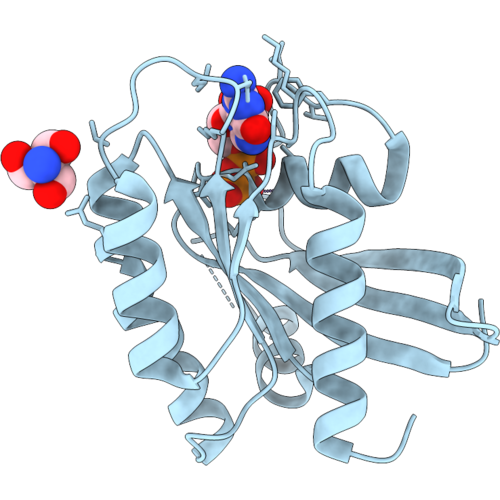 |
Structure Of Gdp-Bound Kras-G12C/Y96C, A Switch-Ii Pocket Inhibitor Resistance Mutant
Organism: Homo sapiens
Method: X-RAY DIFFRACTION Resolution:1.50 Å Release Date: 2026-10-07 Classification: SIGNALING PROTEIN Ligands: MG, GDP, TRS |
Organism: Homo sapiens
Method: X-RAY DIFFRACTION
Release Date: 2026-10-07
Ligands: MG, GDP, TRS
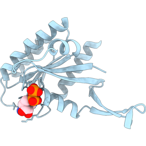 |
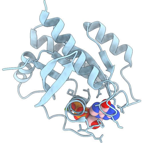
Structure Of Gdp-Bound Kras-G12C/Y96D, A Switch-Ii Pocket Inhibitor Resistance Mutant
Organism: Homo sapiens
Method: X-RAY DIFFRACTION Resolution:1.50 Å Release Date: 2026-10-07 Classification: SIGNALING PROTEIN Ligands: GDP |
Organism: Homo sapiens
Method: X-RAY DIFFRACTION
Release Date: 2026-10-07
Ligands: GDP
 |
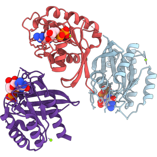
Structure Of Gdp-Bound Kras-G12C/Q99L, A Switch-Ii Pocket Inhibitor Resistance Mutant
Organism: Homo sapiens
Method: X-RAY DIFFRACTION Resolution:1.16 Å Release Date: 2026-10-07 Classification: SIGNALING PROTEIN Ligands: MG, GDP |
Organism: Homo sapiens
Method: X-RAY DIFFRACTION
Release Date: 2026-10-07
Ligands: MG, GDP
 |
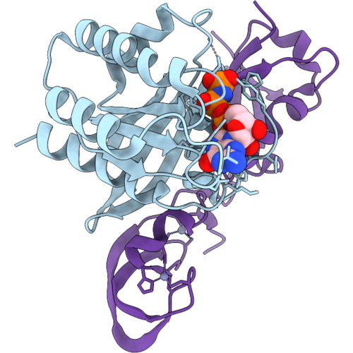
Structure Of Gdp-Bound Kras-G12C/R68S, A Switch-Ii Pocket Inhibitor Resistance Mutant
Organism: Homo sapiens
Method: X-RAY DIFFRACTION Resolution:1.65 Å Release Date: 2026-10-07 Classification: SIGNALING PROTEIN Ligands: GDP, MG |
Organism: Homo sapiens
Method: X-RAY DIFFRACTION
Release Date: 2026-10-07
Ligands: GDP, MG
 |
Structure Of Gmppnp-Bound Kras-G12C/Y96D, A Switch-Ii Pocket Inhibitor Resistance Mutant, In Complex With The Raf1 Rbd-Crd
Organism: Homo sapiens
Method: X-RAY DIFFRACTION Resolution:2.50 Å Release Date: 2026-10-07 Classification: SIGNALING PROTEIN Ligands: GNP, MG, ZN Modified Residue(s): CAF |
Organism: Homo sapiens
Method: X-RAY DIFFRACTION
Release Date: 2026-10-07
Ligands: GNP, MG, ZN
 |
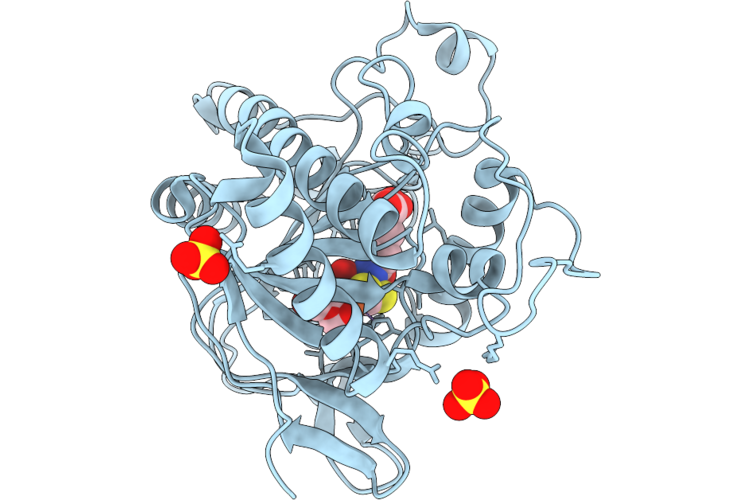
Structure Of Cdk11-Cyclin L-Sap30Bp In Complex With Iacs-054647
Organism: Homo sapiens
Method: ELECTRON MICROSCOPY Resolution:2.90 Å Release Date: 2026-08-19 Classification: TRANSFERASE Ligands: A1KC9 Modified Residue(s): TPO |
Organism: Homo sapiens
Method: ELECTRON MICROSCOPY
Release Date: 2026-08-19
Ligands: A1KC9
 |
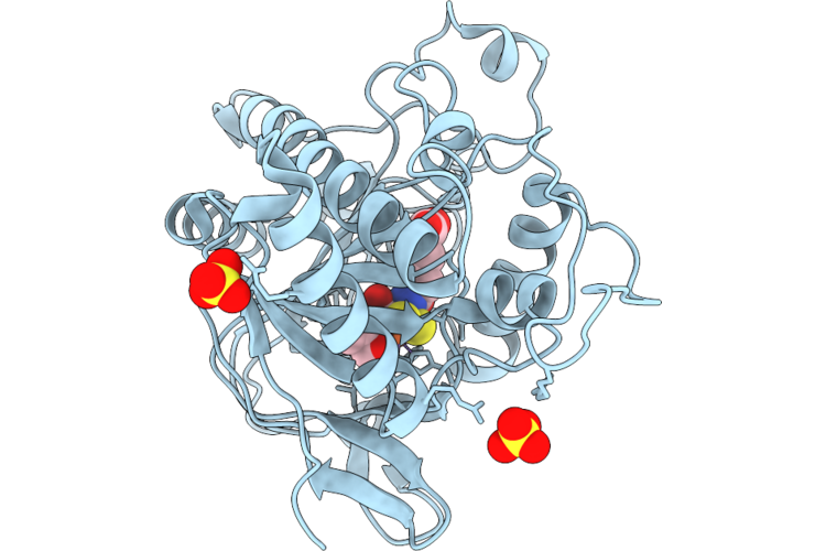
Isopenicillin N Synthase Co-Crystallised With Fe And Acdv After 6S O2 Exposure
Organism: Aspergillus nidulans fgsc a4
Method: X-RAY DIFFRACTION Resolution:1.50 Å Release Date: 2026-08-12 Classification: OXIDOREDUCTASE Ligands: A1IZG, A1I0E, SO4, OXY, FE, IO0, ACV |
Organism: Aspergillus nidulans fgsc a4
Method: X-RAY DIFFRACTION
Release Date: 2026-08-12
Ligands: A1IZG, A1I0E, SO4, OXY, FE, IO0, ACV
 |
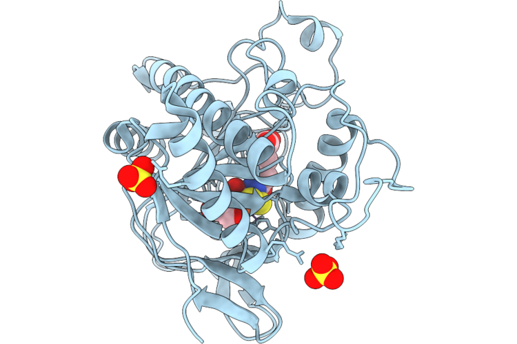
Isopenicillin N Synthase Co-Crystallised With Fe And Acdv After 8S O2 Exposure
Organism: Aspergillus nidulans fgsc a4
Method: X-RAY DIFFRACTION Resolution:1.60 Å Release Date: 2026-08-12 Classification: OXIDOREDUCTASE Ligands: A1IZG, IO0, OXY, A1I0E, SO4, FE |
Organism: Aspergillus nidulans fgsc a4
Method: X-RAY DIFFRACTION
Release Date: 2026-08-12
Ligands: A1IZG, IO0, OXY, A1I0E, SO4, FE
 |
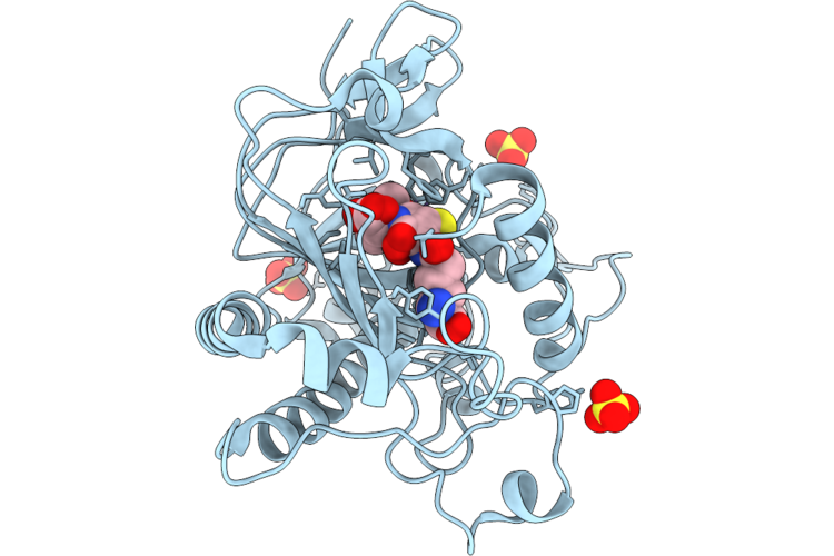
Isopenicillin N Synthase Co-Crystallised With Fe And Acdv After 10S O2 Exposure
Organism: Aspergillus nidulans fgsc a4
Method: X-RAY DIFFRACTION Resolution:1.70 Å Release Date: 2026-08-12 Classification: OXIDOREDUCTASE Ligands: IO0, OXY, A1I0E, A1IZG, SO4, FE |
Organism: Aspergillus nidulans fgsc a4
Method: X-RAY DIFFRACTION
Release Date: 2026-08-12
Ligands: IO0, OXY, A1I0E, A1IZG, SO4, FE
 |
Isopenicillin N Synthase Co-Crystallised With Fe And Acdv After 10S O2 Exposure
Organism: Aspergillus nidulans fgsc a4
Method: X-RAY DIFFRACTION Resolution:1.68 Å Release Date: 2026-08-12 Classification: OXIDOREDUCTASE Ligands: SO4, FE, IP1, A1I0E, A1IZG, IO0, OXY |
Organism: Aspergillus nidulans fgsc a4
Method: X-RAY DIFFRACTION
Release Date: 2026-08-12
Ligands: SO4, FE, IP1, A1I0E, A1IZG, IO0, OXY
 |
Crystal Structure Of The Hpk1 Kinase Domain In Complex With Inhibitor 5B
Organism: Homo sapiens
Method: X-RAY DIFFRACTION Resolution:1.42 Å Release Date: 2026-08-05 Classification: TRANSFERASE Ligands: A1KCT, EDO |
Organism: Homo sapiens
Method: X-RAY DIFFRACTION
Release Date: 2026-08-05
Ligands: A1KCT, EDO
 |
Crystal Structure Of The Hpk1 Kinase Domain In Complex With Inhibitor 6B
Organism: Homo sapiens
Method: X-RAY DIFFRACTION Resolution:1.31 Å Release Date: 2026-08-05 Classification: TRANSFERASE Ligands: A1KCQ, EDO |
Organism: Homo sapiens
Method: X-RAY DIFFRACTION
Release Date: 2026-08-05
Ligands: A1KCQ, EDO
 |
Crystal Structure Of The Hpk1 Kinase Domain In Complex With Inhibitor 12
Organism: Homo sapiens
Method: X-RAY DIFFRACTION Resolution:1.39 Å Release Date: 2026-08-05 Classification: TRANSFERASE Ligands: A1KCR, EDO |
Organism: Homo sapiens
Method: X-RAY DIFFRACTION
Release Date: 2026-08-05
Ligands: A1KCR, EDO
 |
Crystal Structure Of Gmppnp Bound Kras-Y71H In Complex With Rbd Domain Of Craf(Raf1)
Organism: Homo sapiens
Method: X-RAY DIFFRACTION Resolution:1.68 Å Release Date: 2026-05-20 Classification: ONCOPROTEIN Ligands: GNP, MG |
Organism: Homo sapiens
Method: X-RAY DIFFRACTION
Release Date: 2026-05-20
Ligands: GNP, MG
 |
Crystal Structure Of Gmppnp Bound Kras G12C In Complex With Cypa And Rmc-4791
Organism: Homo sapiens
Method: X-RAY DIFFRACTION Resolution:1.57 Å Release Date: 2026-05-20 Classification: ONCOPROTEIN Ligands: MG, GNP, A1CYT |
Organism: Homo sapiens
Method: X-RAY DIFFRACTION
Release Date: 2026-05-20
Ligands: MG, GNP, A1CYT
 |
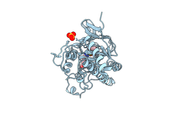
Isopenicillin N Synthase In Complex With Fe And Ipn
Organism: Aspergillus nidulans fgsc a4
Method: X-RAY DIFFRACTION Resolution:1.79 Å Release Date: 2026-04-08 Classification: OXIDOREDUCTASE Ligands: FE, SO4, IP1 |
Organism: Aspergillus nidulans fgsc a4
Method: X-RAY DIFFRACTION
Release Date: 2026-04-08
Ligands: FE, SO4, IP1
 |
Isopenicillin N Synthase In Complex With Fe And Ipn Using Tr-Sfx
Organism: Aspergillus nidulans fgsc a4
Method: X-RAY DIFFRACTION Resolution:1.70 Å Release Date: 2026-04-08 Classification: OXIDOREDUCTASE Ligands: FE2, SO4, IP1 |
Organism: Aspergillus nidulans fgsc a4
Method: X-RAY DIFFRACTION
Release Date: 2026-04-08
Ligands: FE2, SO4, IP1