

Search Count: 1,955
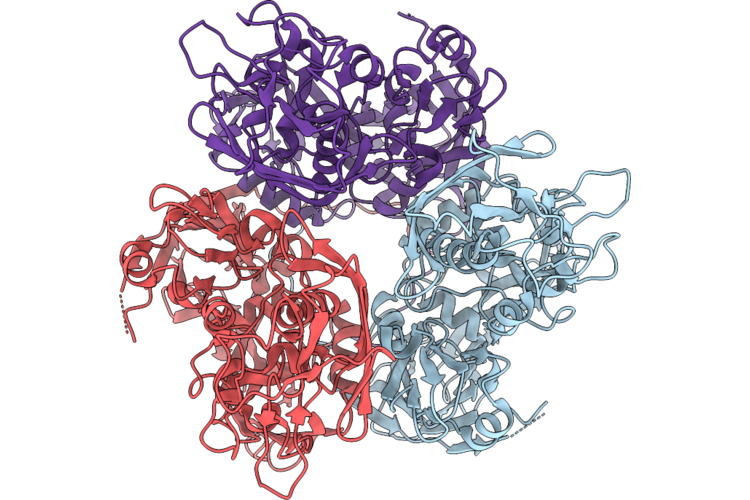 |
Cryo-Em Structure Of The A17(1-16) Peptide-Bound N-Terminal 17 Residue Truncated D13 Trimer From Vaccinia Virus
Organism: Orthopoxvirus vaccinia
Method: ELECTRON MICROSCOPY Resolution:3.04 Å Release Date: 2026-08-05 Classification: VIRAL PROTEIN |
Organism: Orthopoxvirus vaccinia
Method: ELECTRON MICROSCOPY
Release Date: 2026-08-05
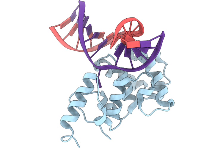 |
Structure Of Mbd4 R512K/D560N On A T:G Mispair
Organism: Homo sapiens, Synthetic construct
Method: X-RAY DIFFRACTION Resolution:1.57 Å Release Date: 2026-08-05 Classification: DNA BINDING PROTEIN |
Organism: Homo sapiens, Synthetic construct
Method: X-RAY DIFFRACTION
Release Date: 2026-08-05
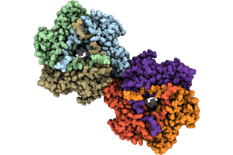 |
Two Interacting D13 Trimers At Mode I Interface In Twister Assembly
Organism: Orthopoxvirus vaccinia
Method: ELECTRON MICROSCOPY Release Date: 2026-08-05 Classification: VIRAL PROTEIN |
Organism: Orthopoxvirus vaccinia
Method: ELECTRON MICROSCOPY
Release Date: 2026-08-05
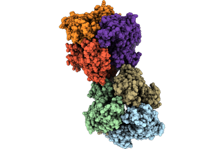 |
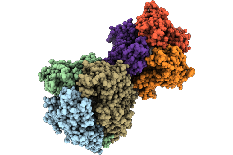
Two Interacting D13 Trimers At Mode Ii Interface In Twister Assembly
Organism: Orthopoxvirus vaccinia
Method: ELECTRON MICROSCOPY Resolution:3.20 Å Release Date: 2026-08-05 Classification: VIRAL PROTEIN |
Organism: Orthopoxvirus vaccinia
Method: ELECTRON MICROSCOPY
Release Date: 2026-08-05
 |
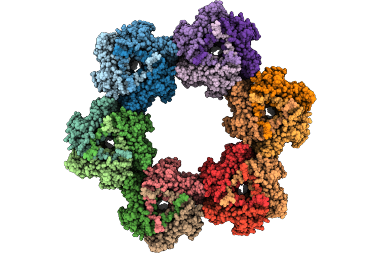
Two Interacting D13 Trimers At Mode Iii Interface In Twister Assembly
Organism: Orthopoxvirus vaccinia
Method: ELECTRON MICROSCOPY Resolution:3.10 Å Release Date: 2026-08-05 Classification: VIRAL PROTEIN |
Organism: Orthopoxvirus vaccinia
Method: ELECTRON MICROSCOPY
Release Date: 2026-08-05
 |
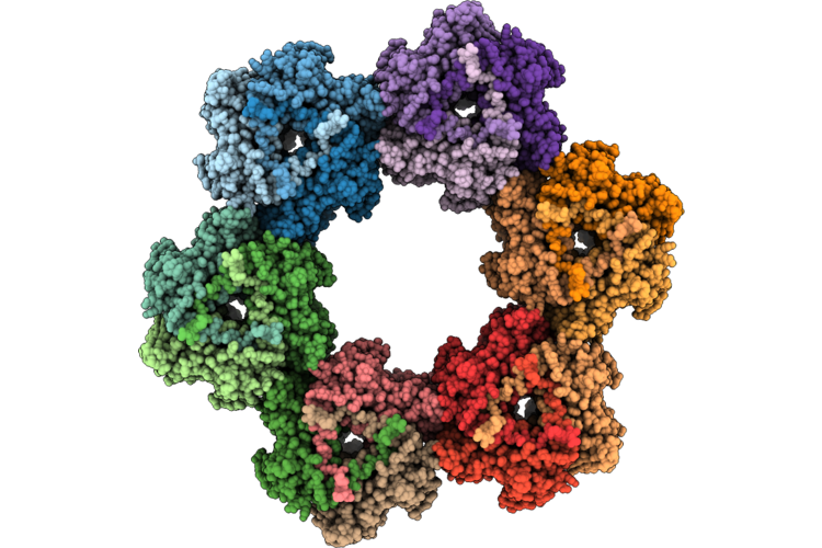
Cryo-Em Structure Of His6-Tagged D13 Assembled Into Scaffold-Like Particles From Vaccinia Virus
Organism: Orthopoxvirus vaccinia
Method: ELECTRON MICROSCOPY Resolution:2.88 Å Release Date: 2026-08-05 Classification: VIRAL PROTEIN |
Organism: Orthopoxvirus vaccinia
Method: ELECTRON MICROSCOPY
Release Date: 2026-08-05
 |
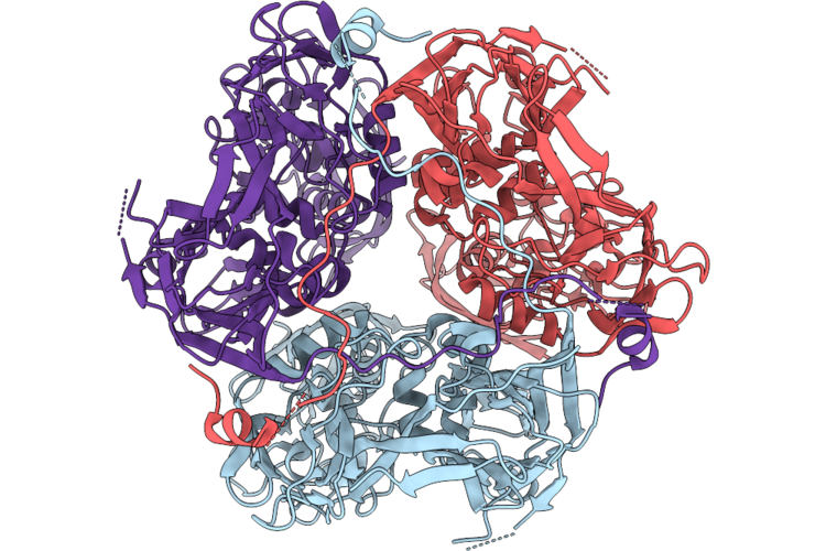
Cryo-Em Structure Of Egfp-Tagged D13 Assembled Into Scaffold-Like Particles From Vaccinia Virus
Organism: Orthopoxvirus vaccinia
Method: ELECTRON MICROSCOPY Resolution:3.70 Å Release Date: 2026-08-05 Classification: VIRAL PROTEIN |
Organism: Orthopoxvirus vaccinia
Method: ELECTRON MICROSCOPY
Release Date: 2026-08-05
 |
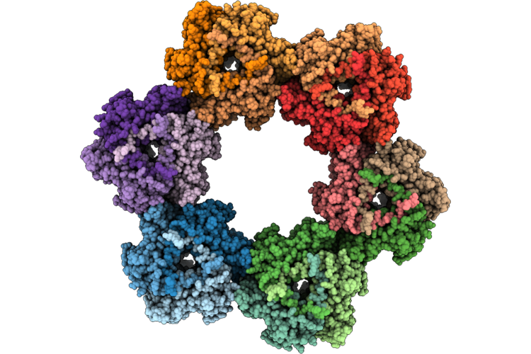
Cryo-Em Structure Of A17(1-16) Peptide-Bound D13 Trimer From Vaccinia Virus
Organism: Orthopoxvirus vaccinia
Method: ELECTRON MICROSCOPY Resolution:2.56 Å Release Date: 2026-08-05 Classification: VIRAL PROTEIN |
Organism: Orthopoxvirus vaccinia
Method: ELECTRON MICROSCOPY
Release Date: 2026-08-05
 |
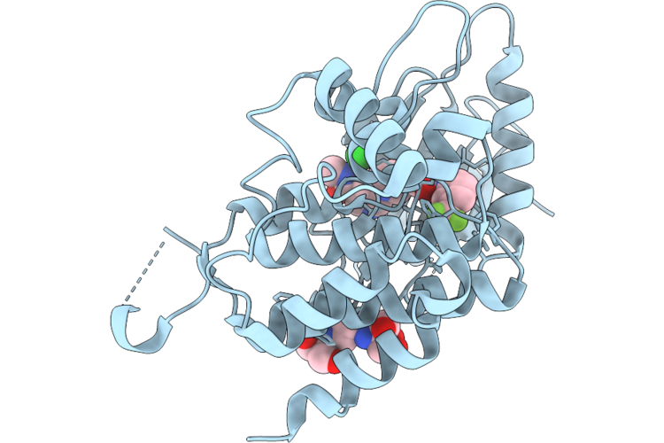
Cryo-Em Structure Of A17(1-16) Peptide-Bound D13 Assembled Into Scaffold-Like Particles From Vaccinia Virus
Organism: Orthopoxvirus vaccinia
Method: ELECTRON MICROSCOPY Resolution:3.63 Å Release Date: 2026-08-05 Classification: VIRAL PROTEIN |
Organism: Orthopoxvirus vaccinia
Method: ELECTRON MICROSCOPY
Release Date: 2026-08-05
 |
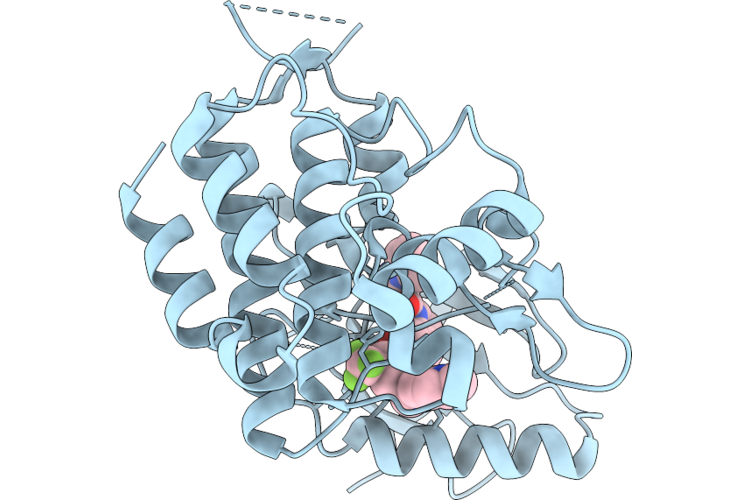
Human Ddr1 In Complex With A Pyrrolidine Inhibitor
Organism: Homo sapiens
Method: X-RAY DIFFRACTION Resolution:2.12 Å Release Date: 2026-07-29 Classification: TRANSFERASE Ligands: A1JN1, CL, B3P |
Organism: Homo sapiens
Method: X-RAY DIFFRACTION
Release Date: 2026-07-29
Ligands: A1JN1, CL, B3P
 |
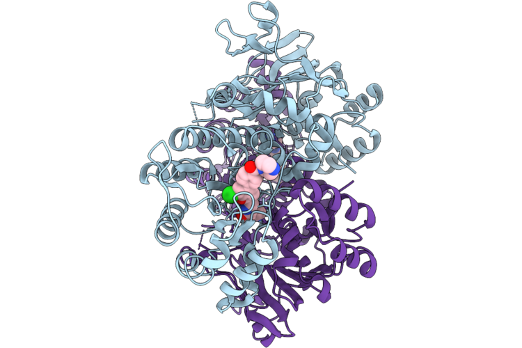
Human Ddr1 In Complex With A Indoline Inhibitor
Organism: Homo sapiens
Method: X-RAY DIFFRACTION Resolution:2.59 Å Release Date: 2026-07-29 Classification: TRANSFERASE Ligands: A1JPO, CL |
Organism: Homo sapiens
Method: X-RAY DIFFRACTION
Release Date: 2026-07-29
Ligands: A1JPO, CL
 |
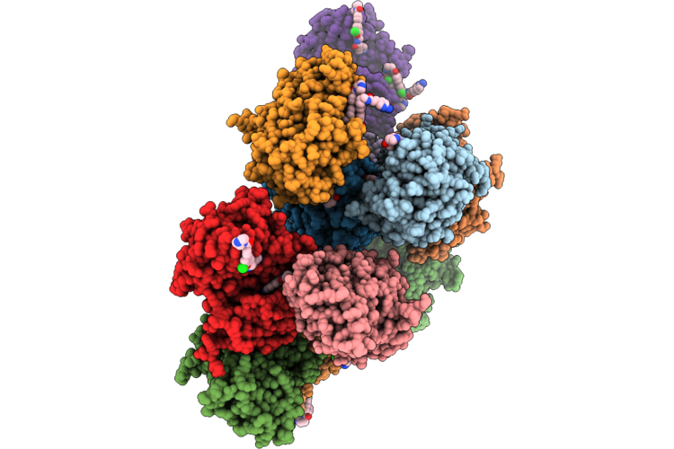
Crystal Structure Of Nampt-Kpt-7523 Complex
Organism: Homo sapiens
Method: X-RAY DIFFRACTION Resolution:1.45 Å Release Date: 2026-07-29 Classification: TRANSFERASE Ligands: A1MBG |
Organism: Homo sapiens
Method: X-RAY DIFFRACTION
Release Date: 2026-07-29
Ligands: A1MBG
 |
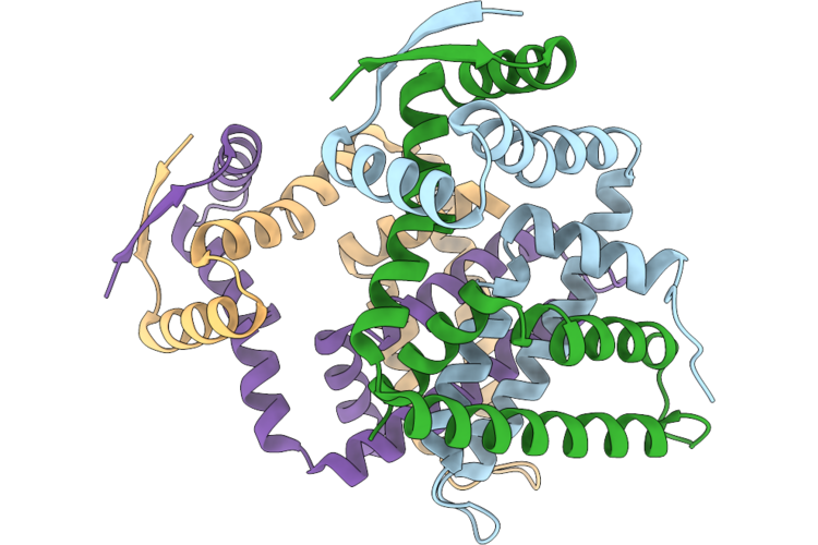
Crystal Structure Of Pak4-Kpt-7523 Complex
Organism: Homo sapiens
Method: X-RAY DIFFRACTION Resolution:2.20 Å Release Date: 2026-07-29 Classification: STRUCTURAL PROTEIN Ligands: A1MBG |
Organism: Homo sapiens
Method: X-RAY DIFFRACTION
Release Date: 2026-07-29
Ligands: A1MBG
 |
Crystal Structure Of Acriia19 From Staphylococcus Pseudintermedius
Organism: Staphylococcus pseudintermedius
Method: X-RAY DIFFRACTION Resolution:1.98 Å Release Date: 2026-07-08 Classification: IMMUNE SYSTEM |
Organism: Staphylococcus pseudintermedius
Method: X-RAY DIFFRACTION
Release Date: 2026-07-08
 |
Crystal Structure Of Haspin (Gsg2) With An Atp-Mimicking Competitive Inhibitor
Organism: Homo sapiens
Method: X-RAY DIFFRACTION Resolution:1.73 Å Release Date: 2026-06-24 Classification: TRANSFERASE Ligands: A1MFS |
Organism: Homo sapiens
Method: X-RAY DIFFRACTION
Release Date: 2026-06-24
Ligands: A1MFS
 |
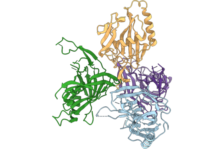
Crystal Structure Of Haspin (Gsg2) With An Atp-Mimicking Inhibitor Lj-5242
Organism: Homo sapiens
Method: X-RAY DIFFRACTION Resolution:1.88 Å Release Date: 2026-06-24 Classification: TRANSFERASE Ligands: A1MFT |
Organism: Homo sapiens
Method: X-RAY DIFFRACTION
Release Date: 2026-06-24
Ligands: A1MFT
 |
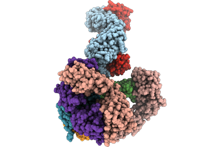
Crystal Structure Of Cas5Ic From Moraxella Bovoculi
Organism: Moraxella bovoculi
Method: X-RAY DIFFRACTION Resolution:3.33 Å Release Date: 2026-06-17 Classification: IMMUNE SYSTEM |
Organism: Moraxella bovoculi
Method: X-RAY DIFFRACTION
Release Date: 2026-06-17
 |
X-Ray Crystal Structure Of A High-Affinity Monoclonal Antibody Sequesters Xylazine
Organism: Homo sapiens
Method: X-RAY DIFFRACTION Resolution:1.99 Å Release Date: 2026-06-10 Classification: IMMUNE SYSTEM Ligands: A1C4R, CL |
Organism: Homo sapiens
Method: X-RAY DIFFRACTION
Release Date: 2026-06-10
Ligands: A1C4R, CL
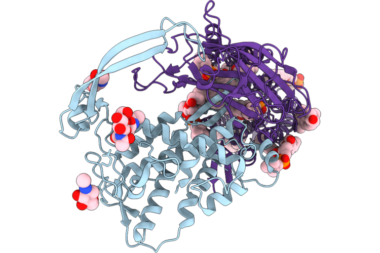 |
Cryo-Em Structure Of Human Wntless In Complex With Wnt5A At 1:1 Stoichiometry
Organism: Homo sapiens
Method: ELECTRON MICROSCOPY Release Date: 2026-06-10 Classification: MEMBRANE PROTEIN Ligands: PAM, NAG, PCW, CLR, LPE |
Organism: Homo sapiens
Method: ELECTRON MICROSCOPY
Release Date: 2026-06-10
Ligands: PAM, NAG, PCW, CLR, LPE
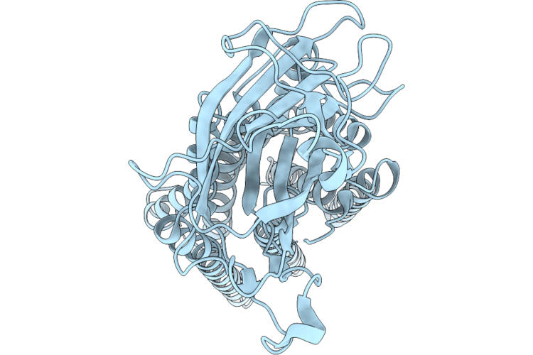 |
Cryo-Em Structure Of Human Wntless In Its Apo State
Organism: Homo sapiens
Method: ELECTRON MICROSCOPY Release Date: 2026-06-10 Classification: MEMBRANE PROTEIN |
Organism: Homo sapiens
Method: ELECTRON MICROSCOPY
Release Date: 2026-06-10