

Search Count: 156
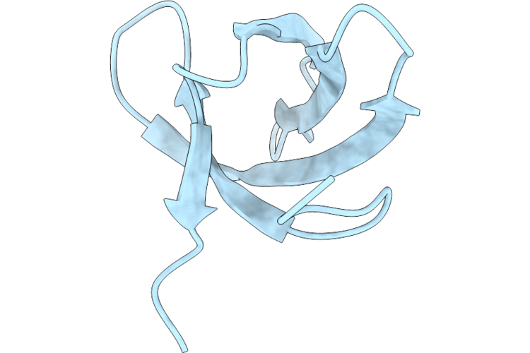 |
Crystal Structure Of Human Phf20L1(85-142)
Organism: Homo sapiens
Method: X-RAY DIFFRACTION Resolution:2.31 Å Release Date: 2026-08-12 Classification: PROTEIN BINDING |
Organism: Homo sapiens
Method: X-RAY DIFFRACTION
Release Date: 2026-08-12
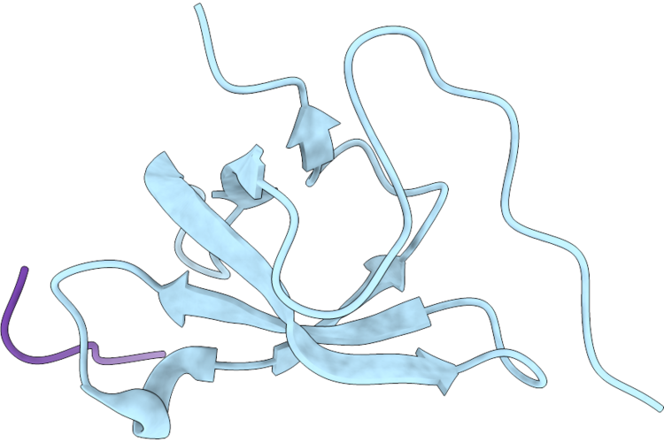 |
Crystal Structure Of Human Phf20L1(1-80) In Complex With H3K36Me
Organism: Homo sapiens
Method: X-RAY DIFFRACTION Resolution:1.34 Å Release Date: 2026-08-12 Classification: PROTEIN BINDING |
Organism: Homo sapiens
Method: X-RAY DIFFRACTION
Release Date: 2026-08-12
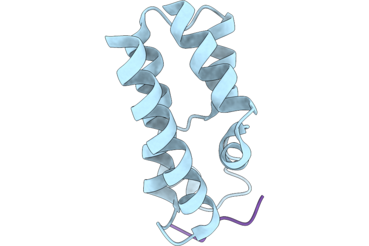 |
Crystal Structure Of Baz2A In Complex With An S-Hdag_K72Ac Peptide
Organism: Homo sapiens, Hepatitis delta virus
Method: X-RAY DIFFRACTION Resolution:1.61 Å Release Date: 2026-08-12 Classification: PROTEIN BINDING |
Organism: Homo sapiens, Hepatitis delta virus
Method: X-RAY DIFFRACTION
Release Date: 2026-08-12
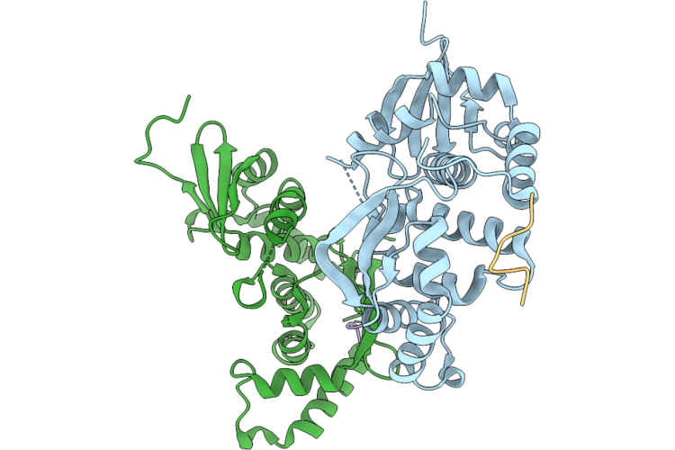 |
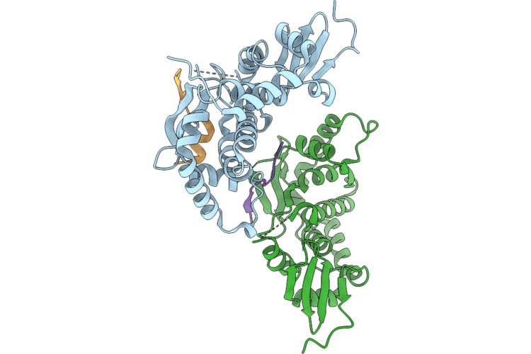
Crystal Structure Of The Mlh1 Protein Bound To The Fan1 Peptide
Organism: Homo sapiens
Method: X-RAY DIFFRACTION Resolution:3.05 Å Release Date: 2026-08-12 Classification: PROTEIN BINDING |
Organism: Homo sapiens
Method: X-RAY DIFFRACTION
Release Date: 2026-08-12
 |
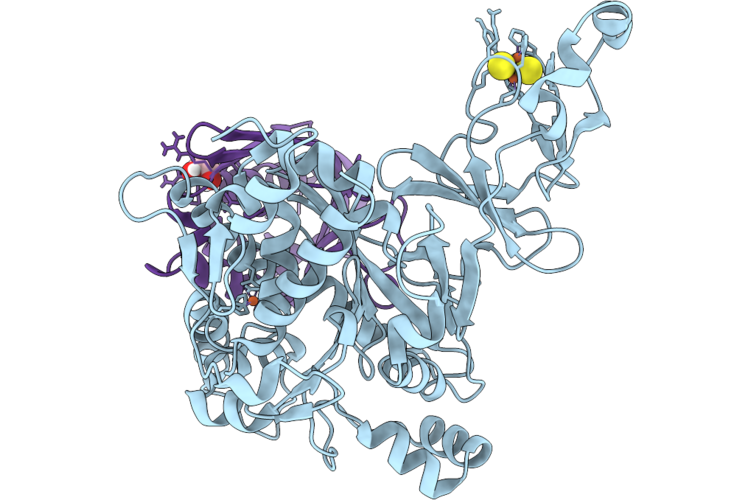
Crystal Structure Of The Mlh1 Protein Bound To The Fan1 Peptide
Organism: Homo sapiens
Method: X-RAY DIFFRACTION Resolution:2.28 Å Release Date: 2026-08-12 Classification: PROTEIN BINDING |
Organism: Homo sapiens
Method: X-RAY DIFFRACTION
Release Date: 2026-08-12
 |
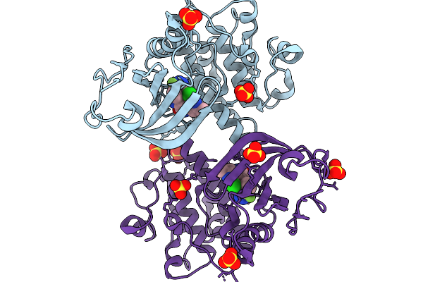
Hexameric Rieske Non-Heme Iron Dioxygenase
Organism: Variovorax paradoxus
Method: X-RAY DIFFRACTION Resolution:1.28 Å Release Date: 2026-05-20 Classification: METAL BINDING PROTEIN Ligands: FES, FE2, EDO |
Organism: Variovorax paradoxus
Method: X-RAY DIFFRACTION
Release Date: 2026-05-20
Ligands: FES, FE2, EDO
 |
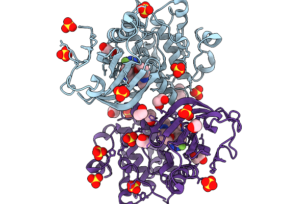
Crystal Structure Of Human Pkmyt1 Protein Kinase Domain With Naphthyridinone Inhibitor Compound 11
Organism: Homo sapiens
Method: X-RAY DIFFRACTION Resolution:1.74 Å Release Date: 2026-03-25 Classification: TRANSFERASE Ligands: A1E5A, SO4 |
Organism: Homo sapiens
Method: X-RAY DIFFRACTION
Release Date: 2026-03-25
Ligands: A1E5A, SO4
 |
Crystal Structure Of Human Pkmyt1 Protein Kinase Domain With Naphthyridinone Inhibitor Compound 16
Organism: Homo sapiens
Method: X-RAY DIFFRACTION Resolution:1.59 Å Release Date: 2026-03-25 Classification: TRANSFERASE Ligands: A1E5J, DMS, EDO, SO4 |
Organism: Homo sapiens
Method: X-RAY DIFFRACTION
Release Date: 2026-03-25
Ligands: A1E5J, DMS, EDO, SO4
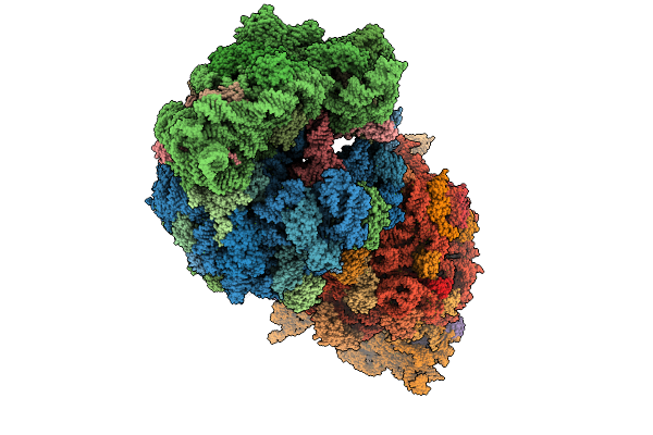 |
Crystal Structure Of The Wild-Type Thermus Thermophilus 70S Ribosome In Complex With O-Cresomycin, Mrna, Deacylated A-Site Trnaphe, Aminoacylated P-Site Fmet-Trnamet, And Deacylated E-Site Trnaphe At 2.50A Resolution
Organism: Escherichia coli, Escherichia phage t4, Thermus thermophilus hb8
Method: X-RAY DIFFRACTION Resolution:2.50 Å Release Date: 2025-08-27 Classification: RIBOSOME Ligands: MG, K, A1A1F, ZN, SF4 |
Organism: Escherichia coli, Escherichia phage t4, Thermus thermophilus hb8
Method: X-RAY DIFFRACTION
Release Date: 2025-08-27
Ligands: MG, K, A1A1F, ZN, SF4
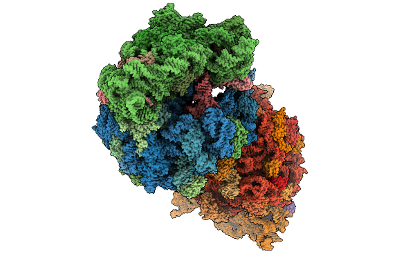 |
Crystal Structure Of The Wild-Type Thermus Thermophilus 70S Ribosome In Complex With C-Cresomycin, Mrna, Deacylated A-Site Trnaphe, Aminoacylated P-Site Fmet-Trnamet, And Deacylated E-Site Trnaphe At 2.50A Resolution
Organism: Escherichia coli, Escherichia phage t4, Thermus thermophilus hb8
Method: X-RAY DIFFRACTION Resolution:2.50 Å Release Date: 2025-08-27 Classification: RIBOSOME Ligands: MG, K, A1A1J, ZN, SF4 |
Organism: Escherichia coli, Escherichia phage t4, Thermus thermophilus hb8
Method: X-RAY DIFFRACTION
Release Date: 2025-08-27
Ligands: MG, K, A1A1J, ZN, SF4
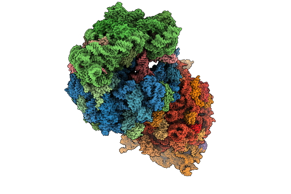 |
Crystal Structure Of The Wild-Type Thermus Thermophilus 70S Ribosome In Complex With Se-Cresomycin, Mrna, Deacylated A-Site Trnaphe, Aminoacylated P-Site Fmet-Trnamet, And Deacylated E-Site Trnaphe At 2.45A Resolution
Organism: Escherichia coli, Escherichia phage t4, Thermus thermophilus hb8
Method: X-RAY DIFFRACTION Resolution:2.45 Å Release Date: 2025-08-27 Classification: RIBOSOME Ligands: MG, K, A1A1K, ZN, SF4 |
Organism: Escherichia coli, Escherichia phage t4, Thermus thermophilus hb8
Method: X-RAY DIFFRACTION
Release Date: 2025-08-27
Ligands: MG, K, A1A1K, ZN, SF4
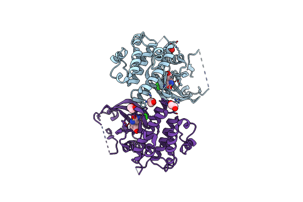 |
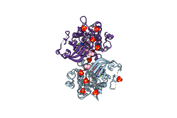
Crystal Structure Of Human Pkmyt1 Protein Kinase Domain With Naphthyridinone Inhibitor Compound 6
Organism: Homo sapiens
Method: X-RAY DIFFRACTION Resolution:2.17 Å Release Date: 2025-05-14 Classification: TRANSFERASE Ligands: A1EJW, EDO, CL |
Organism: Homo sapiens
Method: X-RAY DIFFRACTION
Release Date: 2025-05-14
Ligands: A1EJW, EDO, CL
 |
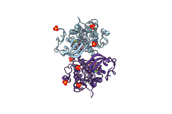
Crystal Structure Of Human Pkmyt1 Protein Kinase Domain With Naphthyridinone Inhibitor Compound 27
Organism: Homo sapiens
Method: X-RAY DIFFRACTION Resolution:2.06 Å Release Date: 2025-05-14 Classification: TRANSFERASE Ligands: A1EJX, SO4 |
Organism: Homo sapiens
Method: X-RAY DIFFRACTION
Release Date: 2025-05-14
Ligands: A1EJX, SO4
 |
Crystal Structure Of Human Pkmyt1 Protein Kinase Domain With Naphthyridinone Inhibitor Compound 40
Organism: Homo sapiens
Method: X-RAY DIFFRACTION Resolution:2.03 Å Release Date: 2025-04-16 Classification: TRANSFERASE Ligands: A1EJU, SO4 |
Organism: Homo sapiens
Method: X-RAY DIFFRACTION
Release Date: 2025-04-16
Ligands: A1EJU, SO4
 |
Crystal Structure Of Human Pkmyt1 Protein Kinase Domain With Naphthyridinone Inhibitor Compound 11
Organism: Homo sapiens
Method: X-RAY DIFFRACTION Resolution:2.04 Å Release Date: 2025-04-16 Classification: TRANSFERASE Ligands: TLA, A1EJV, EDO |
Organism: Homo sapiens
Method: X-RAY DIFFRACTION
Release Date: 2025-04-16
Ligands: TLA, A1EJV, EDO
 |
Structure Of The Red Fluorescent Protein Mscarlet3-H At Ph 4.5
Organism: Discosoma sp.
Method: X-RAY DIFFRACTION Resolution:1.46 Å Release Date: 2025-02-19 Classification: FLUORESCENT PROTEIN |
Organism: Discosoma sp.
Method: X-RAY DIFFRACTION
Release Date: 2025-02-19
 |
Structure Of The Red Fluorescent Protein Mscarlet-H At Ph8.5
Organism: Discosoma sp.
Method: X-RAY DIFFRACTION Resolution:1.82 Å Release Date: 2025-02-19 Classification: FLUORESCENT PROTEIN |
Organism: Discosoma sp.
Method: X-RAY DIFFRACTION
Release Date: 2025-02-19
 |
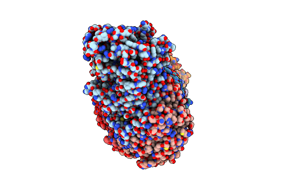
Structure Of Mavs-Card Filament
Organism: Homo sapiens
Method: ELECTRON MICROSCOPY Release Date: 2024-10-16 Classification: IMMUNE SYSTEM |
Organism: Homo sapiens
Method: ELECTRON MICROSCOPY
Release Date: 2024-10-16
 |
Cryo-Em Structure Of Crispr-Csm Effector Complex From Mycobacterium Canettii
Organism: Mycobacterium canettii
Method: ELECTRON MICROSCOPY Release Date: 2024-07-31 Classification: RNA BINDING PROTEIN/RNA |
Organism: Mycobacterium canettii
Method: ELECTRON MICROSCOPY
Release Date: 2024-07-31
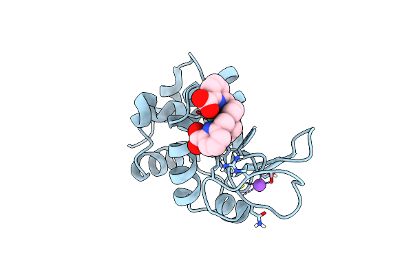 |
Crystal Structure Of Hen Egg White Lysozyme Co-Crystallized With 10 Mm Tbxo4
Organism: Gallus gallus
Method: X-RAY DIFFRACTION Resolution:1.70 Å Release Date: 2024-07-17 Classification: HYDROLASE Ligands: TB, 7MT, CL, NA |
Organism: Gallus gallus
Method: X-RAY DIFFRACTION
Release Date: 2024-07-17
Ligands: TB, 7MT, CL, NA