

Search Count: 193
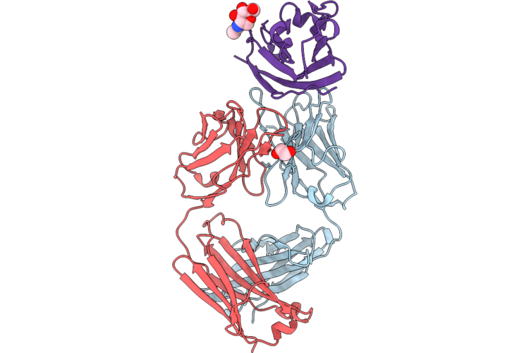 |
Structure Of Fabs1Ce2_P2A In Complex With The N-Terminal Domain Of Pd-L1
Organism: Homo sapiens
Method: X-RAY DIFFRACTION Resolution:2.22 Å Release Date: 2026-06-03 Classification: IMMUNE SYSTEM Ligands: EDO, CL, NAG |
Organism: Homo sapiens
Method: X-RAY DIFFRACTION
Release Date: 2026-06-03
Ligands: EDO, CL, NAG
 |
Structure Of Fabs1Ce2_Er-3C In Complex With The Extracellular Region Of Egfr
Organism: Homo sapiens
Method: X-RAY DIFFRACTION Resolution:2.40 Å Release Date: 2026-06-03 Classification: SIGNALING PROTEIN Ligands: NAG, SCN, CL, NA, K, PEG, PGE, EDO |
Organism: Homo sapiens
Method: X-RAY DIFFRACTION
Release Date: 2026-06-03
Ligands: NAG, SCN, CL, NA, K, PEG, PGE, EDO
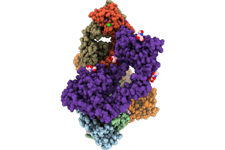 |
Structure Of Fabs1Ce2_Er-2A In Complex With The Extracellular Region Of Egfr
Organism: Homo sapiens
Method: X-RAY DIFFRACTION Resolution:3.06 Å Release Date: 2026-06-03 Classification: SIGNALING PROTEIN Ligands: PEG, NA, CL, EDO, NAG |
Organism: Homo sapiens
Method: X-RAY DIFFRACTION
Release Date: 2026-06-03
Ligands: PEG, NA, CL, EDO, NAG
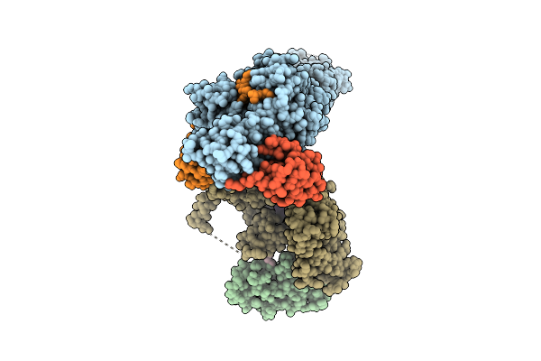 |
Structure Of Rbr Binding E2 Variant Crosslinked With Nedd8-Cul5-Rbx2 Bound Arih2 And Ub
Organism: Homo sapiens, Synthetic construct
Method: ELECTRON MICROSCOPY Resolution:2.97 Å Release Date: 2025-12-24 Classification: LIGASE Ligands: ZN, SY8 |
Organism: Homo sapiens, Synthetic construct
Method: ELECTRON MICROSCOPY
Release Date: 2025-12-24
Ligands: ZN, SY8
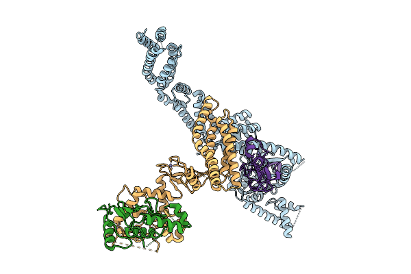 |
Structure Of Rbr E2 Variant Binding To Cul5-Rbx2 Bound Arih2
Organism: Synthetic construct, Homo sapiens
Method: ELECTRON MICROSCOPY Resolution:3.06 Å Release Date: 2025-12-24 Classification: LIGASE Ligands: ZN |
Organism: Synthetic construct, Homo sapiens
Method: ELECTRON MICROSCOPY
Release Date: 2025-12-24
Ligands: ZN
 |
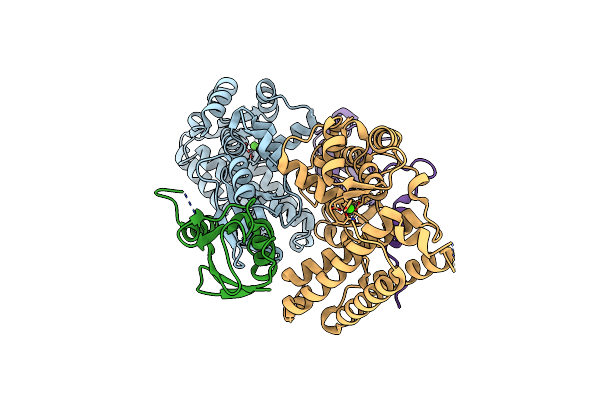
Crystal Structure Of A High Affinity Vl-Vh Tetrabody For The Erythropoietin Receptor
Organism: Homo sapiens
Method: X-RAY DIFFRACTION Resolution:1.96 Å Release Date: 2025-10-01 Classification: SIGNALING PROTEIN Ligands: NHE, CL, SO4, NA |
Organism: Homo sapiens
Method: X-RAY DIFFRACTION
Release Date: 2025-10-01
Ligands: NHE, CL, SO4, NA
 |
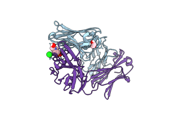
Structure Of Cbl-Tkbd Bound To Ubiquitin-Fused Cblock Peptide
Organism: Homo sapiens
Method: X-RAY DIFFRACTION Resolution:2.02 Å Release Date: 2025-05-14 Classification: LIGASE Ligands: CA |
Organism: Homo sapiens
Method: X-RAY DIFFRACTION
Release Date: 2025-05-14
Ligands: CA
 |
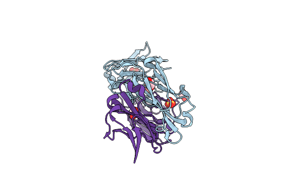
Anti-Phosphohistidine Fab Hsc44.Ck.Elbow.20.N32F
Organism: Oryctolagus cuniculus
Method: X-RAY DIFFRACTION Resolution:1.84 Å Release Date: 2025-03-19 Classification: IMMUNE SYSTEM Ligands: EPE, EDO, CL |
Organism: Oryctolagus cuniculus
Method: X-RAY DIFFRACTION
Release Date: 2025-03-19
Ligands: EPE, EDO, CL
 |
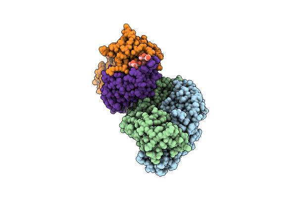
Anti-Phosphohistidine Fab Hsc44.Ck.20.N32F
Organism: Oryctolagus cuniculus
Method: X-RAY DIFFRACTION Resolution:1.94 Å Release Date: 2025-03-19 Classification: IMMUNE SYSTEM Ligands: EDO, SO4 |
Organism: Oryctolagus cuniculus
Method: X-RAY DIFFRACTION
Release Date: 2025-03-19
Ligands: EDO, SO4
 |
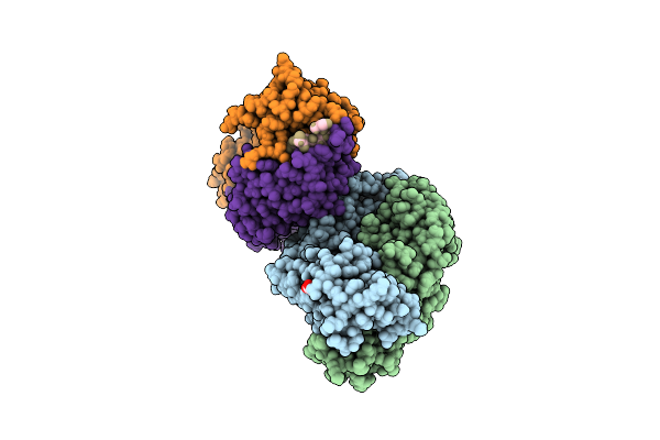
Anti-Phosphohistidine Fab Hsc44.Ck.20.N32F With 3Phis Peptide
Organism: Oryctolagus cuniculus, Synthetic construct
Method: X-RAY DIFFRACTION Resolution:2.09 Å Release Date: 2025-03-19 Classification: IMMUNE SYSTEM Ligands: EDO, ACT |
Organism: Oryctolagus cuniculus, Synthetic construct
Method: X-RAY DIFFRACTION
Release Date: 2025-03-19
Ligands: EDO, ACT
 |
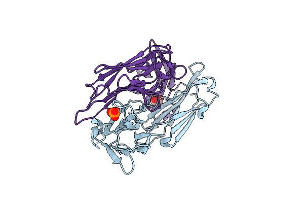
Anti-Phosphohistidine Fab Hsc44.Ck.20.N32F With 3Ptza Peptide
Organism: Oryctolagus cuniculus, Synthetic construct
Method: X-RAY DIFFRACTION Resolution:1.98 Å Release Date: 2025-03-19 Classification: IMMUNE SYSTEM Ligands: EDO, ACT |
Organism: Oryctolagus cuniculus, Synthetic construct
Method: X-RAY DIFFRACTION
Release Date: 2025-03-19
Ligands: EDO, ACT
 |
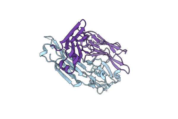
Anti-Phosphohistidine Fab Hsc44.Ck.20.Elbow Bound To Phosphate
Organism: Oryctolagus cuniculus
Method: X-RAY DIFFRACTION Resolution:2.40 Å Release Date: 2025-03-19 Classification: IMMUNE SYSTEM Ligands: EDO, PO4 |
Organism: Oryctolagus cuniculus
Method: X-RAY DIFFRACTION
Release Date: 2025-03-19
Ligands: EDO, PO4
 |
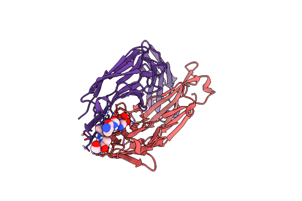
Anti-Phosphohistidine Fab Hsc44.Ck.20.Elbow Bound To Glu H1
Organism: Oryctolagus cuniculus
Method: X-RAY DIFFRACTION Resolution:2.45 Å Release Date: 2025-03-19 Classification: IMMUNE SYSTEM |
Organism: Oryctolagus cuniculus
Method: X-RAY DIFFRACTION
Release Date: 2025-03-19
 |
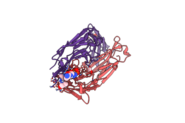
Anti-Phosphohistidine Fab Hsc44.Ck.20 With 3Phis Peptide
Organism: Oryctolagus cuniculus, Synthetic construct
Method: X-RAY DIFFRACTION Resolution:1.90 Å Release Date: 2025-03-19 Classification: IMMUNE SYSTEM |
Organism: Oryctolagus cuniculus, Synthetic construct
Method: X-RAY DIFFRACTION
Release Date: 2025-03-19
 |
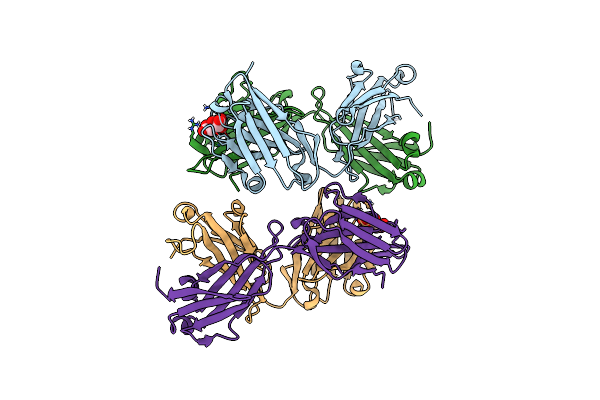
Anti-Phosphohistidine Fab Hsc44.Ck.20 With 3Ptza Peptide
Organism: Oryctolagus cuniculus, Synthetic construct
Method: X-RAY DIFFRACTION Resolution:1.85 Å Release Date: 2025-03-19 Classification: IMMUNE SYSTEM Ligands: EDO |
Organism: Oryctolagus cuniculus, Synthetic construct
Method: X-RAY DIFFRACTION
Release Date: 2025-03-19
Ligands: EDO
 |
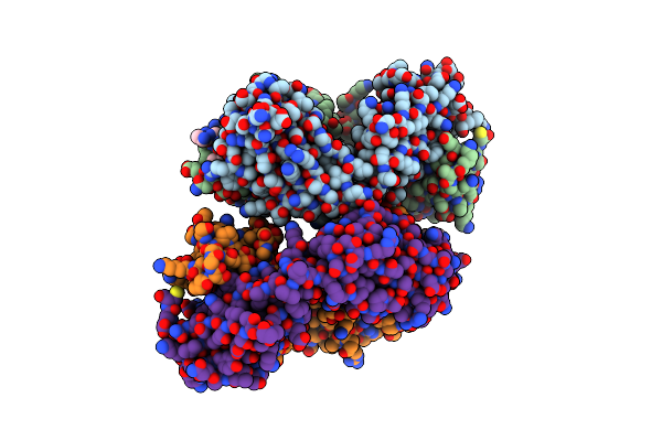
Anti-Phosphohistidine Fab Hsc44.Ck
Organism: Oryctolagus cuniculus
Method: X-RAY DIFFRACTION Resolution:1.75 Å Release Date: 2025-03-19 Classification: IMMUNE SYSTEM Ligands: CIT |
Organism: Oryctolagus cuniculus
Method: X-RAY DIFFRACTION
Release Date: 2025-03-19
Ligands: CIT
 |
Anti-Phosphohistidine Fab Hsc44.Ck With 3Phis Peptide
Organism: Oryctolagus cuniculus, Synthetic construct
Method: X-RAY DIFFRACTION Resolution:2.20 Å Release Date: 2025-03-19 Classification: IMMUNE SYSTEM |
Organism: Oryctolagus cuniculus, Synthetic construct
Method: X-RAY DIFFRACTION
Release Date: 2025-03-19
 |
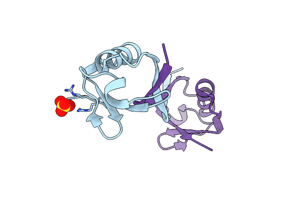
Anti-Phosphohistidine Fab Hsc44.Ck With 3Ptza Peptide
Organism: Oryctolagus cuniculus, Synthetic construct
Method: X-RAY DIFFRACTION Resolution:1.95 Å Release Date: 2025-03-19 Classification: IMMUNE SYSTEM Ligands: EDO |
Organism: Oryctolagus cuniculus, Synthetic construct
Method: X-RAY DIFFRACTION
Release Date: 2025-03-19
Ligands: EDO
 |
Structure Of A Dimeric Ubiquitin Variant (Ubv3) That Inhibits The Protease (Pro) From Turnip Yellow Mosaic Virus (Tymv)
Organism: Synthetic construct
Method: X-RAY DIFFRACTION Resolution:1.84 Å Release Date: 2025-01-22 Classification: BIOSYNTHETIC PROTEIN Ligands: SO4 |
Organism: Synthetic construct
Method: X-RAY DIFFRACTION
Release Date: 2025-01-22
Ligands: SO4
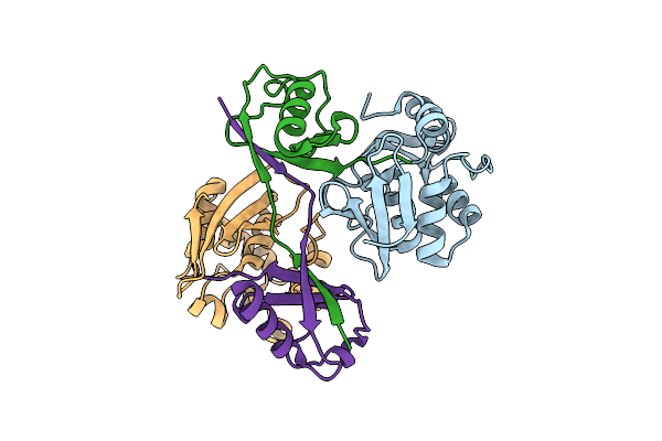 |
Turnip Yellow Mosaic Virus (Tymv) Protease (Pro) Bound To A Ubiquitin Variant (Ubv3)
Organism: Turnip yellow mosaic virus, Synthetic construct
Method: X-RAY DIFFRACTION Resolution:2.80 Å Release Date: 2025-01-22 Classification: VIRAL PROTEIN |
Organism: Turnip yellow mosaic virus, Synthetic construct
Method: X-RAY DIFFRACTION
Release Date: 2025-01-22