
Search Count: 458
 |
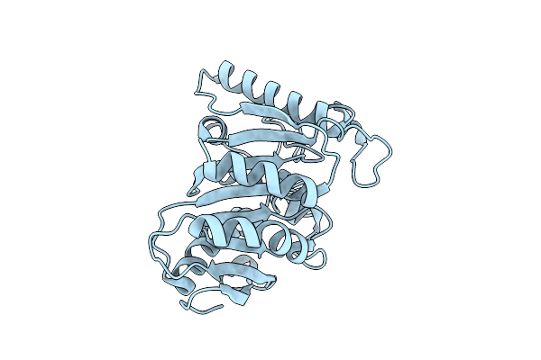
A Misfolded Structure Of An Fad-Binding Protein Ct375
Organism: Chlamydia trachomatis d/uw-3/cx
Method: X-RAY DIFFRACTION Resolution:2.30 Å Release Date: 2026-07-01 Classification: OXIDOREDUCTASE |
Organism: Chlamydia trachomatis d/uw-3/cx
Method: X-RAY DIFFRACTION
Release Date: 2026-07-01
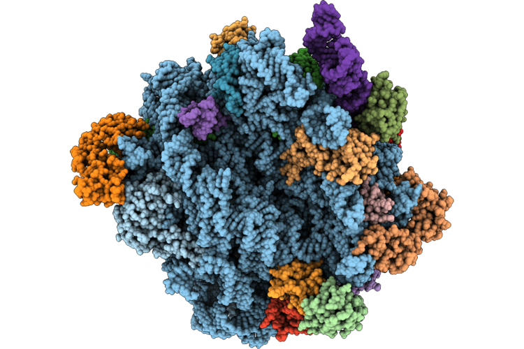 |
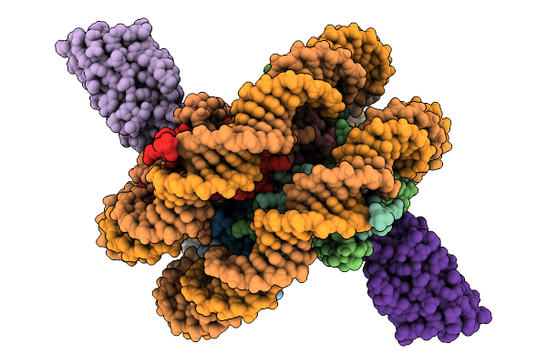
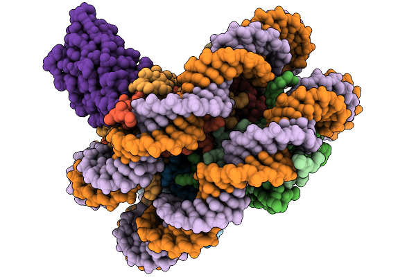
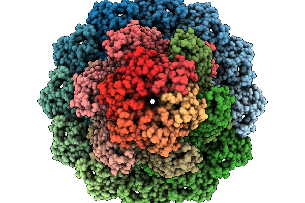
50S Ribosomal Subunit With E-Trna And Rsfs From The Alphaproteobacteria Asaia Platycodi
Organism: Asaia platycodi jcm 25414
Method: ELECTRON MICROSCOPY Release Date: 2026-06-03 Classification: RIBOSOME |
Organism: Asaia platycodi jcm 25414
Method: ELECTRON MICROSCOPY
Release Date: 2026-06-03
 |
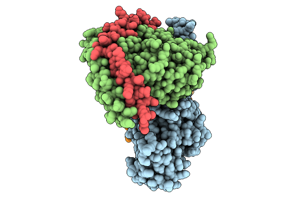
Crystal Structure Of Human Mait A-F7 Tcr-Mr1*02-5-Op-Ru Complex
Organism: Homo sapiens
Method: X-RAY DIFFRACTION Resolution:2.30 Å Release Date: 2026-05-13 Classification: IMMUNE SYSTEM Ligands: Q87, ACT, CL, GOL, NA |
Organism: Homo sapiens
Method: X-RAY DIFFRACTION
Release Date: 2026-05-13
Ligands: Q87, ACT, CL, GOL, NA
 |
Crystal Structure Of Human Mait A-F7 Tcr-Mr1*03-5-Op-Ru Complex
Organism: Homo sapiens
Method: X-RAY DIFFRACTION Resolution:2.10 Å Release Date: 2026-05-13 Classification: IMMUNE SYSTEM Ligands: ACY, Q87, GOL, CL, NA |
Organism: Homo sapiens
Method: X-RAY DIFFRACTION
Release Date: 2026-05-13
Ligands: ACY, Q87, GOL, CL, NA
 |
Crystal Structure Of Human Mait A-F7 Tcr-Mr1*04 Complex
Organism: Homo sapiens
Method: X-RAY DIFFRACTION Resolution:3.00 Å Release Date: 2026-05-13 Classification: IMMUNE SYSTEM Ligands: GOL |
Organism: Homo sapiens
Method: X-RAY DIFFRACTION
Release Date: 2026-05-13
Ligands: GOL
 |
Crystal Structure Of Human Mait A-F7 Tcr-Mr1*05-5-Op-Ru Complex
Organism: Homo sapiens
Method: X-RAY DIFFRACTION Resolution:2.40 Å Release Date: 2026-05-13 Classification: IMMUNE SYSTEM Ligands: Q87, GOL, NA |
Organism: Homo sapiens
Method: X-RAY DIFFRACTION
Release Date: 2026-05-13
Ligands: Q87, GOL, NA
 |
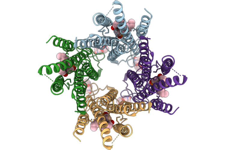
Structure Of Human Kcnq1-Cam-Pip2 Intermediate State
Organism: Homo sapiens
Method: ELECTRON MICROSCOPY Resolution:3.47 Å Release Date: 2026-05-06 Classification: MEMBRANE PROTEIN Ligands: PT5, CA |
Organism: Homo sapiens
Method: ELECTRON MICROSCOPY
Release Date: 2026-05-06
Ligands: PT5, CA
 |
Cryo-Em Structure Of Human Lipid Phosphate Phosphatase 2
Organism: Homo sapiens
Method: ELECTRON MICROSCOPY Resolution:2.90 Å Release Date: 2026-05-06 Classification: HYDROLASE Ligands: CPL, NAG |
Organism: Homo sapiens
Method: ELECTRON MICROSCOPY
Release Date: 2026-05-06
Ligands: CPL, NAG
 |
Structure Determination Of Pedobacter Sp. Kp-2 Pahz1
Organism: Pedobacter sp. kp-2
Method: X-RAY DIFFRACTION Resolution:2.00 Å Release Date: 2026-03-11 Classification: HYDROLASE |
Organism: Pedobacter sp. kp-2
Method: X-RAY DIFFRACTION
Release Date: 2026-03-11
 |
Competition For Different Elements Of The Nucleosome Acidic Patch Yields Distinct Functional Outcomes. Vhh 1B2
Organism: Homo sapiens, Artificial sequences
Method: ELECTRON MICROSCOPY Resolution:3.17 Å Release Date: 2026-03-04 Classification: DE NOVO PROTEIN |
Organism: Homo sapiens, Artificial sequences
Method: ELECTRON MICROSCOPY
Release Date: 2026-03-04
 |
Competition For Different Elements Of The Nucleosome Acidic Patch Yields Distinct Functional Outcomes. Vhh 1G1
Organism: Artificial sequences, Homo sapiens
Method: ELECTRON MICROSCOPY Resolution:3.10 Å Release Date: 2026-03-04 Classification: DE NOVO PROTEIN |
Organism: Artificial sequences, Homo sapiens
Method: ELECTRON MICROSCOPY
Release Date: 2026-03-04
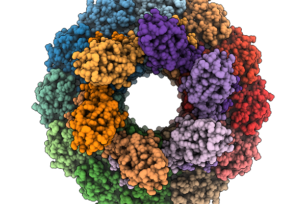 |
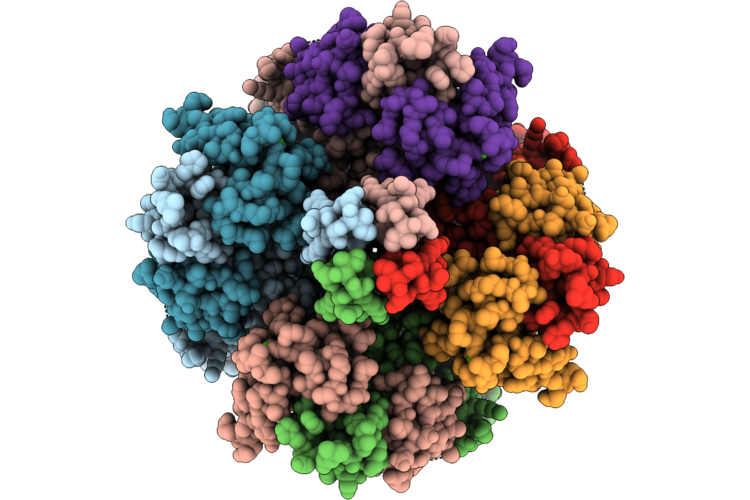
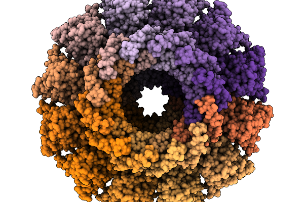
Structure Of Bacteriophage T4 Neck Protein Gp13 And Gp14 Assembled In Vitro In C6 Symmetry
Organism: Escherichia phage t4
Method: ELECTRON MICROSCOPY Release Date: 2026-02-18 Classification: VIRAL PROTEIN |
Organism: Escherichia phage t4
Method: ELECTRON MICROSCOPY
Release Date: 2026-02-18
 |
Structure Of Bacteriophage T4 Neck Protein Gp13 And Gp14 And Hfq Assembled In Vitro In C6 Symmetry
Organism: Escherichia phage t4
Method: ELECTRON MICROSCOPY Release Date: 2026-02-18 Classification: VIRAL PROTEIN |
Organism: Escherichia phage t4
Method: ELECTRON MICROSCOPY
Release Date: 2026-02-18
 |
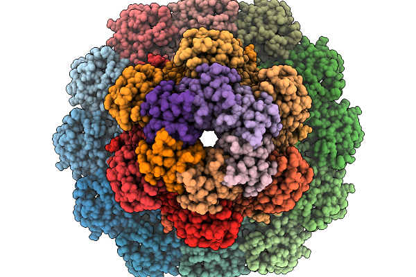
Structure Of Bacteriophage T4 Protal-Neck Protein Gp20-Gp13-Gp14-Hfq Assembled In Vitro In C6 Symmetry
Organism: Escherichia phage t4
Method: ELECTRON MICROSCOPY Resolution:2.91 Å Release Date: 2026-02-18 Classification: VIRAL PROTEIN |
Organism: Escherichia phage t4
Method: ELECTRON MICROSCOPY
Release Date: 2026-02-18
 |
Structure Of Bacteriophage T4 Protal-Neck Mismatch Complex Gp20-Gp14-Gp13 Assembled In Vitro In C6 Symmetry
Organism: Escherichia phage t4
Method: ELECTRON MICROSCOPY Release Date: 2026-02-18 Classification: VIRAL PROTEIN |
Organism: Escherichia phage t4
Method: ELECTRON MICROSCOPY
Release Date: 2026-02-18
 |
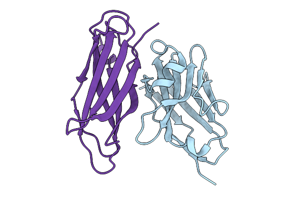
Crystal Structure Of Malaria Transmission-Blocking Antigen Pfs48/45 C-Terminal Domain In Complex With Nanobody B2
Organism: Plasmodium falciparum 3d7, Vicugna pacos
Method: X-RAY DIFFRACTION Resolution:2.50 Å Release Date: 2026-01-21 Classification: PROTEIN BINDING Ligands: SO4, EPE |
Organism: Plasmodium falciparum 3d7, Vicugna pacos
Method: X-RAY DIFFRACTION
Release Date: 2026-01-21
Ligands: SO4, EPE
 |
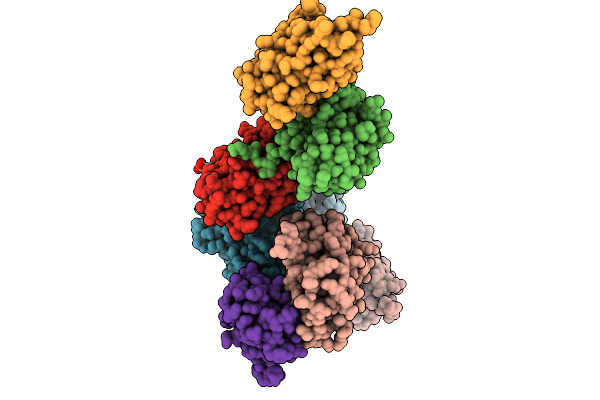
Positive Allosteric Modulator(Bms986187)-Bound Delta-Opioid Receptor-Gi Complex
Organism: Homo sapiens, Rattus norvegicus, Bos taurus, Synthetic construct
Method: ELECTRON MICROSCOPY Resolution:2.97 Å Release Date: 2026-01-07 Classification: MEMBRANE PROTEIN Ligands: A1D6B, A1D6F |
Organism: Homo sapiens, Rattus norvegicus, Bos taurus, Synthetic construct
Method: ELECTRON MICROSCOPY
Release Date: 2026-01-07
Ligands: A1D6B, A1D6F
 |
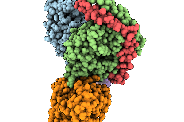
Gi-Bound Kappa Opioid Receptor In Complex With Dynorphin And Positive Allosteric Modulator Mpam-15
Organism: Homo sapiens, Rattus norvegicus, Bos taurus
Method: ELECTRON MICROSCOPY Resolution:2.90 Å Release Date: 2025-12-31 Classification: MEMBRANE PROTEIN Ligands: CLR, A1D6C |
Organism: Homo sapiens, Rattus norvegicus, Bos taurus
Method: ELECTRON MICROSCOPY
Release Date: 2025-12-31
Ligands: CLR, A1D6C
 |
Crystal Structure Of Malaria Transmission-Blocking Antigen Pfhap2 Domain 3 In Complex With Nanobody Wnb 334
Organism: Vicugna pacos, Plasmodium falciparum
Method: X-RAY DIFFRACTION Resolution:2.80 Å Release Date: 2025-12-31 Classification: PROTEIN BINDING |
Organism: Vicugna pacos, Plasmodium falciparum
Method: X-RAY DIFFRACTION
Release Date: 2025-12-31
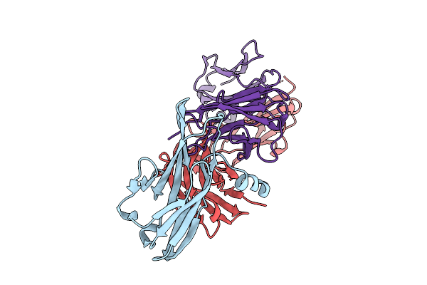 |
Crystal Structure Of Pilra In Complex With Fab Portion Of Antagonist Antibody
Organism: Homo sapiens
Method: X-RAY DIFFRACTION Resolution:2.58 Å Release Date: 2025-12-17 Classification: SIGNALING PROTEIN |
Organism: Homo sapiens
Method: X-RAY DIFFRACTION
Release Date: 2025-12-17