
Search Count: 56
All
Selected
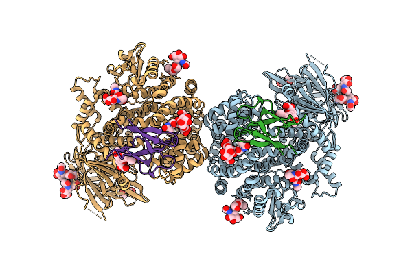 |
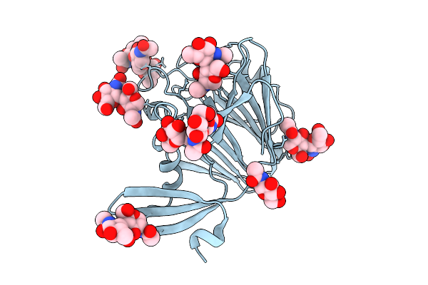
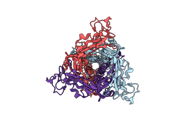
Molecular Basis Of Pathogenicity Of The Recently Emerged Fcov-23 Coronavirus. Complex Of Fapn With Fcov-23 Rbd
Organism: Felis catus, Feline coronavirus
Method: ELECTRON MICROSCOPY Release Date: 2025-07-09 Classification: VIRAL PROTEIN/HYDROLASE Ligands: NAG, ZN |
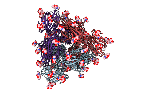 |
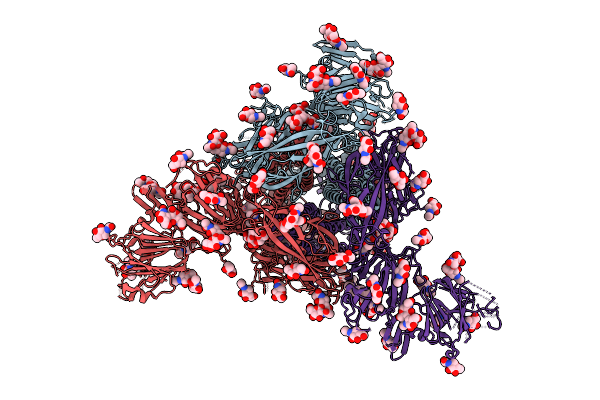
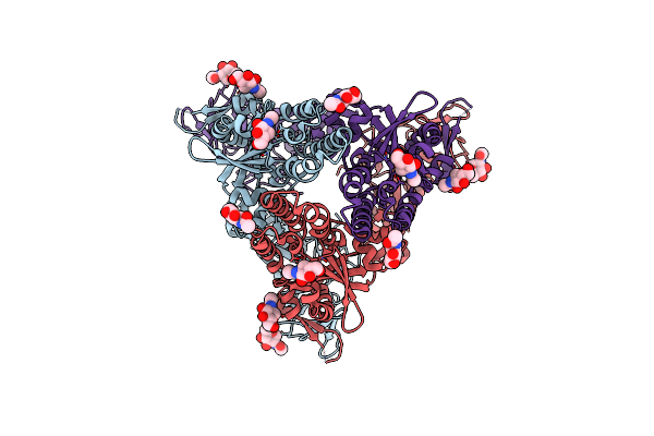
Molecular Basis Of Pathogenicity Of The Recently Emerged Fcov-23 Coronavirus. Fcov-23 S Short
Organism: Feline coronavirus
Method: ELECTRON MICROSCOPY Release Date: 2025-07-09 Classification: VIRAL PROTEIN Ligands: NAG, PAM |
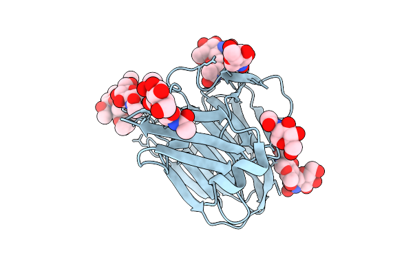 |
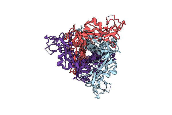
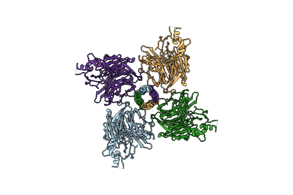
Molecular Basis Of Pathogenicity Of The Recently Emerged Fcov-23 Coronavirus. Fcov-23 S Do In Proximal Conformation (Local Refinement)
Organism: Feline coronavirus
Method: ELECTRON MICROSCOPY Release Date: 2025-07-09 Classification: VIRAL PROTEIN Ligands: NAG |
 |
Molecular Basis Of Pathogenicity Of The Recently Emerged Fcov-23 Coronavirus. Fcov-23 S Long With Do In Swung-Out Conformation
Organism: Feline coronavirus
Method: ELECTRON MICROSCOPY Release Date: 2025-07-09 Classification: VIRAL PROTEIN Ligands: NAG, PAM |
 |
Molecular Basis Of Pathogenicity Of The Recently Emerged Fcov-23 Coronavirus. Fcov-23 S Long Domain 0 In Swung-Out Conformation (Local Refinement)
Organism: Feline coronavirus
Method: ELECTRON MICROSCOPY Release Date: 2025-07-09 Classification: VIRAL PROTEIN Ligands: NAG |
 |
Molecular Basis Of Pathogenicity Of The Recently Emerged Fcov-23 Coronavirus. Fcov-23 S Long With Do In Mixed Conformations (Global Refinement).
Organism: Feline coronavirus
Method: ELECTRON MICROSCOPY Release Date: 2025-07-09 Classification: VIRAL PROTEIN Ligands: NAG, PAM |
 |
Organism: Langya virus
Method: ELECTRON MICROSCOPY Release Date: 2024-05-08 Classification: VIRAL PROTEIN Ligands: NAG |
 |
Organism: Langya virus
Method: ELECTRON MICROSCOPY Release Date: 2024-05-08 Classification: VIRAL PROTEIN Ligands: NAG |
 |
Langya Henipavirus Postfusion F Protein In Complex With The 4G5 Fab, Local Refinement Of The Viral Membrane Distal Region
Organism: Langya virus
Method: ELECTRON MICROSCOPY Release Date: 2024-05-08 Classification: VIRAL PROTEIN Ligands: NAG |
 |
Langya Henipavirus Postfusion F Protein In Complex With 4G5 Fab, Local Refinement Of The Viral Membrane Proximal Region
Organism: Mus musculus, Langya virus
Method: ELECTRON MICROSCOPY Release Date: 2024-05-08 Classification: VIRAL PROTEIN/IMMUNE SYSTEM Ligands: NAG |
 |
Organism: Henipavirus ghanaense, Thermotoga maritima msb8
Method: ELECTRON MICROSCOPY Release Date: 2024-05-01 Classification: VIRAL PROTEIN Ligands: NAG |
 |
Organism: Langya virus
Method: ELECTRON MICROSCOPY Release Date: 2024-05-01 Classification: VIRAL PROTEIN Ligands: NAG, ZN |
 |
Organism: Langya virus
Method: ELECTRON MICROSCOPY Release Date: 2024-05-01 Classification: VIRAL PROTEIN Ligands: NAG |
 |
Organism: Human coronavirus hku1 (isolate n1), Homo sapiens
Method: ELECTRON MICROSCOPY Release Date: 2024-03-06 Classification: VIRAL PROTEIN Ligands: NAG |
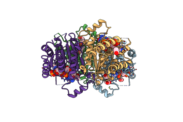 |
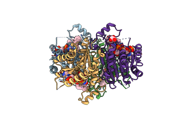
Crystal Structure Of Pteridine Reductase 1 (Ptr1) From Trypanosoma Brucei In Ternary Complex With Cofactor And Inhibitor
Organism: Trypanosoma brucei brucei
Method: X-RAY DIFFRACTION Resolution:2.20 Å Release Date: 2015-01-21 Classification: OXIDOREDUCTASE Ligands: OZJ, NAP, ACT |
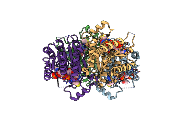 |
Crystal Structure Of Pteridine Reductase 1 (Ptr1) From Trypanosoma Brucei In Ternary Complex With Cofactor And Inhibitor
Organism: Trypanosoma brucei brucei
Method: X-RAY DIFFRACTION Resolution:1.77 Å Release Date: 2015-01-21 Classification: OXIDOREDUCTASE Ligands: NAP, JUO, ACT |
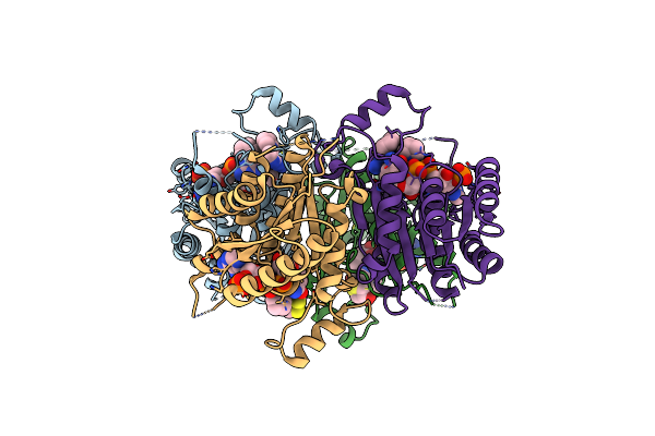 |
Crystal Structure Of Pteridine Reductase 1 (Ptr1) From Trypanosoma Brucei In Ternary Complex With Cofactor And Inhibitor
Organism: Trypanosoma brucei brucei
Method: X-RAY DIFFRACTION Resolution:1.80 Å Release Date: 2015-01-21 Classification: OXIDOREDUCTASE Ligands: NAP, JR2, DTD |
 |
Crystal Structure Of Pteridine Reductase 1 (Ptr1) From Trypanosoma Brucei In Ternary Complex With Cofactor And Inhibitor
Organism: Trypanosoma brucei brucei
Method: X-RAY DIFFRACTION Resolution:1.85 Å Release Date: 2015-01-21 Classification: OXIDOREDUCTASE Ligands: NAP, W8G, ACT |
 |
Crystal Structure Of Pteridine Reductase 1 (Ptr1) From Trypanosoma Brucei In Ternary Complex With Cofactor And Inhibitor
Organism: Trypanosoma brucei brucei
Method: X-RAY DIFFRACTION Resolution:1.88 Å Release Date: 2015-01-21 Classification: OXIDOREDUCTASE Ligands: NAP, XP0, GOL |
 |
Crystal Structure Of Pteridine Reductase 1 (Ptr1) From Trypanosoma Brucei In Ternary Complex With Cofactor And Inhibitor
Organism: Trypanosoma brucei brucei
Method: X-RAY DIFFRACTION Resolution:1.75 Å Release Date: 2015-01-21 Classification: OXIDOREDUCTASE Ligands: NAP, FDB |