

Search Count: 36
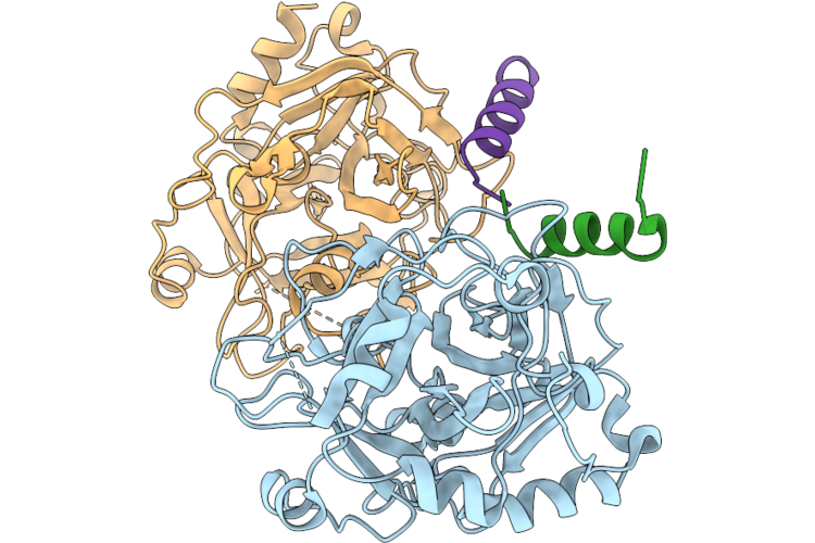 |
Crystal Structure Of Prethrombin-2 With A Peptide Corresponding To The C-Terminus Of The Heavy Chain Of Factor Va
Organism: Homo sapiens
Method: X-RAY DIFFRACTION Resolution:2.80 Å Release Date: 2026-08-12 Classification: BLOOD CLOTTING Modified Residue(s): PTR |
Organism: Homo sapiens
Method: X-RAY DIFFRACTION
Release Date: 2026-08-12
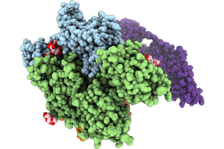 |
Structure Of Human Prothrombinase With Prothrombin
Organism: Homo sapiens
Method: ELECTRON MICROSCOPY Release Date: 2026-08-12 Classification: BLOOD CLOTTING Ligands: CA, NA Modified Residue(s): TYS |
Organism: Homo sapiens
Method: ELECTRON MICROSCOPY
Release Date: 2026-08-12
Ligands: CA, NA
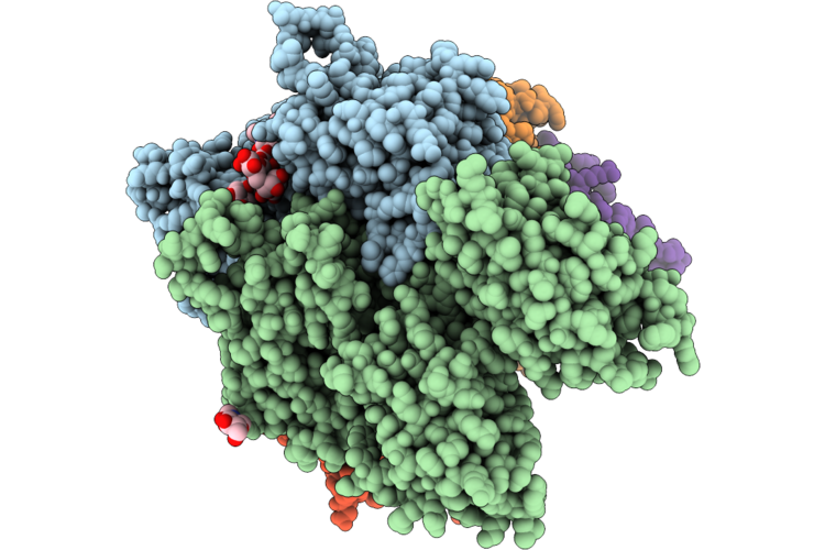 |
Structure Of Human Prothrombinase With Meizothrombin
Organism: Homo sapiens
Method: ELECTRON MICROSCOPY Release Date: 2026-08-12 Classification: BLOOD CLOTTING Ligands: NAG, CA, NA Modified Residue(s): TYS |
Organism: Homo sapiens
Method: ELECTRON MICROSCOPY
Release Date: 2026-08-12
Ligands: NAG, CA, NA
 |
Structure Of The Arabidopsis Thaliana 80S Ribosome In Complex With P- And E-Site Trnas And Mrna
Organism: Arabidopsis thaliana
Method: ELECTRON MICROSCOPY Resolution:1.82 Å Release Date: 2026-02-04 Classification: RIBOSOME Ligands: MG, K, TER, SPD, EPE, ZN Modified Residue(s): PSU, A2M, OMG, HIC, MLY |
Organism: Arabidopsis thaliana
Method: ELECTRON MICROSCOPY
Release Date: 2026-02-04
Ligands: MG, K, TER, SPD, EPE, ZN
 |
Structure Of The Arabidopsis Thaliana 80S Ribosome In Complex With P- And E-Site Trnas, Mrna, And Thermospermine
Organism: Arabidopsis thaliana
Method: ELECTRON MICROSCOPY Resolution:2.20 Å Release Date: 2026-02-04 Classification: RIBOSOME Ligands: TER, MG, K, SPD, EPE, ZN Modified Residue(s): PSU, A2M, OMG, HIC, MLY |
Organism: Arabidopsis thaliana
Method: ELECTRON MICROSCOPY
Release Date: 2026-02-04
Ligands: TER, MG, K, SPD, EPE, ZN
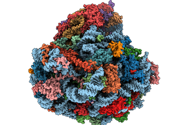 |
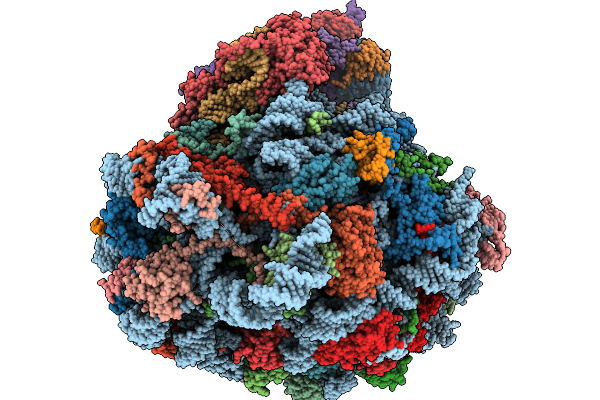
Structure Of The Arabidopsis Thaliana 80S Ribosome Ovac Mutant In Complex With P- And E-Site Trnas, Mrna, And Thermospermine
Organism: Arabidopsis thaliana
Method: ELECTRON MICROSCOPY Resolution:2.25 Å Release Date: 2026-02-04 Classification: RIBOSOME Ligands: TER, MG, K, SPD, EPE, ZN Modified Residue(s): PSU, A2M, OMG, HIC, MLY |
Organism: Arabidopsis thaliana
Method: ELECTRON MICROSCOPY
Release Date: 2026-02-04
Ligands: TER, MG, K, SPD, EPE, ZN
 |
Structure Of The Arabidopsis Thaliana 80S Ribosome Ovac Mutant In Complex With P- And E-Site Trnas And Mrna
Organism: Arabidopsis thaliana
Method: ELECTRON MICROSCOPY Resolution:2.25 Å Release Date: 2026-02-04 Classification: RIBOSOME Ligands: MG, K, TER, SPD, EPE, ZN Modified Residue(s): PSU, A2M, OMG, HIC, MLY |
Organism: Arabidopsis thaliana
Method: ELECTRON MICROSCOPY
Release Date: 2026-02-04
Ligands: MG, K, TER, SPD, EPE, ZN
 |
Coagulation Factor Xa Complex With A2-Loop Peptide
Organism: Homo sapiens, Synthetic construct
Method: X-RAY DIFFRACTION Resolution:1.20 Å Release Date: 2025-11-26 Classification: BLOOD CLOTTING Ligands: CA, NA, CL, IMD, 0GJ Modified Residue(s): PTR |
Organism: Homo sapiens, Synthetic construct
Method: X-RAY DIFFRACTION
Release Date: 2025-11-26
Ligands: CA, NA, CL, IMD, 0GJ
 |
A 3.3 Angstrom Cryo-Em Structure Of An Engineered High-Affinity Human Prothrombinase Complex
Organism: Homo sapiens
Method: ELECTRON MICROSCOPY Release Date: 2025-11-26 Classification: BLOOD CLOTTING Ligands: CA, NA, 0GJ, CU Modified Residue(s): TYS |
Organism: Homo sapiens
Method: ELECTRON MICROSCOPY
Release Date: 2025-11-26
Ligands: CA, NA, 0GJ, CU
 |
Structure Of The Human 1-Phosphatidylinositol 4,5-Bisphosphate Phosphodiesterase Gamma-2 (Plcg2) Protein
Organism: Homo sapiens
Method: ELECTRON MICROSCOPY Resolution:3.50 Å Release Date: 2025-03-26 Classification: LIPID BINDING PROTEIN |
Organism: Homo sapiens
Method: ELECTRON MICROSCOPY
Release Date: 2025-03-26
 |
Crystal Structure Of Apo-Pptt From Mycobacterium Tuberculosis
Organism: Mycobacterium tuberculosis
Method: X-RAY DIFFRACTION Resolution:1.70 Å Release Date: 2024-06-19 Classification: TRANSFERASE Ligands: ACT, MG |
Organism: Mycobacterium tuberculosis
Method: X-RAY DIFFRACTION
Release Date: 2024-06-19
Ligands: ACT, MG
 |
Crystal Structure Of Pptt-Acp From Mycobacterium Tuberculosis
Organism: Mycobacterium tuberculosis, Mycobacterium tuberculosis h37rv
Method: X-RAY DIFFRACTION Resolution:2.50 Å Release Date: 2024-06-19 Classification: TRANSFERASE Ligands: COA, MN, IPA Modified Residue(s): MSE |
Organism: Mycobacterium tuberculosis, Mycobacterium tuberculosis h37rv
Method: X-RAY DIFFRACTION
Release Date: 2024-06-19
Ligands: COA, MN, IPA
 |
Crystal Structure Of Pptt-Adp From Mycobacterium Tuberculosis
Organism: Mycobacterium tuberculosis
Method: X-RAY DIFFRACTION Resolution:2.00 Å Release Date: 2024-06-19 Classification: TRANSFERASE Ligands: A3P, MN |
Organism: Mycobacterium tuberculosis
Method: X-RAY DIFFRACTION
Release Date: 2024-06-19
Ligands: A3P, MN
 |
Human Mature Large Subunit Of The Ribosome With Eif6 And Homoharringtonine Bound
Organism: Homo sapiens
Method: ELECTRON MICROSCOPY Resolution:1.67 Å Release Date: 2023-03-08 Classification: TRANSLATION Ligands: MG, K, NA, HMT, SPM, EPE, ZN Modified Residue(s): A2M, OMG, 1MA, OMC, PSU, OMU, 5MC, 6MZ, UR3 |
Organism: Homo sapiens
Method: ELECTRON MICROSCOPY
Release Date: 2023-03-08
Ligands: MG, K, NA, HMT, SPM, EPE, ZN
 |
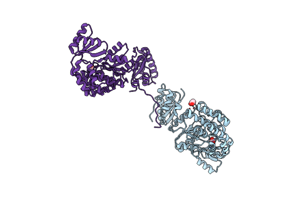
Eif6-Bound Large Subunit Of The Human Ribosome
Organism: Homo sapiens
Method: ELECTRON MICROSCOPY Resolution:2.40 Å Release Date: 2022-10-05 Classification: TRANSLATION Ligands: MG, ZN, K, NA, BR |
Organism: Homo sapiens
Method: ELECTRON MICROSCOPY
Release Date: 2022-10-05
Ligands: MG, ZN, K, NA, BR
 |
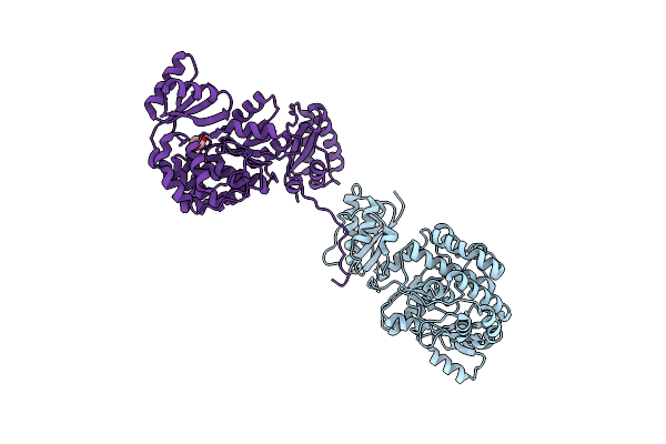
Structure Of The S726F Mutant Of Acyltransferase Domain Of Mycocerosic Acid Synthase From Mycobacterium Tuberculosis
Organism: Mycobacterium bovis (strain atcc baa-935 / af2122/97)
Method: X-RAY DIFFRACTION Resolution:2.80 Å Release Date: 2020-12-16 Classification: TRANSFERASE Ligands: ACT, GOL |
Organism: Mycobacterium bovis (strain atcc baa-935 / af2122/97)
Method: X-RAY DIFFRACTION
Release Date: 2020-12-16
Ligands: ACT, GOL
 |
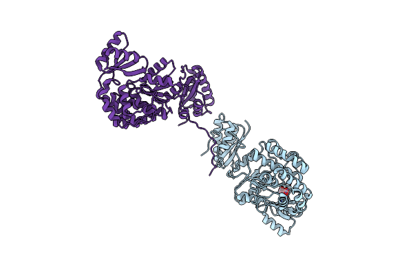
Structure Of The M624V-S726F Mutant Of Acyltransferase Domain Of Mycocerosic Acid Synthase From Mycobacterium Tuberculosis Soaked With Methylmalonyl Coenzyme A
Organism: Mycobacterium bovis (strain atcc baa-935 / af2122/97)
Method: X-RAY DIFFRACTION Resolution:2.80 Å Release Date: 2020-12-16 Classification: TRANSFERASE Ligands: EDO |
Organism: Mycobacterium bovis (strain atcc baa-935 / af2122/97)
Method: X-RAY DIFFRACTION
Release Date: 2020-12-16
Ligands: EDO
 |
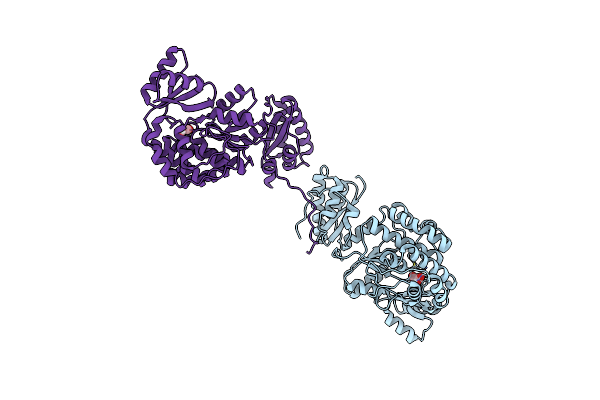
Structure Of The Acyltransferase Domain Of Mycocerosic Acid Synthase From Mycobacterium Tuberculosis Acylated Methylmalonyl-Coenzymea
Organism: Mycobacterium bovis (strain atcc baa-935 / af2122/97)
Method: X-RAY DIFFRACTION Resolution:3.10 Å Release Date: 2020-12-16 Classification: TRANSFERASE Ligands: DXX |
Organism: Mycobacterium bovis (strain atcc baa-935 / af2122/97)
Method: X-RAY DIFFRACTION
Release Date: 2020-12-16
Ligands: DXX
 |
Structure Of The S726F Mutant Of Acyltransferase Domain Of Mycocerosic Acid Synthase From Mycobacterium Tuberculosis Acylated With Malonyl-Coenzyme A
Organism: Mycobacterium bovis (strain atcc baa-935 / af2122/97)
Method: X-RAY DIFFRACTION Resolution:2.90 Å Release Date: 2020-12-16 Classification: TRANSFERASE Ligands: MLA |
Organism: Mycobacterium bovis (strain atcc baa-935 / af2122/97)
Method: X-RAY DIFFRACTION
Release Date: 2020-12-16
Ligands: MLA
 |
Structure Of The S726F Mutant Of Acyltransferase Domain Of Mycocerosic Acid Synthase From Mycobacterium Tuberculosis Acylated With Methylmalonyl-Coenzyme A
Organism: Mycobacterium bovis (strain atcc baa-935 / af2122/97)
Method: X-RAY DIFFRACTION Resolution:3.10 Å Release Date: 2020-12-16 Classification: TRANSFERASE Ligands: DXX, EDO |
Organism: Mycobacterium bovis (strain atcc baa-935 / af2122/97)
Method: X-RAY DIFFRACTION
Release Date: 2020-12-16
Ligands: DXX, EDO