
Search Count: 2,601
All
Selected
 |
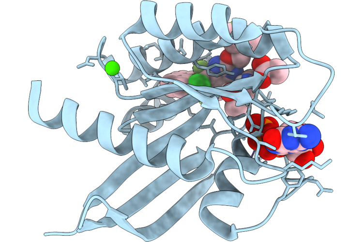
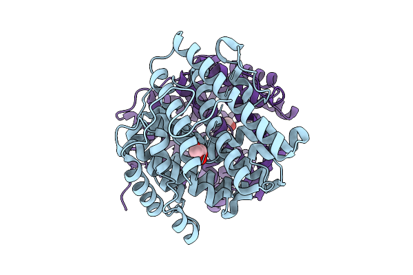
Organism: Homo sapiens
Method: X-RAY DIFFRACTION Resolution:1.60 Å Release Date: 2026-04-22 Classification: SIGNALING PROTEIN Ligands: MG, GDP, A1JU5 |
 |
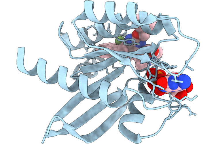
Organism: Homo sapiens
Method: X-RAY DIFFRACTION Resolution:1.65 Å Release Date: 2026-04-22 Classification: SIGNALING PROTEIN Ligands: GDP, MG, A1JU6 |
 |
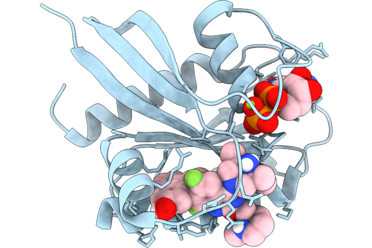
Organism: Homo sapiens
Method: X-RAY DIFFRACTION Resolution:1.54 Å Release Date: 2026-04-22 Classification: SIGNALING PROTEIN Ligands: MG, GDP, CA, A1JU7 |
 |
Organism: Homo sapiens
Method: X-RAY DIFFRACTION Resolution:1.60 Å Release Date: 2026-04-22 Classification: SIGNALING PROTEIN Ligands: MG, GDP, A1JU8 |
 |
Organism: Homo sapiens
Method: X-RAY DIFFRACTION Resolution:1.30 Å Release Date: 2026-04-22 Classification: SIGNALING PROTEIN Ligands: GDP, MG, CA, ACT, A1JU3 |
 |
Organism: Homo sapiens
Method: X-RAY DIFFRACTION Resolution:1.35 Å Release Date: 2026-04-22 Classification: SIGNALING PROTEIN Ligands: MG, GDP, A1JU9 |
 |
Organism: Homo sapiens
Method: X-RAY DIFFRACTION Resolution:1.63 Å Release Date: 2026-04-22 Classification: SIGNALING PROTEIN Ligands: GDP, MG, A1JU2 |
 |
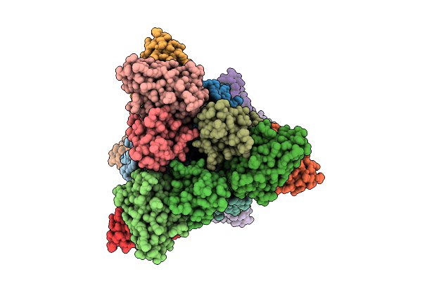
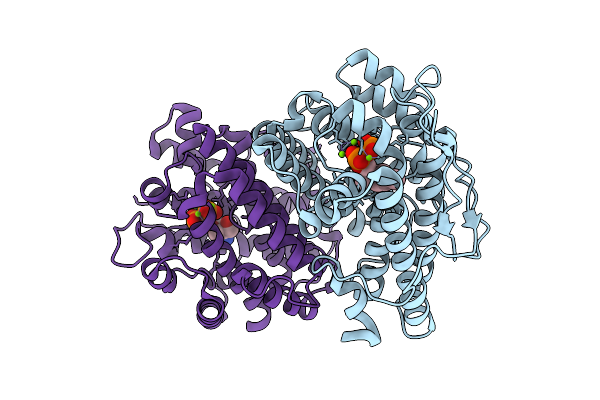
Structure Of The Plasmodium Falciparum 20S Proteasome In Complex With A Beta5-Selective Covalent Syringolin Analogue Inhibitor.
Organism: Plasmodium falciparum 3d7
Method: ELECTRON MICROSCOPY Resolution:2.70 Å Release Date: 2026-04-15 Classification: HYDROLASE Ligands: A1CY6 |
 |
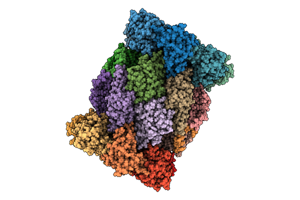
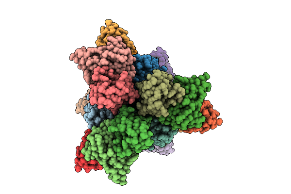
Structure Of The Human 20S Proteasome In Complex With A Beta5-Selective Covalent Syringolin Analogue Inhibitor.
Organism: Homo sapiens
Method: ELECTRON MICROSCOPY Resolution:2.60 Å Release Date: 2026-04-15 Classification: HYDROLASE Ligands: A1CY5 |
 |
Organism: Homo sapiens
Method: ELECTRON MICROSCOPY Release Date: 2026-04-01 Classification: MEMBRANE PROTEIN Ligands: NAG, A1EQU, NA |
 |
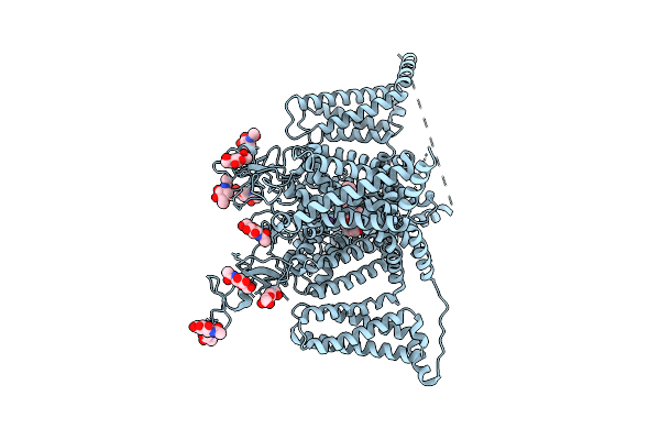
Organism: Homo sapiens
Method: ELECTRON MICROSCOPY Resolution:3.00 Å Release Date: 2026-03-25 Classification: MEMBRANE PROTEIN Ligands: NAG, Y01, LPE, PCW, A1E26 |
 |
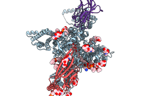
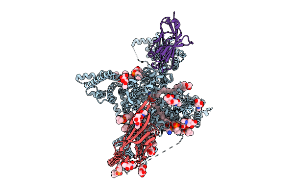
Cryo-Em Structure Of Sudan Ebolavirus Gp Bound By Three Neutralizing Antibodies 545S, 523S And 294S
Organism: Sudan ebolavirus, Homo sapiens
Method: ELECTRON MICROSCOPY Release Date: 2026-03-18 Classification: VIRAL PROTEIN/IMMUNE SYSTEM |
 |
Cryo-Em Structure Of Sudan Ebolavirus Gp Bound By Three Neutralizing Antibodies 316L, 523S And 294S
Organism: Sudan ebolavirus, Macaca mulatta, Homo sapiens
Method: ELECTRON MICROSCOPY Release Date: 2026-03-18 Classification: VIRAL PROTEIN/IMMUNE SYSTEM |
 |
Crystal Structure Of Terpeniod Cyclase Spsods From Rhizobacterium Serratia Plymuthica
Organism: Serratia plymuthica 4rx13
Method: X-RAY DIFFRACTION Resolution:2.22 Å Release Date: 2026-03-18 Classification: LYASE Ligands: GOL |
 |
Organism: Serratia plymuthica 4rx13
Method: X-RAY DIFFRACTION Resolution:1.87 Å Release Date: 2026-03-18 Classification: LYASE Ligands: POP, MG, TRS |
 |
Organism: Serratia plymuthica 4rx13
Method: X-RAY DIFFRACTION Resolution:1.74 Å Release Date: 2026-03-18 Classification: LYASE Ligands: GPP, MG, TRS, POP |
 |
The Structure Of Nav1.7 With Veratridine Standing Near The Ifm Motif (Site I)
Organism: Homo sapiens
Method: ELECTRON MICROSCOPY Resolution:2.70 Å Release Date: 2026-03-11 Classification: MEMBRANE PROTEIN Ligands: NAG, LPE, A1E26, Y01, PCW, P3X |
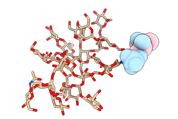 |
Tlp-2A, A Glycofibril Obtained From A Karst Cave From Guilin City, Guangxi Zhuang Autonomous Region, China
Organism: Unidentified
Method: ELECTRON MICROSCOPY Release Date: 2026-02-18 Classification: PROTEIN FIBRIL |
 |
Tlp-2F, A Glycofibril Obtained From A Karst Cave From Guilin City, Guangxi Zhuang Autonomous Region, China
Organism: Unidentified
Method: ELECTRON MICROSCOPY Release Date: 2026-02-18 Classification: UNKNOWN FUNCTION |
 |
Tlp-2G, A Glycofibril Obtained From A Karst Cave From Guilin City, Guangxi Zhuang Autonomous Region, China
Organism: Unidentified
Method: ELECTRON MICROSCOPY Release Date: 2026-02-18 Classification: UNKNOWN FUNCTION Ligands: HOH |