

Search Count: 1,050
 |
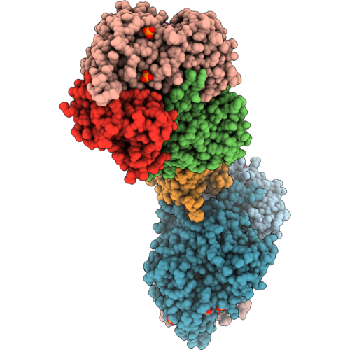
Therapeutic Antibody With Captured Antigen
Organism: Homo sapiens
Method: X-RAY DIFFRACTION Resolution:2.60 Å Release Date: 2026-09-30 Classification: BLOOD CLOTTING Ligands: MG, NAG, EDO |
Organism: Homo sapiens
Method: X-RAY DIFFRACTION
Release Date: 2026-09-30
Ligands: MG, NAG, EDO
 |
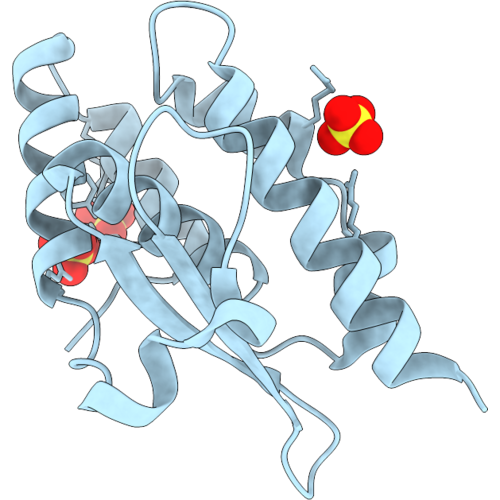
B. Subtilis Sufs Wt - E. Coli Sufe Variant With Deletion Of Tqhl (Residues 112-115)
Organism: Bacillus subtilis subsp. subtilis str. 168, Escherichia coli k-12
Method: X-RAY DIFFRACTION Resolution:2.00 Å Release Date: 2026-09-02 Classification: BIOSYNTHETIC PROTEIN Ligands: PO4 |
Organism: Bacillus subtilis subsp. subtilis str. 168, Escherichia coli k-12
Method: X-RAY DIFFRACTION
Release Date: 2026-09-02
Ligands: PO4
 |
E. Coli Sufe With Deletion Of Tqhl (Residues 112-115)
Organism: Escherichia coli k-12
Method: X-RAY DIFFRACTION Resolution:1.53 Å Release Date: 2026-09-02 Classification: BIOSYNTHETIC PROTEIN Ligands: SO4 |
Organism: Escherichia coli k-12
Method: X-RAY DIFFRACTION
Release Date: 2026-09-02
Ligands: SO4
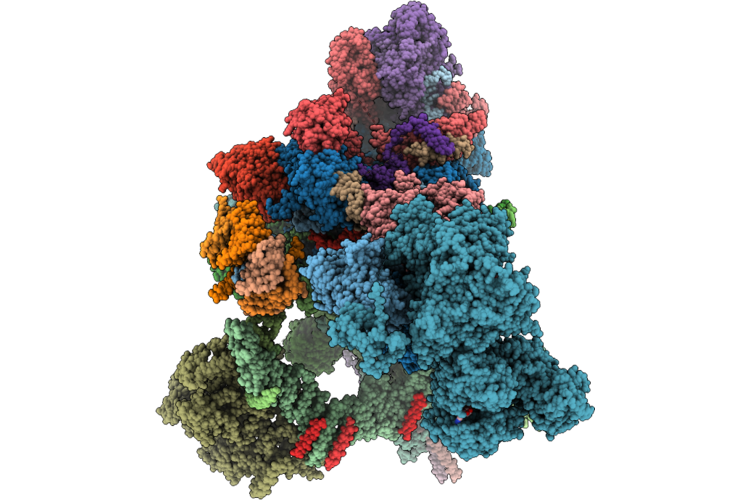 |
Human Minor Spliceosome Branching-Completed C Complex (After Step-I)
Method: ELECTRON MICROSCOPY
Release Date: 2026-08-12 Classification: SPLICING Ligands: MG, K, IHP, GTP, ADP, ZN, ATP |
Method: ELECTRON MICROSCOPY
Release Date: 2026-08-12
Ligands: MG, K, IHP, GTP, ADP, ZN, ATP
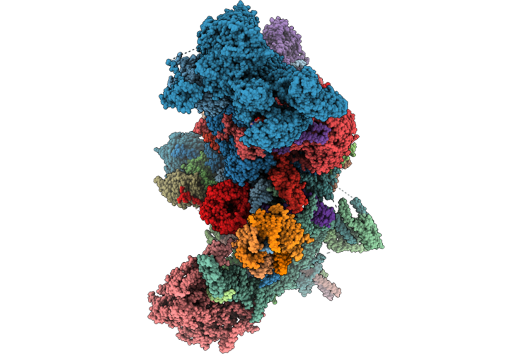 |
Human Minor Spliceosome Exon-Ligation-Ready C* Complex (Prior To Step-Ii)
Organism: Homo sapiens
Method: ELECTRON MICROSCOPY Release Date: 2026-08-12 Classification: SPLICING Ligands: MG, K, IHP, GTP, ADP, ZN, ATP |
Organism: Homo sapiens
Method: ELECTRON MICROSCOPY
Release Date: 2026-08-12
Ligands: MG, K, IHP, GTP, ADP, ZN, ATP
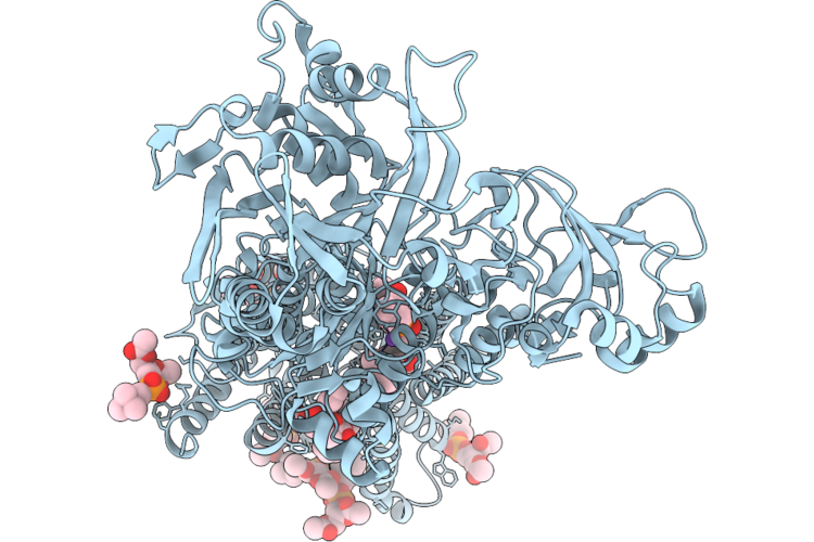 |
Crystal Structure Of Sr Ca2+-Atpase In E2(Tg) With Bound Ahq
Organism: Oryctolagus cuniculus
Method: X-RAY DIFFRACTION Resolution:2.12 Å Release Date: 2026-07-15 Classification: MEMBRANE PROTEIN |
Organism: Oryctolagus cuniculus
Method: X-RAY DIFFRACTION
Release Date: 2026-07-15
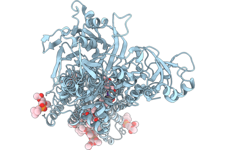 |
Crystal Structure Of Sr Ca2+-Atpase In E2(Tg) With Bound B1Hq
Organism: Oryctolagus cuniculus
Method: X-RAY DIFFRACTION Resolution:2.10 Å Release Date: 2026-07-15 Classification: MEMBRANE PROTEIN |
Organism: Oryctolagus cuniculus
Method: X-RAY DIFFRACTION
Release Date: 2026-07-15
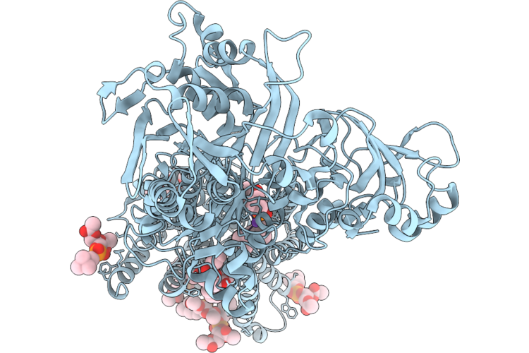 |
Crystal Structure Of Sr Ca2+-Atpase In E2(Tg) With Bound Ba
Organism: Oryctolagus cuniculus
Method: X-RAY DIFFRACTION Resolution:2.50 Å Release Date: 2026-07-15 Classification: MEMBRANE PROTEIN |
Organism: Oryctolagus cuniculus
Method: X-RAY DIFFRACTION
Release Date: 2026-07-15
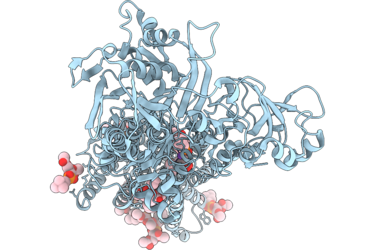 |
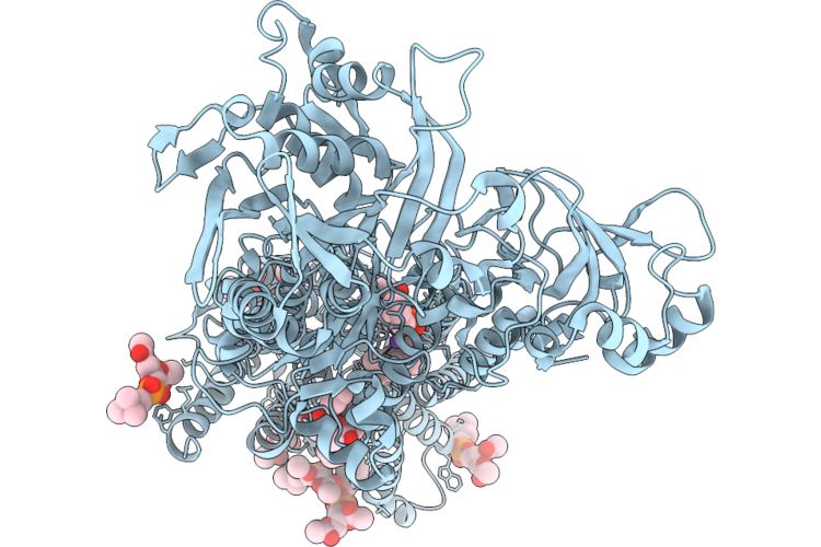
Crystal Structure Of Sr Ca2+-Atpase In E2(Tg) With Bound Bc
Organism: Oryctolagus cuniculus
Method: X-RAY DIFFRACTION Resolution:2.60 Å Release Date: 2026-07-15 Classification: MEMBRANE PROTEIN |
Organism: Oryctolagus cuniculus
Method: X-RAY DIFFRACTION
Release Date: 2026-07-15
 |
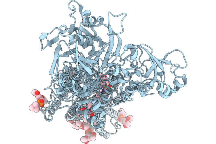
Crystal Structure Of Sr Ca2+-Atpase In E2(Tg) With Bound Bmp
Organism: Oryctolagus cuniculus
Method: X-RAY DIFFRACTION Resolution:2.10 Å Release Date: 2026-07-15 Classification: MEMBRANE PROTEIN |
Organism: Oryctolagus cuniculus
Method: X-RAY DIFFRACTION
Release Date: 2026-07-15
 |
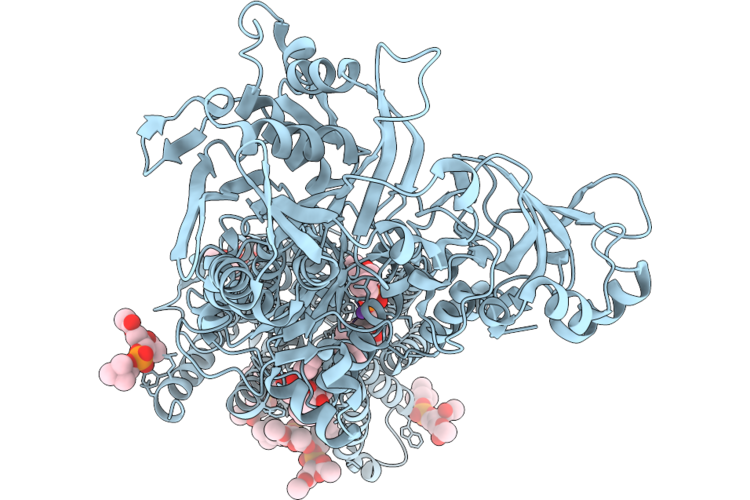
Crystal Structure Of Sr Ca2+-Atpase In E2(Tg) With Bound Ipp
Organism: Oryctolagus cuniculus
Method: X-RAY DIFFRACTION Resolution:2.00 Å Release Date: 2026-07-15 Classification: MEMBRANE PROTEIN |
Organism: Oryctolagus cuniculus
Method: X-RAY DIFFRACTION
Release Date: 2026-07-15
 |
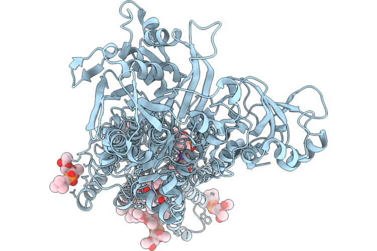
Crystal Structure Of Sr Ca2+-Atpase In E2(Tg) With Bound Mpq
Organism: Oryctolagus cuniculus
Method: X-RAY DIFFRACTION Resolution:2.20 Å Release Date: 2026-07-15 Classification: MEMBRANE PROTEIN |
Organism: Oryctolagus cuniculus
Method: X-RAY DIFFRACTION
Release Date: 2026-07-15
 |
Crystal Structure Of Sr Ca2+-Atpase In E2(Tg) With Bound Phq
Organism: Oryctolagus cuniculus
Method: X-RAY DIFFRACTION Resolution:2.20 Å Release Date: 2026-07-15 Classification: MEMBRANE PROTEIN |
Organism: Oryctolagus cuniculus
Method: X-RAY DIFFRACTION
Release Date: 2026-07-15
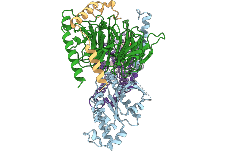 |
Cryo-Em Structure Of Bms-986187-Bound Mor-Gi1 Complex
Organism: Oplophorus gracilirostris, Homo sapiens
Method: ELECTRON MICROSCOPY Resolution:3.04 Å Release Date: 2026-07-08 Classification: MEMBRANE PROTEIN Ligands: A1D6B, A1ESG |
Organism: Oplophorus gracilirostris, Homo sapiens
Method: ELECTRON MICROSCOPY
Release Date: 2026-07-08
Ligands: A1D6B, A1ESG
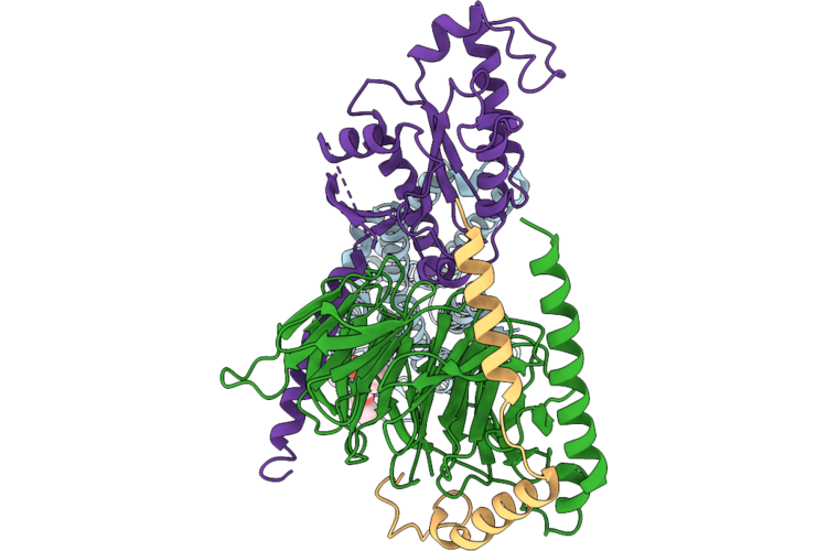 |
Cryo-Em Structure Of Bms986187 Bound Dor-Gi Complex
Organism: Escherichia coli, Homo sapiens, Synthetic construct
Method: ELECTRON MICROSCOPY Resolution:2.79 Å Release Date: 2026-07-08 Classification: MEMBRANE PROTEIN Ligands: A1D6B |
Organism: Escherichia coli, Homo sapiens, Synthetic construct
Method: ELECTRON MICROSCOPY
Release Date: 2026-07-08
Ligands: A1D6B
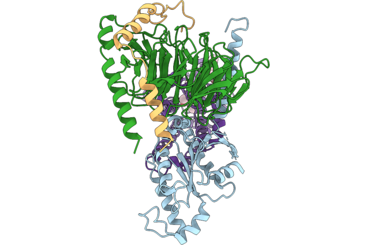 |
Cryo-Em Structure Of Asimadoline-Bms-986187-Bound Kor-Gi1 Complex
Organism: Oplophorus gracilirostris, Homo sapiens, Escherichia coli, Synthetic construct
Method: ELECTRON MICROSCOPY Resolution:2.58 Å Release Date: 2026-07-08 Classification: MEMBRANE PROTEIN Ligands: A1E0F, A1D6B |
Organism: Oplophorus gracilirostris, Homo sapiens, Escherichia coli, Synthetic construct
Method: ELECTRON MICROSCOPY
Release Date: 2026-07-08
Ligands: A1E0F, A1D6B
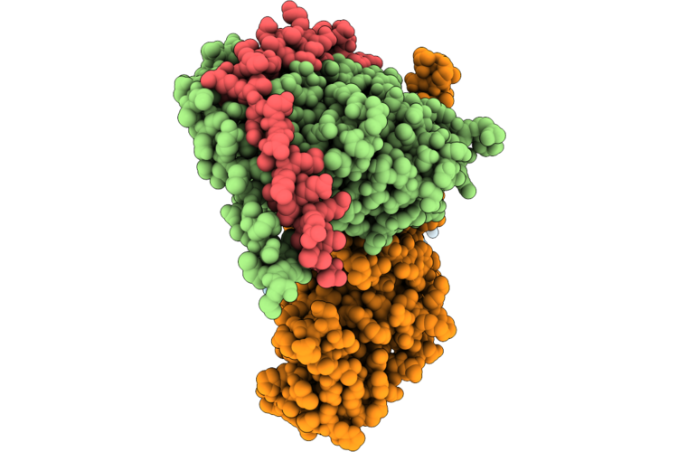 |
Cryo-Em Structure Of Leu-Enkephalin-Bms-986187-Bound Dor-Gi2 Complex
Organism: Escherichia coli, Homo sapiens, Synthetic construct
Method: ELECTRON MICROSCOPY Release Date: 2026-07-08 Classification: MEMBRANE PROTEIN Ligands: A1D6B |
Organism: Escherichia coli, Homo sapiens, Synthetic construct
Method: ELECTRON MICROSCOPY
Release Date: 2026-07-08
Ligands: A1D6B
 |
Structural Analysis Of A Plant Glycoside Hydrolase Family 116 Glucosyl Ceramidase By Cryogenic Electron Microscopy (Cryo-Em)
Organism: Arabidopsis thaliana
Method: ELECTRON MICROSCOPY Release Date: 2026-04-29 Classification: HYDROLASE |
Organism: Arabidopsis thaliana
Method: ELECTRON MICROSCOPY
Release Date: 2026-04-29
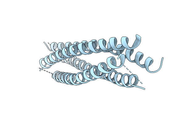 |
Crystal Structure Of Syn-F4-Link, A Covalent Dimer Of Syn-F4 (K4T)
Organism: Synthetic construct
Method: X-RAY DIFFRACTION Resolution:2.30 Å Release Date: 2026-04-15 Classification: DE NOVO PROTEIN |
Organism: Synthetic construct
Method: X-RAY DIFFRACTION
Release Date: 2026-04-15
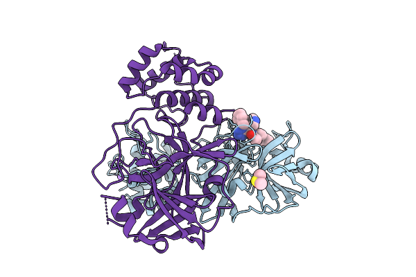 |
Crystal Structure Of Sars-Cov-2 Main Protease In Complex With Rs222C
Organism: Severe acute respiratory syndrome coronavirus 2
Method: X-RAY DIFFRACTION Resolution:2.04 Å Release Date: 2026-03-18 Classification: VIRAL PROTEIN Ligands: DMS, NA, A1I6A |
Organism: Severe acute respiratory syndrome coronavirus 2
Method: X-RAY DIFFRACTION
Release Date: 2026-03-18
Ligands: DMS, NA, A1I6A