9WIZ image
Deposition Date 2025-08-30
Release Date 2026-04-29
Last Version Date 2026-04-29
Entry Detail
PDB ID:
9WIZ
Keywords:
Title:
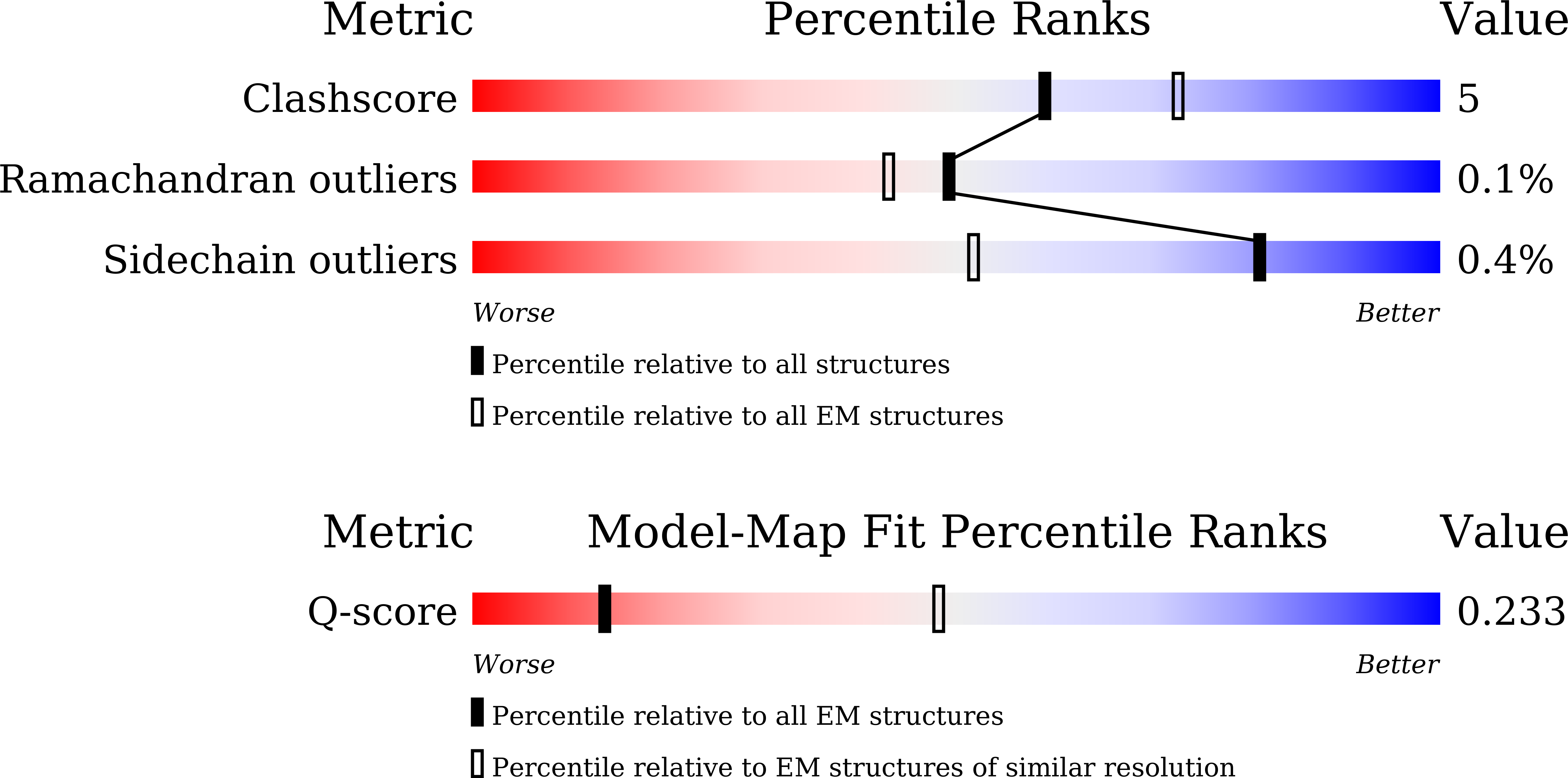
Structural Analysis of a Plant Glycoside Hydrolase Family 116 Glucosyl Ceramidase by Cryogenic Electron Microscopy (Cryo-EM)
Biological Source:
Source Organism(s):
Expression System(s):
Method Details:
Experimental Method:
Resolution:
4.90 Å
Aggregation State:
PARTICLE
Reconstruction Method:
SINGLE PARTICLE
Macromolecular Entities
Structures with similar UniProt ID
Protein Blast
Polymer Type:polypeptide(L)
Molecule:Non-lysosomal glucosylceramid
Chain IDs:A, B, C, D, E (auth: F), F (auth: E)
Chain Length:925
Number of Molecules:6
Biological Source:Arabidopsis thaliana
Ligand Molecules
Primary Citation
Structural analysis of the plant glycoside hydrolase family 116 glucosylceramidase AtGCD3 by cryogenic electron microscopy.
Int.J.Biol.Macromol. 350 150988 150988 (2026)
PMID: 41720384 DOI: 10.1016/j.ijbiomac.2026.150988

Abstact

Glucosylceramide (GlcCer) is a common glycosphingolipid that accumulates in cells in response to Gaucher disease, diabetes, and skin disorders in humans and is also found in plants. In animals, GlcCer is catabolized by glucosylcerebrosidase 1 and 2 (GBA1 and GBA2). GBA1 is a lysosomal enzyme in glycoside hydrolase (GH) family GH30, while GBA2 is a membrane-associated cytoplasmic protein in family GH116. Currently, there are no experimental structures of eukaryotic GH116 homologues. Although the bacterial TxGH116 beta-glucosidase structure was determined by X-ray crystallography, TxGH116 does not hydrolyze glucosylceramides, unlike the animal and plant enzymes. Therefore, we have investigated the structure of plant GH116 (AtGCD3) by cryogenic electron microscopy (Cryo-EM) single-particle analysis. The recombinant AtGCD3 protein was produced in Escherichia coli and purified by immobilized-metal affinity chromatography followed by size-exclusion chromatography. The Cryo-EM structure revealed a unique hexameric arrangement, composed of a dimer of trimers. Hydrophobic interactions and hydrogen bonds stabilize each trimer at the trimer interface. The two trimers stack face-to-face with a slight twist, with salt bridges and hydrogen bonding at their interface. Two alpha-helices not found in previously described GH116 structures cover the active site, forming two hydrophobic channels that may be involved in glucosylceramide binding. Molecular dynamics simulations showed that glucosylceramide can bind stably in the active site with its lipid tails in these channels. This first eukaryotic structure of a GH116 enzyme generates a template for improved modeling of human GBA2, with implications for treating human diseases, such as Gaucher disease and hereditary spastic paraplegia.

Legend

Protein

Chemical

Disease

Primary Citation of related structures
Feedback Form
Name
Email
Institute
Feedback