
Search Count: 20,326
All
Selected
 |
Organism: Homo sapiens
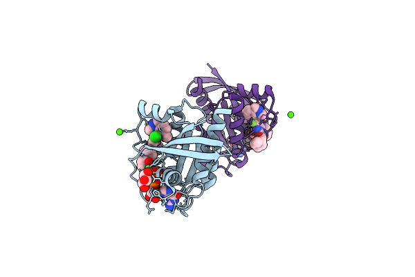
Method: X-RAY DIFFRACTION Resolution:1.50 Å Release Date: 2026-04-15 Classification: ONCOPROTEIN Ligands: CA, A1C6Y, GOL, GDP |
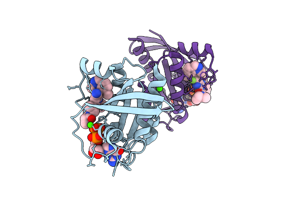 |
Organism: Homo sapiens
Method: X-RAY DIFFRACTION Resolution:1.52 Å Release Date: 2026-04-15 Classification: ONCOPROTEIN Ligands: GDP, A1C60, CA |
 |
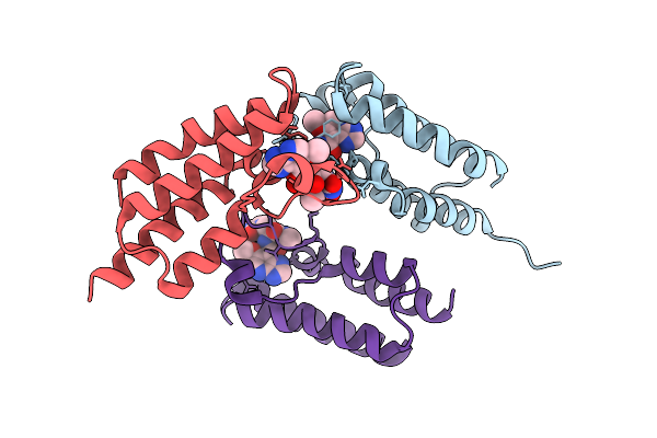
Androgen Receptor Ligand-Binding Domain In Complex With 11-Ketodihydrotestosterone
Organism: Homo sapiens
Method: X-RAY DIFFRACTION Resolution:1.91 Å Release Date: 2026-04-15 Classification: DNA BINDING PROTEIN Ligands: A1IQ9, SPD, SO4, GOL, IMD |
 |
Organism: Streptomyces lividans 1326
Method: X-RAY DIFFRACTION Resolution:1.90 Å Release Date: 2026-04-15 Classification: OXIDOREDUCTASE Ligands: HEM, MG, O |
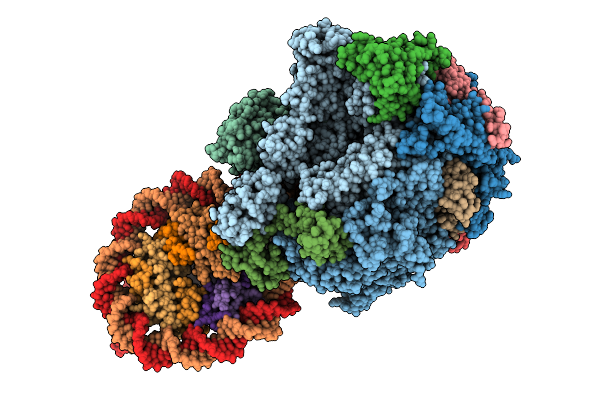 |
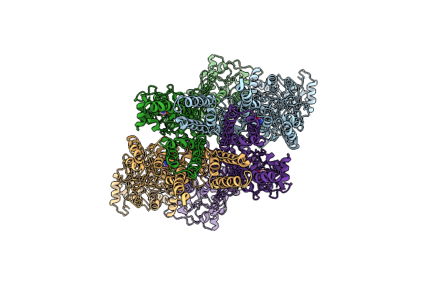
Rna Polymerase Ii Elongation Complex Stalled At Shl(-4) Of The H3-H4 Octasome
Organism: Homo sapiens, Synthetic construct, Komagataella phaffii gs115
Method: ELECTRON MICROSCOPY Resolution:3.62 Å Release Date: 2026-04-15 Classification: TRANSCRIPTION/RNA/DNA |
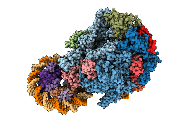 |
Rna Polymerase Ii Elongation Complex Stalled At Shl(-0.5) Of The H3-H4 Octasome (Tetrasome)
Organism: Homo sapiens, Synthetic construct, Komagataella phaffii gs115
Method: ELECTRON MICROSCOPY Resolution:6.54 Å Release Date: 2026-04-15 Classification: TRANSCRIPTION/RNA/DNA |
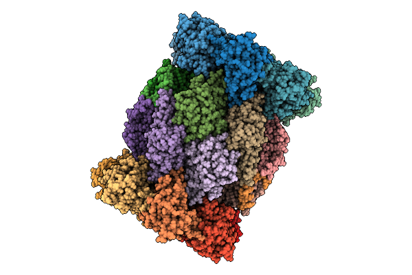 |
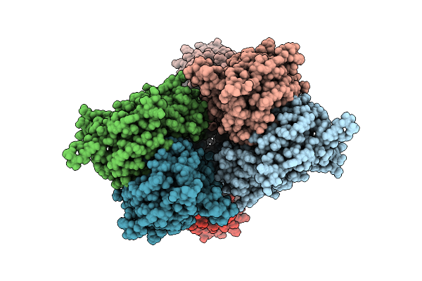
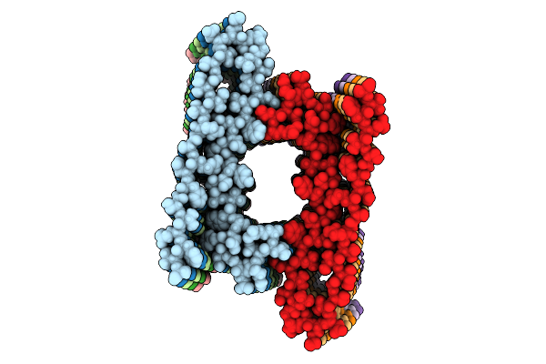
Structure Of The Plasmodium Falciparum 20S Proteasome In Complex With A Beta5-Selective Covalent Syringolin Analogue Inhibitor.
Organism: Plasmodium falciparum 3d7
Method: ELECTRON MICROSCOPY Resolution:2.70 Å Release Date: 2026-04-15 Classification: HYDROLASE Ligands: A1CY6 |
 |
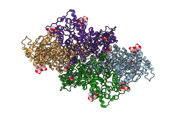
Structure Of The Human 20S Proteasome In Complex With A Beta5-Selective Covalent Syringolin Analogue Inhibitor.
Organism: Homo sapiens
Method: ELECTRON MICROSCOPY Resolution:2.60 Å Release Date: 2026-04-15 Classification: HYDROLASE Ligands: A1CY5 |
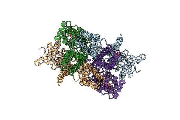 |
Organism: Rattus norvegicus
Method: ELECTRON MICROSCOPY Release Date: 2026-04-08 Classification: MEMBRANE PROTEIN Ligands: ZK1, NA |
 |
Composite Map Of Glua1/A2 In The Activated State, In Complex With Positive Allosteric Modulator (R,R)-2B And Agonist Glutamate (Atd-Lbd-Tmd)
Organism: Rattus norvegicus
Method: ELECTRON MICROSCOPY Release Date: 2026-04-08 Classification: MEMBRANE PROTEIN Ligands: GLU, NA, FWF |
 |
Heteromeric Glua1/A2-Cnih1 In The Activated State, Composite Map Of Lbd-Tmd
Organism: Rattus norvegicus, Homo sapiens
Method: ELECTRON MICROSCOPY Release Date: 2026-04-08 Classification: MEMBRANE PROTEIN Ligands: GLU, POV, FWF, NA |
 |
Heteromeric Glua1/A2 In The Desensitized State, Composite Map Of Atd-Lbd-Tmd
Organism: Rattus norvegicus
Method: ELECTRON MICROSCOPY Release Date: 2026-04-08 Classification: MEMBRANE PROTEIN Ligands: QUS |
 |
Streptavidin K121M With A Thiophenol Cofactor As Artificial Hydrogen Atom Transferase
Organism: Streptomyces avidinii
Method: X-RAY DIFFRACTION Resolution:2.00 Å Release Date: 2026-04-08 Classification: TRANSFERASE Ligands: A1I8C, EDO |
 |
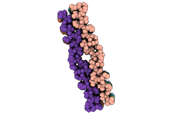
Population B Fibril Generated From The Heterotypic Interaction Of Abeta40 And Medin.
Organism: Homo sapiens
Method: ELECTRON MICROSCOPY Release Date: 2026-04-08 Classification: PROTEIN FIBRIL |
 |
Population A Fibril Generated From The Heterotypic Interaction Of Abeta40 And Medin.
Organism: Homo sapiens
Method: ELECTRON MICROSCOPY Resolution:3.10 Å Release Date: 2026-04-08 Classification: PROTEIN FIBRIL |
 |
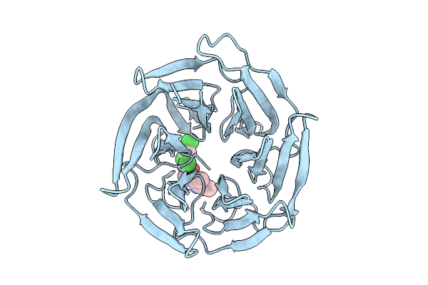
Crystal Structure Of Bromodomain From Plasmodium Falciparum Gcn5 Complexed With A Ligand
Organism: Plasmodium falciparum 3d7
Method: X-RAY DIFFRACTION Resolution:1.80 Å Release Date: 2026-04-08 Classification: TRANSFERASE Ligands: A1JWP, CL |
 |
Medin Fibril Generated From The Heterotypic Interaction Of Abeta40 And Medin.
Organism: Homo sapiens
Method: ELECTRON MICROSCOPY Resolution:2.70 Å Release Date: 2026-04-08 Classification: PROTEIN FIBRIL |
 |
Organism: Homo sapiens
Method: X-RAY DIFFRACTION Resolution:2.70 Å Release Date: 2026-04-08 Classification: LIGASE Ligands: A1CTS |
 |
Organism: Homo sapiens
Method: X-RAY DIFFRACTION Resolution:1.27 Å Release Date: 2026-04-08 Classification: LIGASE Ligands: A1CTR |
 |
Organism: Homo sapiens
Method: X-RAY DIFFRACTION Resolution:1.27 Å Release Date: 2026-04-08 Classification: LIGASE Ligands: A1CTQ |