

Search Count: 1,064
 |
Apo Pdz Domain From Mouse Shank3 (570-664)
Organism: Mus musculus
Method: X-RAY DIFFRACTION Resolution:1.26 Å Release Date: 2026-09-30 Classification: PROTEIN BINDING Ligands: GOL |
Organism: Mus musculus
Method: X-RAY DIFFRACTION
Release Date: 2026-09-30
Ligands: GOL
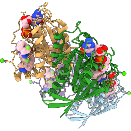 |
Kras G12V In Complex With Gdp And A Macrocyclic Inhibitor (Compound 20).
Organism: Homo sapiens
Method: X-RAY DIFFRACTION Resolution:2.10 Å Release Date: 2026-09-09 Classification: HYDROLASE Ligands: MG, A1DB0, GDP, CA |
Organism: Homo sapiens
Method: X-RAY DIFFRACTION
Release Date: 2026-09-09
Ligands: MG, A1DB0, GDP, CA
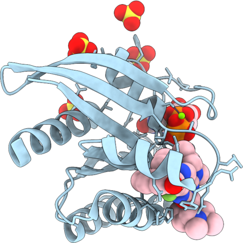 |
Kras G12V In Complex With Gdp And A Macrocyclic Inhibitor (Compound 12).
Organism: Homo sapiens
Method: X-RAY DIFFRACTION Resolution:1.43 Å Release Date: 2026-09-09 Classification: HYDROLASE Ligands: MG, A1DBZ, GDP, SO4 |
Organism: Homo sapiens
Method: X-RAY DIFFRACTION
Release Date: 2026-09-09
Ligands: MG, A1DBZ, GDP, SO4
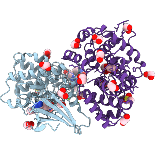 |
Crystal Structure Of Selective Inhibitor 16 Bound At The Active Site Of Cdk2
Organism: Homo sapiens
Method: X-RAY DIFFRACTION Resolution:1.59 Å Release Date: 2026-09-02 Classification: SIGNALING PROTEIN Ligands: MG, EDO, CL, DMS, A1DEQ, IPA |
Organism: Homo sapiens
Method: X-RAY DIFFRACTION
Release Date: 2026-09-02
Ligands: MG, EDO, CL, DMS, A1DEQ, IPA
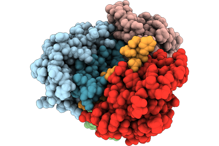 |
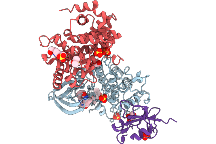
Shank3 Pdz (570-664) In Complex With An Internal Pdz Binding Motif (843-864) In Densin-180
Organism: Mus musculus, Rattus norvegicus
Method: X-RAY DIFFRACTION Resolution:1.98 Å Release Date: 2026-07-22 Classification: PROTEIN BINDING |
Organism: Mus musculus, Rattus norvegicus
Method: X-RAY DIFFRACTION
Release Date: 2026-07-22
 |
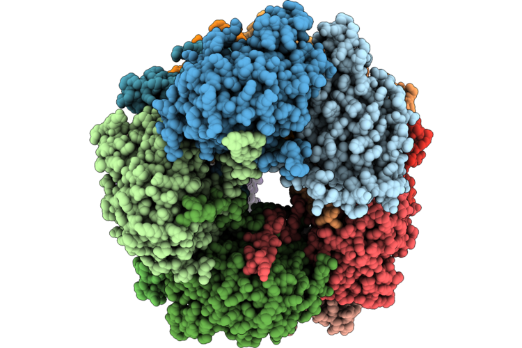
Crystal Structure Of Selective Inhibitor 16 Bound At The Active Site Of Cdk1
Organism: Homo sapiens
Method: X-RAY DIFFRACTION Resolution:2.40 Å Release Date: 2026-07-08 Classification: SIGNALING PROTEIN Ligands: A1DEQ, A1DJL, GOL, SO4, DMS, EPE |
Organism: Homo sapiens
Method: X-RAY DIFFRACTION
Release Date: 2026-07-08
Ligands: A1DEQ, A1DJL, GOL, SO4, DMS, EPE
 |
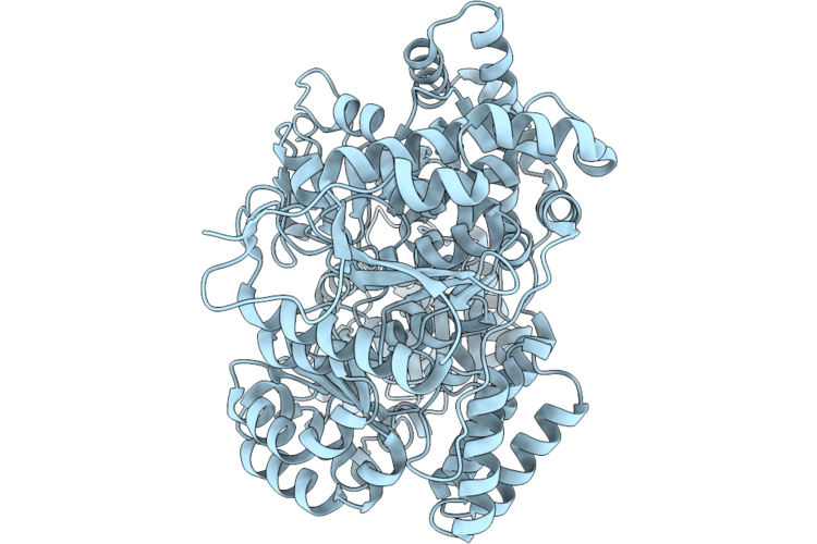
15-Subunit Assembly Of Cyanide Dihydratase From Stutzerimonas Stutzeri (Pseudomonas Stutzeri Ak1)
Organism: Stutzerimonas
Method: ELECTRON MICROSCOPY Release Date: 2026-07-08 Classification: HYDROLASE |
Organism: Stutzerimonas
Method: ELECTRON MICROSCOPY
Release Date: 2026-07-08
 |
Hna Monomer
Organism: Sinorhizobium meliloti
Method: ELECTRON MICROSCOPY Release Date: 2026-06-03 Classification: IMMUNE SYSTEM |
Organism: Sinorhizobium meliloti
Method: ELECTRON MICROSCOPY
Release Date: 2026-06-03
 |
Kras G13D Mutant In Complex With Gdp And Compound 6.
Organism: Homo sapiens
Method: X-RAY DIFFRACTION Resolution:1.70 Å Release Date: 2026-06-03 Classification: ONCOPROTEIN Ligands: A1C3J, GDP, MG, PO4 |
Organism: Homo sapiens
Method: X-RAY DIFFRACTION
Release Date: 2026-06-03
Ligands: A1C3J, GDP, MG, PO4
 |
Kras G12V Mutant In Complex With Gdp And Compound 12.
Organism: Homo sapiens
Method: X-RAY DIFFRACTION Resolution:1.68 Å Release Date: 2026-06-03 Classification: ONCOPROTEIN Ligands: MG, A1C3N, GDP, SO4 |
Organism: Homo sapiens
Method: X-RAY DIFFRACTION
Release Date: 2026-06-03
Ligands: MG, A1C3N, GDP, SO4
 |
Kras G12V Mutant In Complex With Gdp And Compound 8.
Organism: Homo sapiens
Method: X-RAY DIFFRACTION Resolution:1.91 Å Release Date: 2026-06-03 Classification: ONCOPROTEIN Ligands: MG, A1C3O, GDP |
Organism: Homo sapiens
Method: X-RAY DIFFRACTION
Release Date: 2026-06-03
Ligands: MG, A1C3O, GDP
 |
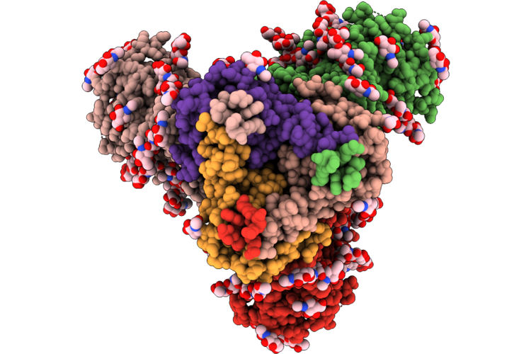
Cryo-Em Structure Of Shiv-Elicited Ce79-1571 Fab In Complex With Hiv Env Trimer Q23-Sct27
Organism: Macaca mulatta, Human immunodeficiency virus
Method: ELECTRON MICROSCOPY Resolution:3.30 Å Release Date: 2026-06-03 Classification: VIRAL PROTEIN/IMMUNE SYSTEM Ligands: NAG |
Organism: Macaca mulatta, Human immunodeficiency virus
Method: ELECTRON MICROSCOPY
Release Date: 2026-06-03
Ligands: NAG
 |
Cryo-Em Structure Of Shiv-Elicited Cn81-2029 Fab In Complex With Hiv Env Trimer Q23-Sct27
Organism: Macaca mulatta, Human immunodeficiency virus
Method: ELECTRON MICROSCOPY Resolution:2.90 Å Release Date: 2026-06-03 Classification: VIRAL PROTEIN/IMMUNE SYSTEM Ligands: NAG |
Organism: Macaca mulatta, Human immunodeficiency virus
Method: ELECTRON MICROSCOPY
Release Date: 2026-06-03
Ligands: NAG
 |
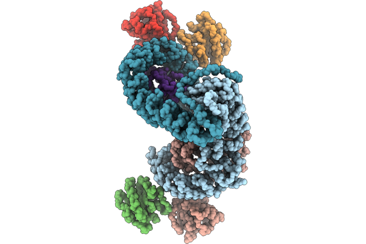
Cryo-Em Structure Of Shiv-Elicited Ci93-1365 Fab In Complex With Hiv Env Trimer Q23-Sct27
Organism: Macaca mulatta, Human immunodeficiency virus
Method: ELECTRON MICROSCOPY Resolution:2.90 Å Release Date: 2026-06-03 Classification: VIRAL PROTEIN/IMMUNE SYSTEM Ligands: NAG |
Organism: Macaca mulatta, Human immunodeficiency virus
Method: ELECTRON MICROSCOPY
Release Date: 2026-06-03
Ligands: NAG
 |
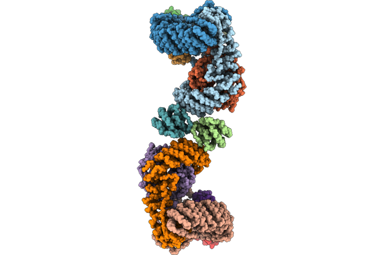
Mammalian Ap3 Complex On Tubular Membranes (Ap3 Centered)
Organism: Pan troglodytes, Homo sapiens, Rattus norvegicus
Method: ELECTRON MICROSCOPY Release Date: 2026-05-27 Classification: ENDOCYTOSIS |
Organism: Pan troglodytes, Homo sapiens, Rattus norvegicus
Method: ELECTRON MICROSCOPY
Release Date: 2026-05-27
 |
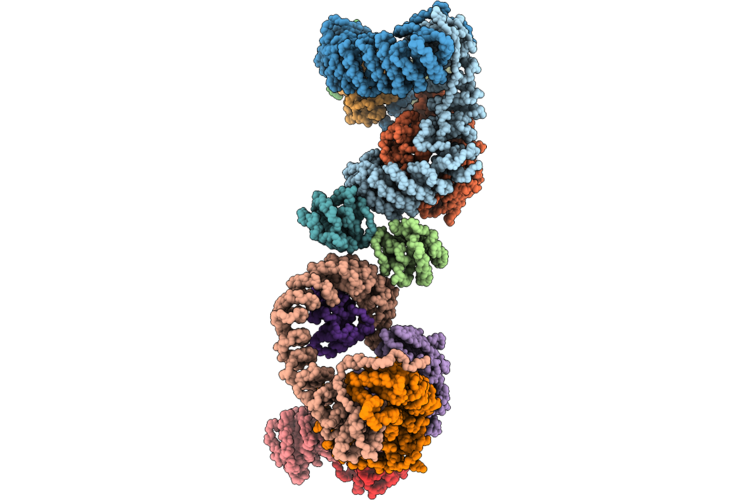
Mammalian Ap3 Complex On Tubular Membranes (Arf1 Centered Beta3-Arf1 Dimer-Beta3 Interface)
Organism: Pan troglodytes, Homo sapiens, Rattus norvegicus
Method: ELECTRON MICROSCOPY Release Date: 2026-05-27 Classification: ENDOCYTOSIS |
Organism: Pan troglodytes, Homo sapiens, Rattus norvegicus
Method: ELECTRON MICROSCOPY
Release Date: 2026-05-27
 |
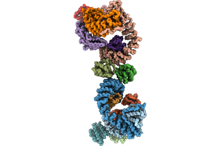
Mammalian Ap3 Complex On Tubular Membranes (Arf1 Centered Beta3-Arf1 Dimer-Delta Interface)
Organism: Pan troglodytes, Homo sapiens, Rattus norvegicus
Method: ELECTRON MICROSCOPY Release Date: 2026-05-27 Classification: ENDOCYTOSIS |
Organism: Pan troglodytes, Homo sapiens, Rattus norvegicus
Method: ELECTRON MICROSCOPY
Release Date: 2026-05-27
 |
Mammalian Ap3 Complex On Tubular Membranes (Arf1 Centered Delta-Arf1 Dimer-Delta3 Interface)
Organism: Pan troglodytes, Homo sapiens, Rattus norvegicus
Method: ELECTRON MICROSCOPY Release Date: 2026-05-27 Classification: ENDOCYTOSIS |
Organism: Pan troglodytes, Homo sapiens, Rattus norvegicus
Method: ELECTRON MICROSCOPY
Release Date: 2026-05-27
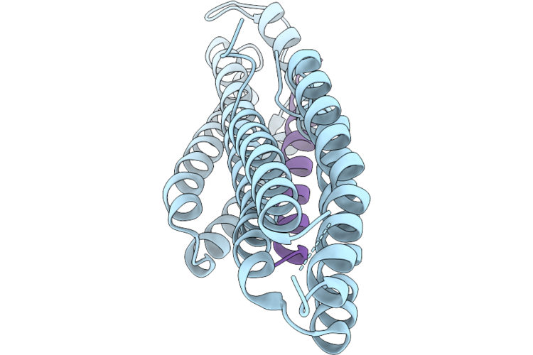 |
Mouse Vinculin Head Domain 1 (Vd1) (Residues 2-258) With A50I Mutation In Complex With Mouse Talin 1 Helix 50 (H50, Residues 2072-2103)
Organism: Mus musculus
Method: X-RAY DIFFRACTION Resolution:1.49 Å Release Date: 2026-05-20 Classification: CELL ADHESION |
Organism: Mus musculus
Method: X-RAY DIFFRACTION
Release Date: 2026-05-20
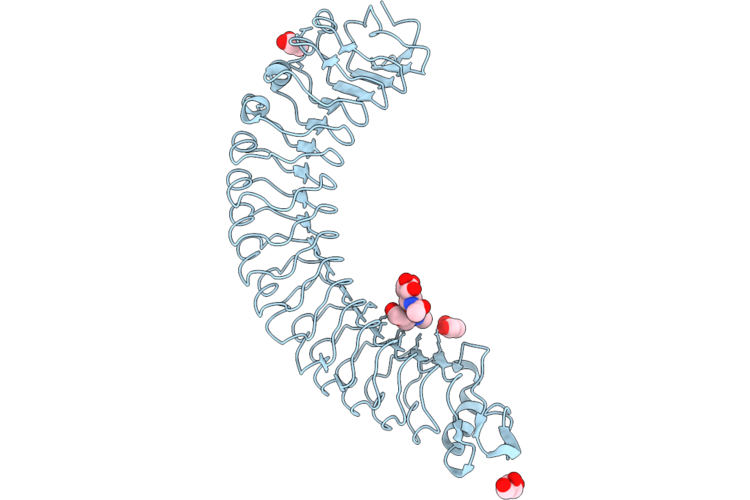 |
Mouse Lrrc15 Extracellular Domain
Organism: Mus musculus
Method: X-RAY DIFFRACTION Resolution:2.26 Å Release Date: 2026-05-13 Classification: SIGNALING PROTEIN Ligands: GOL |
Organism: Mus musculus
Method: X-RAY DIFFRACTION
Release Date: 2026-05-13
Ligands: GOL