

Search Count: 476
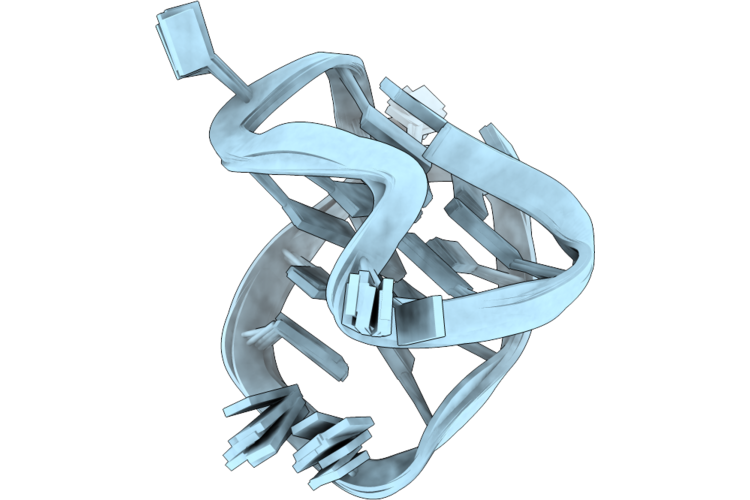 |
Solution Structure Of The Major Pdgfrb Promoter G-Quadruplex With A Broken-Strand G22 Fill-In
|
Organism: Homo sapiens
Method: SOLUTION NMR
Release Date: 2026-08-26
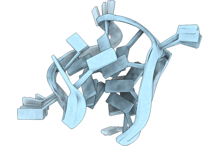 |
Solution Structure Of The Major Pdgfrb Promoter G-Quadruplex With A Broken-Strand G21 Fill-In
|
Organism: Homo sapiens
Method: SOLUTION NMR
Release Date: 2026-08-26
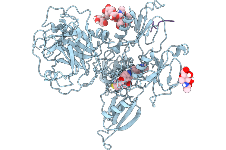 |
The Discovery Of A Novel Anticoagulant Mechanism: Factor Xi Activation Inhibitors
Organism: Homo sapiens
Method: X-RAY DIFFRACTION Resolution:3.22 Å Release Date: 2026-07-29 Classification: HYDROLASE/HYDROLASE INHIBITOR Ligands: A1CJA, NAG |
Organism: Homo sapiens
Method: X-RAY DIFFRACTION
Release Date: 2026-07-29
Ligands: A1CJA, NAG
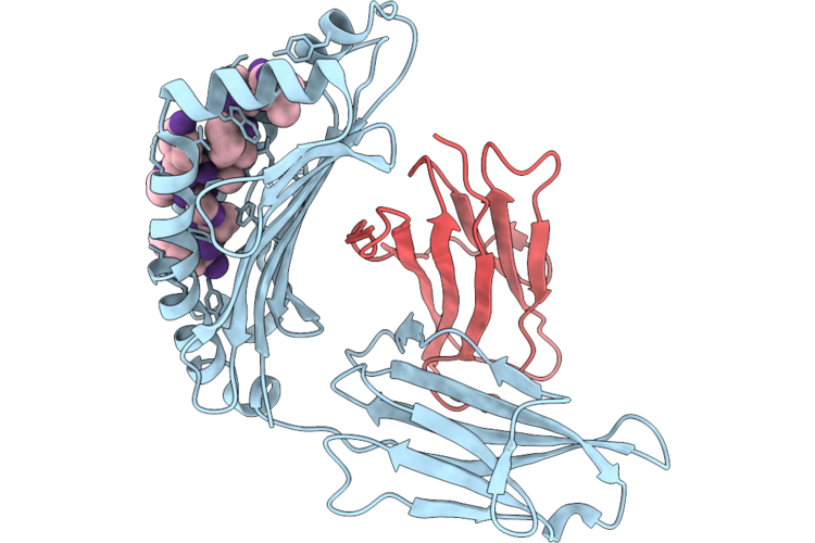 |
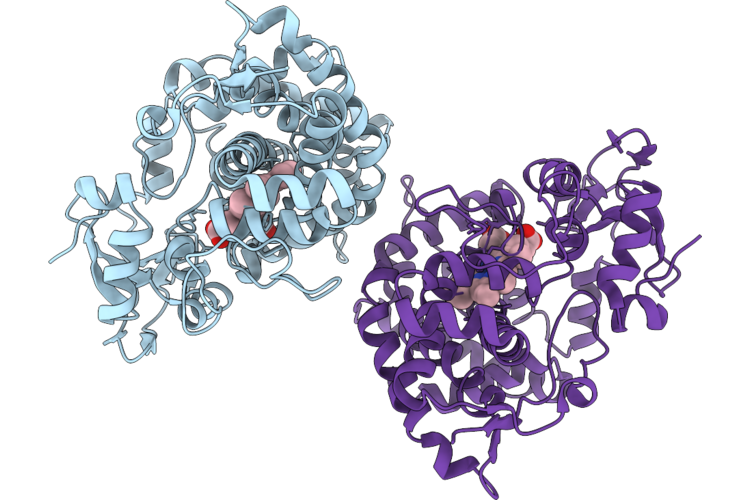
Neoantigen Rac1P29S-Hla-A2
Organism: Homo sapiens
Method: X-RAY DIFFRACTION Resolution:1.80 Å Release Date: 2026-06-17 Classification: IMMUNE SYSTEM |
Organism: Homo sapiens
Method: X-RAY DIFFRACTION
Release Date: 2026-06-17
 |
Crystal Structure Of Avermectin Oxidase
Organism: Streptomyces tubercidicus
Method: X-RAY DIFFRACTION Resolution:3.15 Å Release Date: 2026-05-27 Classification: OXIDOREDUCTASE Ligands: HEM |
Organism: Streptomyces tubercidicus
Method: X-RAY DIFFRACTION
Release Date: 2026-05-27
Ligands: HEM
 |
X-Ray Structure Of Human Pparalpha Ligand Binding Domain-10-Hydroxystearic Acid (10-Hsa) Co-Crystals Obtained By Cross-Seeding
Organism: Homo sapiens
Method: X-RAY DIFFRACTION Resolution:1.46 Å Release Date: 2026-05-27 Classification: TRANSCRIPTION Ligands: GOL, WAD |
Organism: Homo sapiens
Method: X-RAY DIFFRACTION
Release Date: 2026-05-27
Ligands: GOL, WAD
 |
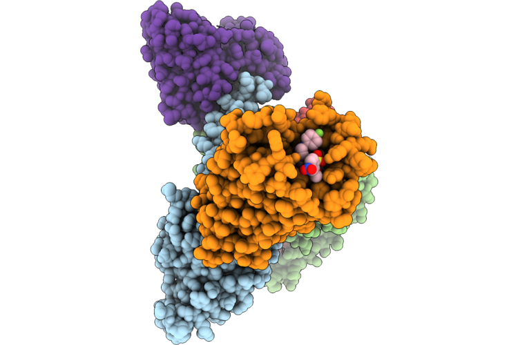
X-Ray Structure Of Human Pparalpha Ligand Binding Domain-10-Oxostearic Acid (10-Oxosa) Co-Crystals Obtained By Cross-Seeding
Organism: Homo sapiens
Method: X-RAY DIFFRACTION Resolution:1.48 Å Release Date: 2026-05-27 Classification: TRANSCRIPTION Ligands: GOL, KTC |
Organism: Homo sapiens
Method: X-RAY DIFFRACTION
Release Date: 2026-05-27
Ligands: GOL, KTC
 |
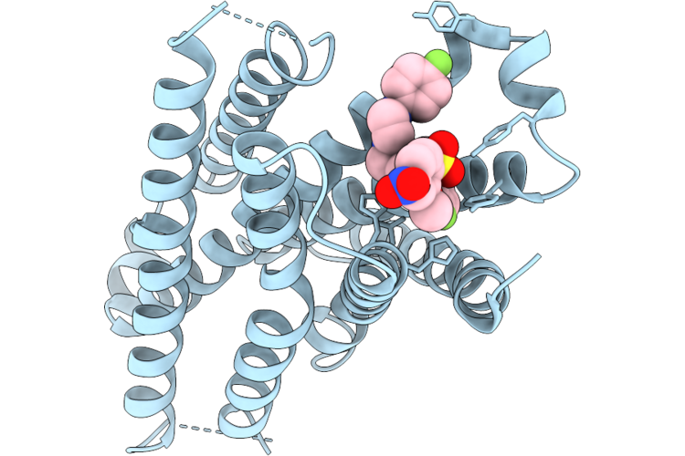
Ar234958 Bound Mas1 Receptor Complex
Organism: Homo sapiens
Method: ELECTRON MICROSCOPY Resolution:3.07 Å Release Date: 2026-05-20 Classification: PEPTIDE BINDING PROTEIN Ligands: A1MCH |
Organism: Homo sapiens
Method: ELECTRON MICROSCOPY
Release Date: 2026-05-20
Ligands: A1MCH
 |
Ar234958 Bound Mas1 Receptor
Organism: Homo sapiens
Method: ELECTRON MICROSCOPY Resolution:3.31 Å Release Date: 2026-05-20 Classification: PEPTIDE BINDING PROTEIN Ligands: A1MCH |
Organism: Homo sapiens
Method: ELECTRON MICROSCOPY
Release Date: 2026-05-20
Ligands: A1MCH
 |
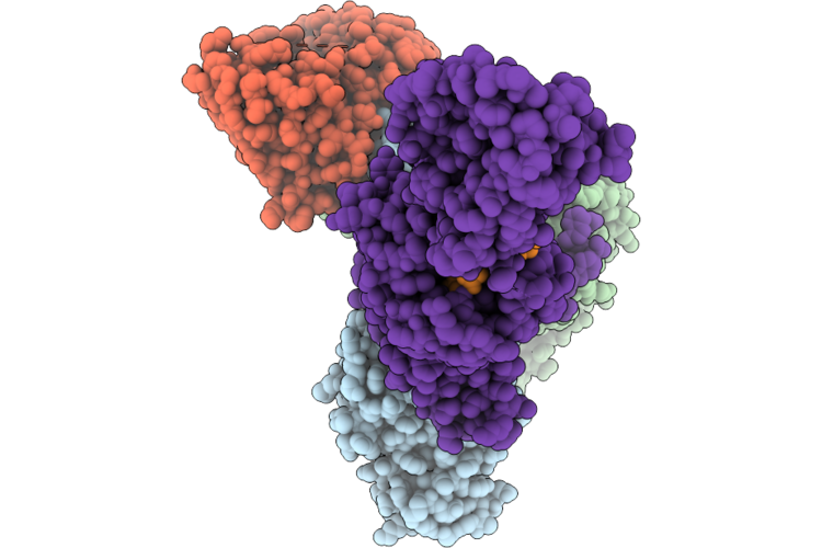
Hybrid-1 Form Of Human Telomere Dna Quadruplex With Wild-Type 5'-Flanking In K+ Solution
|
Organism: Homo sapiens
Method: SOLUTION NMR
Release Date: 2026-05-06
 |
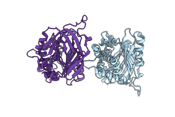
Cryo-Em Structure Of The Glp-1-Bound Human Glp-1R-Gi Complex
Organism: Homo sapiens, Synthetic construct
Method: ELECTRON MICROSCOPY Resolution:3.18 Å Release Date: 2026-05-06 Classification: SIGNALING PROTEIN/IMMUNE SYSTEM |
Organism: Homo sapiens, Synthetic construct
Method: ELECTRON MICROSCOPY
Release Date: 2026-05-06
 |
Crystal Structure Of The Recombinant A1-Antitrypsin F51L/M351V/M358V Triple Mutant
Organism: Homo sapiens
Method: X-RAY DIFFRACTION Resolution:2.60 Å Release Date: 2026-04-15 Classification: RECOMBINATION |
Organism: Homo sapiens
Method: X-RAY DIFFRACTION
Release Date: 2026-04-15
 |
Npff Bound Mas1 Receptor
Organism: Homo sapiens
Method: ELECTRON MICROSCOPY Resolution:2.77 Å Release Date: 2026-04-15 Classification: PEPTIDE BINDING PROTEIN |
Organism: Homo sapiens
Method: ELECTRON MICROSCOPY
Release Date: 2026-04-15
 |
Npff Bound Mas1 Receptor Complex
Organism: Homo sapiens
Method: ELECTRON MICROSCOPY Resolution:2.54 Å Release Date: 2026-04-15 Classification: PEPTIDE BINDING PROTEIN |
Organism: Homo sapiens
Method: ELECTRON MICROSCOPY
Release Date: 2026-04-15
 |
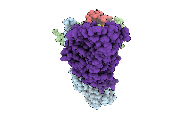
Cryo-Em Structure Of Sars-Cov2 Kp.3.1.1 Spike Protein
Organism: Severe acute respiratory syndrome coronavirus 2
Method: ELECTRON MICROSCOPY Resolution:3.10 Å Release Date: 2026-03-25 Classification: MEMBRANE PROTEIN Ligands: NAG |
Organism: Severe acute respiratory syndrome coronavirus 2
Method: ELECTRON MICROSCOPY
Release Date: 2026-03-25
Ligands: NAG
 |
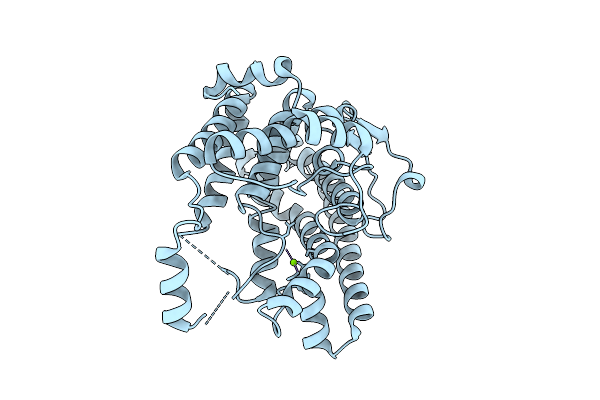
Cryo-Em Structure Of The Glp-1-Bound Human Glp-1R-Gq Complex
Organism: Homo sapiens
Method: ELECTRON MICROSCOPY Resolution:2.92 Å Release Date: 2026-03-25 Classification: SIGNALING PROTEIN/IMMUNE SYSTEM |
Organism: Homo sapiens
Method: ELECTRON MICROSCOPY
Release Date: 2026-03-25
 |
Crystal Structure Of The Tc Domain Of A Bifunctional Sesterterpene Synthase
Organism: Aspergillus stellatus
Method: X-RAY DIFFRACTION Resolution:2.05 Å Release Date: 2026-03-11 Classification: BIOSYNTHETIC PROTEIN Ligands: MG |
Organism: Aspergillus stellatus
Method: X-RAY DIFFRACTION
Release Date: 2026-03-11
Ligands: MG
 |
Cryo-Em Structure Of Evas
Organism: Aspergillus stellatus
Method: ELECTRON MICROSCOPY Release Date: 2026-02-11 Classification: BIOSYNTHETIC PROTEIN |
Organism: Aspergillus stellatus
Method: ELECTRON MICROSCOPY
Release Date: 2026-02-11
 |
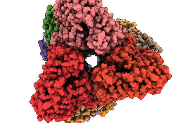
Cryo-Em Structure Of Pbss
Organism: Penicillium brasilianum
Method: ELECTRON MICROSCOPY Release Date: 2026-02-11 Classification: BIOSYNTHETIC PROTEIN |
Organism: Penicillium brasilianum
Method: ELECTRON MICROSCOPY
Release Date: 2026-02-11
 |
Cryo-Em Structure Of The Pt Domain Of Evss
Organism: Aspergillus stellatus
Method: ELECTRON MICROSCOPY Resolution:3.19 Å Release Date: 2026-02-11 Classification: BIOSYNTHETIC PROTEIN |
Organism: Aspergillus stellatus
Method: ELECTRON MICROSCOPY
Release Date: 2026-02-11