
Search Count: 812
All
Selected
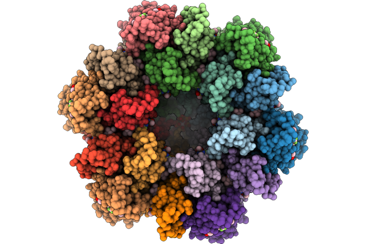 |
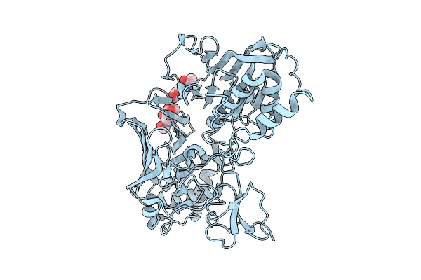
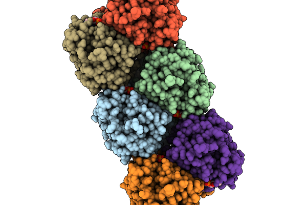
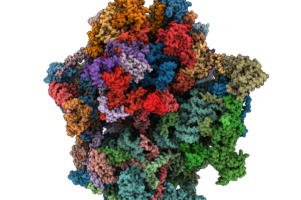
Gag Ca-Sp1 Immature Lattice Bound With Lenacapavir From Enveloped Virus Like Particles
Organism: Human immunodeficiency virus type 1 (new york-5 isolate)
Method: ELECTRON MICROSCOPY Resolution:2.28 Å Release Date: 2026-04-29 Classification: VIRUS LIKE PARTICLE Ligands: QNG, IHP |
 |
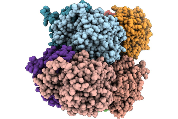
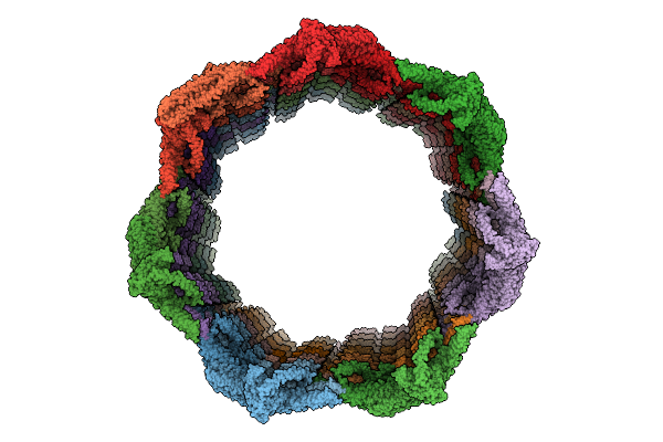
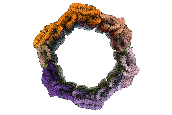
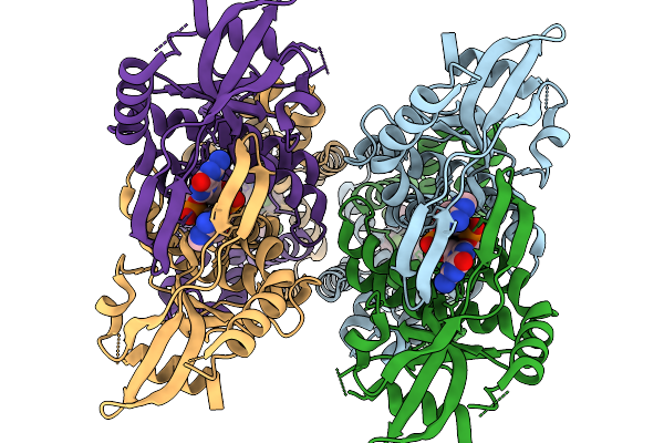
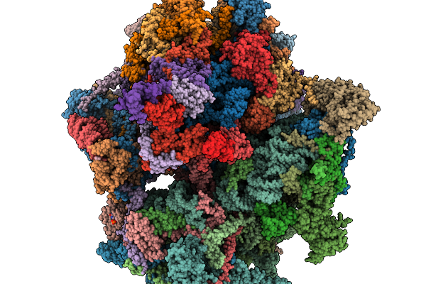
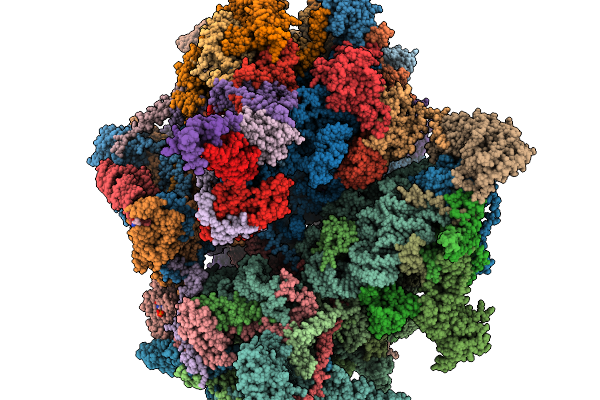
Ca-Sp1 Immature Lattice Assembled In Vitro With Inhibitor Lenacapavir (Dialyzed To 50Nm)
Organism: Human immunodeficiency virus type 1 (new york-5 isolate)
Method: ELECTRON MICROSCOPY Resolution:2.93 Å Release Date: 2026-04-29 Classification: VIRUS LIKE PARTICLE Ligands: QNG, IHP |
 |
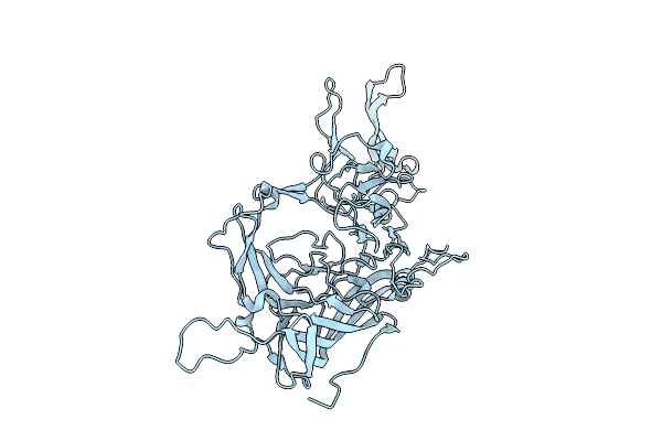
Organism: Acinetobacter baumannii
Method: X-RAY DIFFRACTION Resolution:2.00 Å Release Date: 2026-04-29 Classification: LIPID BINDING PROTEIN |
 |
Organism: Acinetobacter baumannii
Method: X-RAY DIFFRACTION Resolution:2.76 Å Release Date: 2026-04-29 Classification: LIPID BINDING PROTEIN Ligands: 4BW |
 |
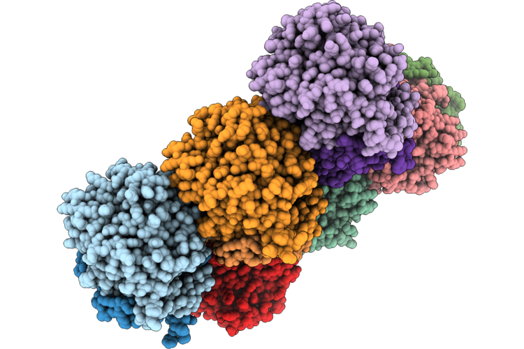
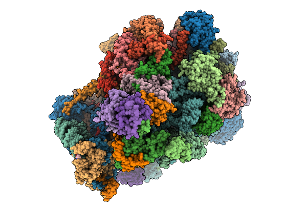
Native Structure Of The Cytoplasmic Lattice (Cpl) Asymmetric Unit From Mouse Mii Eggs
Organism: Mus musculus
Method: ELECTRON MICROSCOPY Resolution:3.50 Å Release Date: 2026-04-08 Classification: CYTOSOLIC PROTEIN Ligands: GTP, MG, ZN, CA |
 |
Organism: Cypridina noctiluca
Method: X-RAY DIFFRACTION Resolution:3.00 Å Release Date: 2026-04-08 Classification: LUMINESCENT PROTEIN Ligands: NAG |
 |
Organism: Cypridina noctiluca
Method: X-RAY DIFFRACTION Resolution:3.14 Å Release Date: 2026-04-08 Classification: LUMINESCENT PROTEIN Ligands: NAG, A1L7I |
 |
Organism: Arabidopsis thaliana
Method: ELECTRON MICROSCOPY Release Date: 2026-03-18 Classification: MEMBRANE PROTEIN |
 |
Organism: Arabidopsis thaliana
Method: ELECTRON MICROSCOPY Release Date: 2026-03-18 Classification: MEMBRANE PROTEIN |
 |
Organism: Adeno-associated virus
Method: ELECTRON MICROSCOPY Release Date: 2026-03-11 Classification: VIRUS |
 |
Organism: Acinetobacter baumannii
Method: ELECTRON MICROSCOPY Release Date: 2026-03-04 Classification: HYDROLASE Ligands: 4BW, PEE |
 |
Organism: Homo sapiens
Method: ELECTRON MICROSCOPY Resolution:2.93 Å Release Date: 2026-02-18 Classification: IMMUNE SYSTEM Ligands: 9IM, 1SY |
 |
Organism: Homo sapiens
Method: ELECTRON MICROSCOPY Release Date: 2026-02-18 Classification: RIBOSOME Ligands: ZN, SPD, MG, PUT, K, SPM, NAD, FES, GDP, ATP, VAL |
 |
Organism: Homo sapiens
Method: ELECTRON MICROSCOPY Release Date: 2026-02-18 Classification: RIBOSOME Ligands: ZN, K, SPD, PUT, MG, VAL, FES, NAD, SPM, ATP, GDP |
 |
Organism: Homo sapiens
Method: ELECTRON MICROSCOPY Release Date: 2026-02-18 Classification: RIBOSOME Ligands: ZN, K, SPD, PUT, MG, VAL, FES, NAD, SPM, ATP, GDP |
 |
Organism: Homo sapiens
Method: ELECTRON MICROSCOPY Resolution:3.02 Å Release Date: 2026-02-18 Classification: RIBOSOME Ligands: ZN, K, SPD, PUT, MG, FES, NAD, SPM, ATP, GDP, VAL |
 |
Organism: Homo sapiens
Method: ELECTRON MICROSCOPY Release Date: 2026-02-18 Classification: RIBOSOME Ligands: ZN, K, SPD, PUT, MG, FES, NAD, SPM, ATP, GDP, VAL |
 |
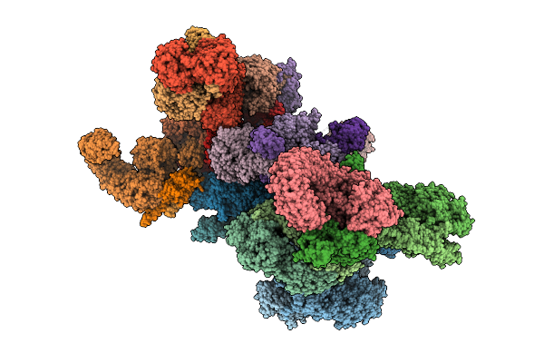
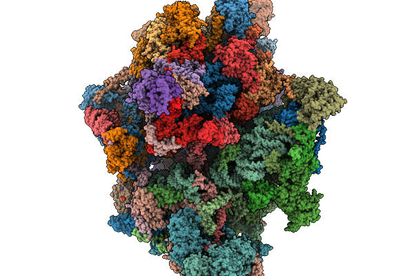
In Situ Structure Of The Human Mitoribosome In The P-E State From Taco1-Knockout Cells
Organism: Homo sapiens
Method: ELECTRON MICROSCOPY Release Date: 2026-02-18 Classification: RIBOSOME Ligands: ZN, K, SPD, PUT, MG, FES, NAD, SPM, ATP, GDP, VAL |
 |
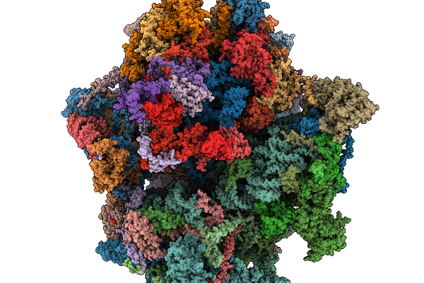
In Situ Structure Of The Human Mitochondrial Large Subunit 39S In Complex With Taco1
Organism: Homo sapiens
Method: ELECTRON MICROSCOPY Release Date: 2026-02-18 Classification: RIBOSOME Ligands: ZN, K, MG, FES, SPD, PUT, VAL |
 |
In Situ Structure Of The Human Mitoribosome Large Subunit 39S In Complex With Ef-Tu
Organism: Homo sapiens
Method: ELECTRON MICROSCOPY Release Date: 2026-02-18 Classification: RIBOSOME Ligands: ZN, K, MG, FES, SPD, PUT, VAL |