

Search Count: 698
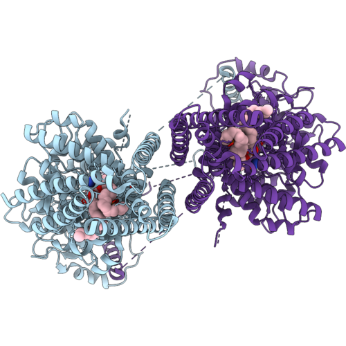 |
Structure Of Class Ii Chitin Synthase, Chs1, From Candida Albicans (Cachs1), State 1 (Post Udp Hydrolysis) After 5 Min Reaction
Organism: Candida albicans
Method: ELECTRON MICROSCOPY Resolution:3.38 Å Release Date: 2026-10-14 Classification: MEMBRANE PROTEIN Ligands: MG, PEE, NAG, UDP |
Organism: Candida albicans
Method: ELECTRON MICROSCOPY
Release Date: 2026-10-14
Ligands: MG, PEE, NAG, UDP
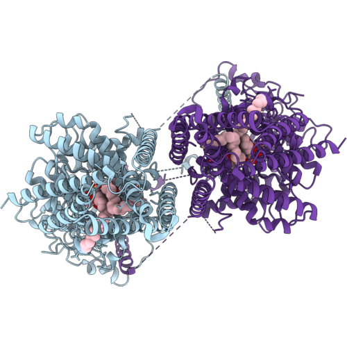 |
Structure Of Class Ii Chitin Synthase, Chs1, From Candida Albicans (Cachs1), State 2 (Pre-Translocation) After 5 Min Reaction
Organism: Candida albicans
Method: ELECTRON MICROSCOPY Release Date: 2026-10-14 Classification: MEMBRANE PROTEIN Ligands: UDP, PEE, MG |
Organism: Candida albicans
Method: ELECTRON MICROSCOPY
Release Date: 2026-10-14
Ligands: UDP, PEE, MG
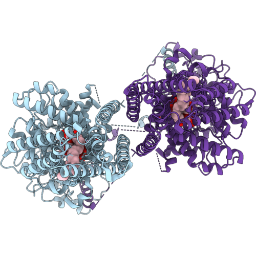 |
Structure Of Class Ii Chitin Synthase, Chs1, From Candida Albicans (Cachs1), State 3 (Post-Translocation) After 5 Min Reaction
Organism: Candida albicans
Method: ELECTRON MICROSCOPY Release Date: 2026-10-14 Classification: MEMBRANE PROTEIN Ligands: UDP, PEE |
Organism: Candida albicans
Method: ELECTRON MICROSCOPY
Release Date: 2026-10-14
Ligands: UDP, PEE
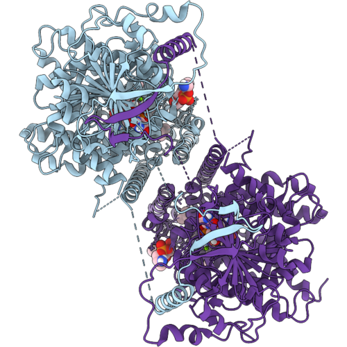 |
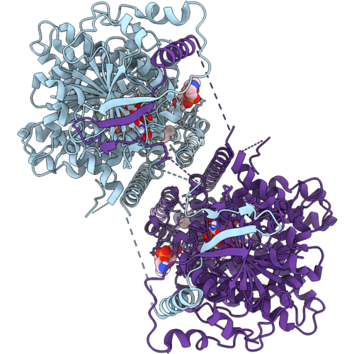
Candida Albicans Chitin Synthase 1 (Cachs1) Intermediate State (Da-2), Bound To Udp And Diynyl Arylamine (Da, Aka Ro-09-3143)
Organism: Candida albicans
Method: ELECTRON MICROSCOPY Resolution:3.02 Å Release Date: 2026-10-14 Classification: MEMBRANE PROTEIN Ligands: PEE, MG, A1DEH, UDP |
Organism: Candida albicans
Method: ELECTRON MICROSCOPY
Release Date: 2026-10-14
Ligands: PEE, MG, A1DEH, UDP
 |
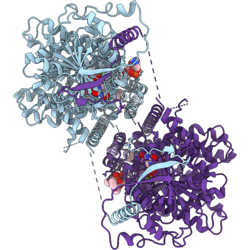
Candida Albicans Chitin Synthase 1 (Cachs1) Intermediate State (Da-3), Bound To Udp And Diynyl Arylamine (Da, Aka Ro-09-3143)
Organism: Candida albicans
Method: ELECTRON MICROSCOPY Resolution:3.00 Å Release Date: 2026-10-14 Classification: MEMBRANE PROTEIN Ligands: PEE, MG, A1DEH, UDP |
Organism: Candida albicans
Method: ELECTRON MICROSCOPY
Release Date: 2026-10-14
Ligands: PEE, MG, A1DEH, UDP
 |
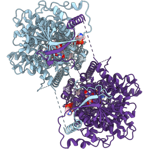
Candida Albicans Chitin Synthase 1 (Cachs1) Intermediate State (Da-4), Bound To Udp And Diynyl Arylamine (Da, Aka Ro-09-3143)
Organism: Candida albicans
Method: ELECTRON MICROSCOPY Resolution:3.10 Å Release Date: 2026-10-14 Classification: MEMBRANE PROTEIN Ligands: PEE, MG, A1DEH, UDP |
Organism: Candida albicans
Method: ELECTRON MICROSCOPY
Release Date: 2026-10-14
Ligands: PEE, MG, A1DEH, UDP
 |
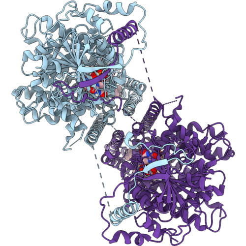
Candida Albicans Chitin Synthase 1 (Cachs1) Intermediate State (Da-5), Bound To Udp And Diynyl Arylamine (Da, Aka Ro-09-3143)
Organism: Candida albicans
Method: ELECTRON MICROSCOPY Resolution:2.85 Å Release Date: 2026-10-14 Classification: MEMBRANE PROTEIN Ligands: PEE, MG, A1DEH, UDP |
Organism: Candida albicans
Method: ELECTRON MICROSCOPY
Release Date: 2026-10-14
Ligands: PEE, MG, A1DEH, UDP
 |
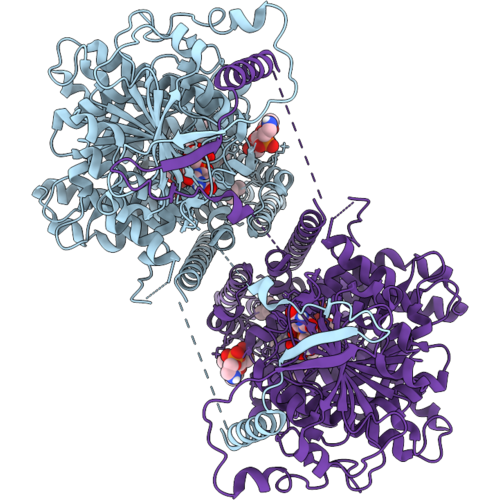
Candida Albicans Chitin Synthase 1 (Cachs1) Intermediate State (Da-6), Bound To Udp And Diynyl Arylamine (Da, Aka Ro-09-3143)
Organism: Candida albicans
Method: ELECTRON MICROSCOPY Resolution:3.29 Å Release Date: 2026-10-14 Classification: MEMBRANE PROTEIN Ligands: A1DEH, PEE, UDP |
Organism: Candida albicans
Method: ELECTRON MICROSCOPY
Release Date: 2026-10-14
Ligands: A1DEH, PEE, UDP
 |
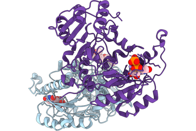
Candida Albicans Chitin Synthase 1 (Cachs1) Intermediate State (Da-7), Bound To Udp And Diynyl Arylamine (Da, Aka Ro-09-3143)
Organism: Candida albicans
Method: ELECTRON MICROSCOPY Resolution:3.10 Å Release Date: 2026-10-14 Classification: MEMBRANE PROTEIN Ligands: PEE, A1DEH, UDP |
Organism: Candida albicans
Method: ELECTRON MICROSCOPY
Release Date: 2026-10-14
Ligands: PEE, A1DEH, UDP
 |
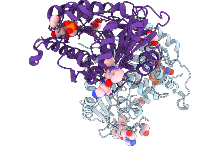
Crystal Structure Of Human Mgat1 In Complex With Udp.
Organism: Homo sapiens
Method: X-RAY DIFFRACTION Resolution:1.55 Å Release Date: 2026-09-23 Classification: TRANSFERASE Ligands: UDP, MN, PG6 |
Organism: Homo sapiens
Method: X-RAY DIFFRACTION
Release Date: 2026-09-23
Ligands: UDP, MN, PG6
 |
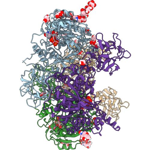
Crystal Structure Of Human Mgat1 In Complex With Tng-9333 And Udp.
Organism: Homo sapiens
Method: X-RAY DIFFRACTION Resolution:2.01 Å Release Date: 2026-09-23 Classification: TRANSFERASE Ligands: MN, UDP, A1C1W, EDO, GOL |
Organism: Homo sapiens
Method: X-RAY DIFFRACTION
Release Date: 2026-09-23
Ligands: MN, UDP, A1C1W, EDO, GOL
 |
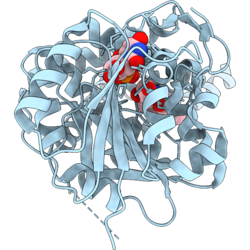
Crystal Structure Of The Mucin-Type O-Glycosyltransferase Pgant5A-4M (A430T, L434N, A437N, S456Y)
Organism: Drosophila melanogaster
Method: X-RAY DIFFRACTION Resolution:3.08 Å Release Date: 2026-09-16 Classification: TRANSFERASE Ligands: UDP, GOL, PG4, EDO, MN, NAG |
Organism: Drosophila melanogaster
Method: X-RAY DIFFRACTION
Release Date: 2026-09-16
Ligands: UDP, GOL, PG4, EDO, MN, NAG
 |
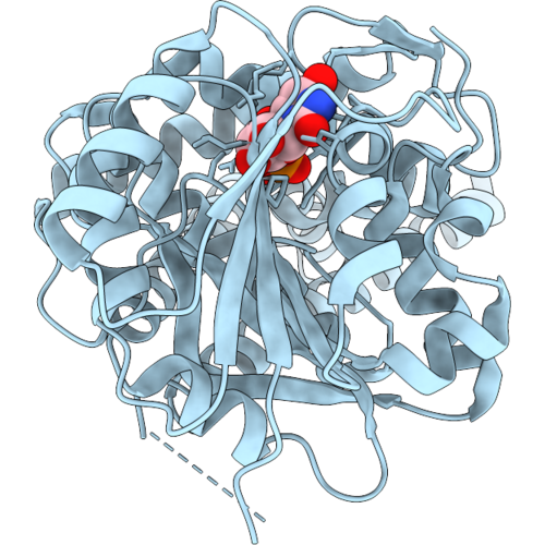
Cryo-Em Structure Of Human Ugcg Bound To Udp And C6-Ceramide
Organism: Homo sapiens
Method: ELECTRON MICROSCOPY Release Date: 2026-09-02 Classification: TRANSFERASE Ligands: 6CM, UDP |
Organism: Homo sapiens
Method: ELECTRON MICROSCOPY
Release Date: 2026-09-02
Ligands: 6CM, UDP
 |
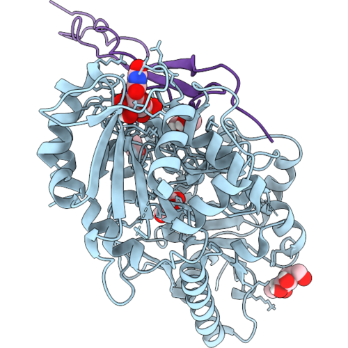
Cryo-Em Structure Of Human Ugcg Bound To Udp
Organism: Homo sapiens
Method: ELECTRON MICROSCOPY Release Date: 2026-09-02 Classification: TRANSFERASE Ligands: UDP |
Organism: Homo sapiens
Method: ELECTRON MICROSCOPY
Release Date: 2026-09-02
Ligands: UDP
 |
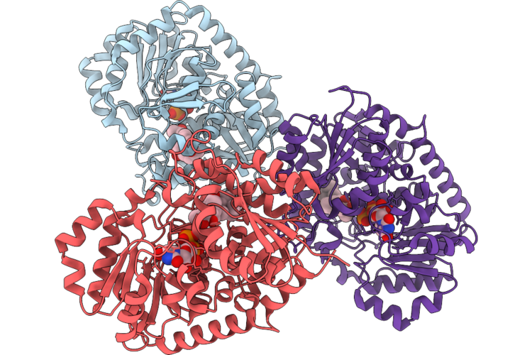
Crystal Structure Of A B1,3-Glucosyltransferase (B3Glct) Reveals An Unusual Mode Of Substrate Recognition By A Two-Domain Gt-A Fold Glycosyltransferase
Organism: Homo sapiens
Method: X-RAY DIFFRACTION Resolution:2.07 Å Release Date: 2026-08-26 Classification: CARBOHYDRATE Ligands: MN, UDP, NAG, GOL, EDO, FUC |
Organism: Homo sapiens
Method: X-RAY DIFFRACTION
Release Date: 2026-08-26
Ligands: MN, UDP, NAG, GOL, EDO, FUC
 |
Crystal Structure Of Sgugt73Am30 In Complex With Mogrol
Organism: Siraitia grosvenorii
Method: X-RAY DIFFRACTION Resolution:2.60 Å Release Date: 2026-08-26 Classification: TRANSFERASE Ligands: UDP, A1E9G |
Organism: Siraitia grosvenorii
Method: X-RAY DIFFRACTION
Release Date: 2026-08-26
Ligands: UDP, A1E9G
 |
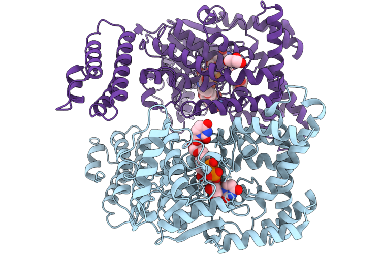
Structure Of Clostridium Difficile Toxin B (Tcdb) Glucosyltransferase In Complex With Udp And Isofagomine Analog 2-4
Organism: Clostridioides difficile
Method: X-RAY DIFFRACTION Resolution:1.85 Å Release Date: 2026-07-08 Classification: TRANSFERASE Ligands: UDP, A1C9K, MN, PEG, EDO, SO4 |
Organism: Clostridioides difficile
Method: X-RAY DIFFRACTION
Release Date: 2026-07-08
Ligands: UDP, A1C9K, MN, PEG, EDO, SO4
 |
Structure Of Clostridium Difficile Toxin B (Tcdb) Glucosyltransferase In Complex With Udp Linked Isofagomine Analog 3-5
Organism: Clostridioides difficile
Method: X-RAY DIFFRACTION Resolution:2.65 Å Release Date: 2026-07-08 Classification: TRANSFERASE Ligands: UDP, A1C96, MN, GOL |
Organism: Clostridioides difficile
Method: X-RAY DIFFRACTION
Release Date: 2026-07-08
Ligands: UDP, A1C96, MN, GOL
 |
P2Y14R-Gi Complex Bound To Udp
Organism: Rattus rattus, Homo sapiens, Escherichia coli, Synthetic construct, Human respiratory syncytial virus, Mus musculus
Method: ELECTRON MICROSCOPY Release Date: 2026-06-10 Classification: SIGNALING PROTEIN Ligands: UDP |
Organism: Rattus rattus, Homo sapiens, Escherichia coli, Synthetic construct, Human respiratory syncytial virus, Mus musculus
Method: ELECTRON MICROSCOPY
Release Date: 2026-06-10
Ligands: UDP
 |
Crystal Structure Of Umpk From S. Aureus In Complex With Utp
Organism: Staphylococcus aureus subsp. aureus nctc 8325
Method: X-RAY DIFFRACTION Resolution:2.30 Å Release Date: 2026-05-27 Classification: TRANSFERASE Ligands: GOL, UTP, UDP, PO4 |
Organism: Staphylococcus aureus subsp. aureus nctc 8325
Method: X-RAY DIFFRACTION
Release Date: 2026-05-27
Ligands: GOL, UTP, UDP, PO4