24XZ image
Deposition Date 2026-03-24
Release Date 2026-06-10
Last Version Date 2026-06-10
Entry Detail
PDB ID:
24XZ
Title:
P2Y14R-Gi complex bound to UDP
Biological Source:
Source Organism(s):
Expression System(s):
Method Details:
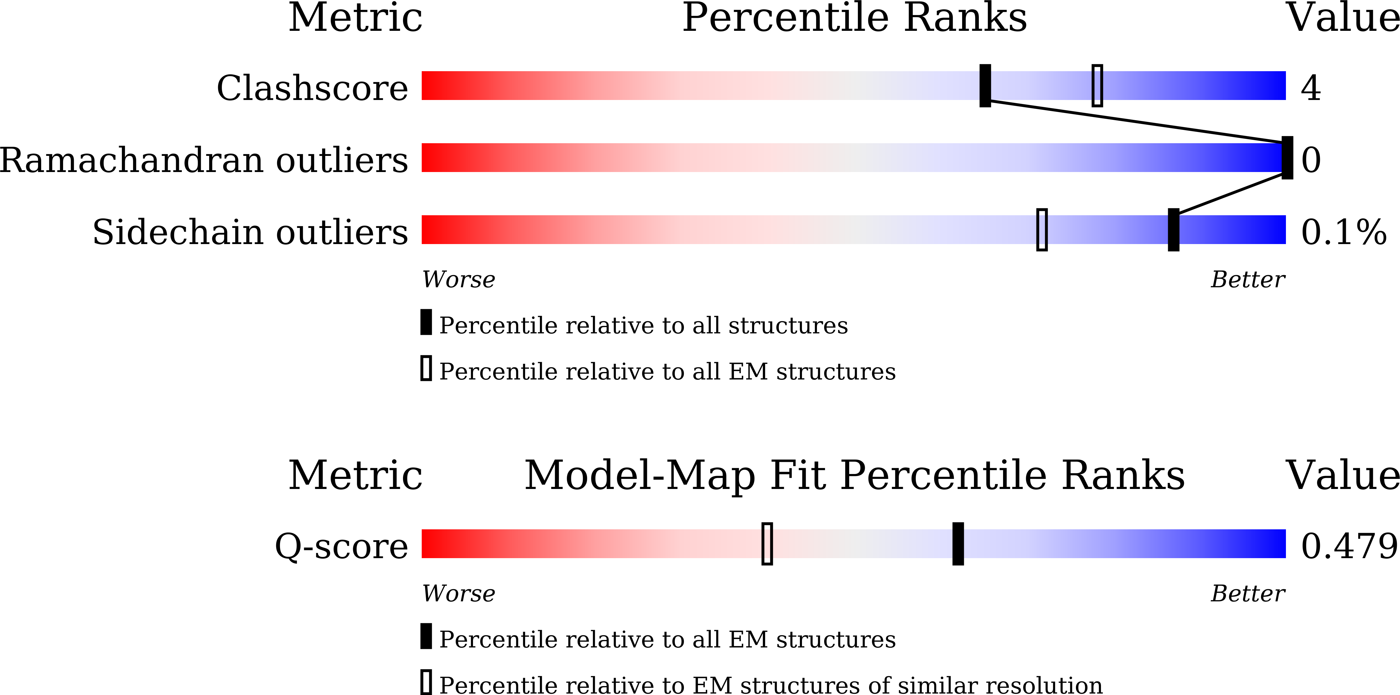
Experimental Method:
Resolution:
2.93 Å
Aggregation State:
PARTICLE
Reconstruction Method:
SINGLE PARTICLE
Macromolecular Entities
Structures with similar UniProt ID
Protein Blast
Polymer Type:polypeptide(L)
Molecule:Guanine nucleotide-binding pr
Gene (Uniprot):GNB1
Chain IDs:A (auth: B)
Chain Length:375
Number of Molecules:1
Biological Source:Rattus rattus
Structures with similar UniProt ID
Protein Blast
Polymer Type:polypeptide(L)
Molecule:Guanine nucleotide-binding pr
Gene (Uniprot):GNAI1
Chain IDs:B (auth: G), D (auth: A)
Chain Length:433
Number of Molecules:2
Biological Source:Homo sapiens
Protein Blast
Polymer Type:polypeptide(L)
Molecule:Soluble cytochrome b562,P2Y p
Gene (Uniprot):cybC, P2RY14
Mutagens:mutation
Chain IDs:C (auth: R)
Chain Length:919
Number of Molecules:1
Biological Source:Escherichia coli, Homo sapiens, synthetic construct, human respiratory syncytial virus
Protein Blast
Polymer Type:polypeptide(L)
Molecule:scFv16
Chain IDs:E (auth: S)
Chain Length:260
Number of Molecules:1
Biological Source:Mus musculus
Ligand Molecules
Primary Citation
Structural insights into the ligand and G protein recognition by P2Y 13 R.
Biochem.Biophys.Res.Commun. 827 153976 153976 (2026)
PMID: 42208230 DOI: 10.1016/j.bbrc.2026.153976

Abstact

P2Y purinergic receptors are GPCRs that recognize extracellular nucleotides to mediate diverse physiological processes. Among 12-like subfamily members, P2Y(13)R has well-documented roles in neuroprotection and cholesterol metabolism. Notably, P2Y(13)R displays robust activity toward the G(q) pathway in addition to its canonical G(i) coupling, yet the structural basis for its ligand recognition and G protein selectivity has remained unclear. Here, we present the cryo-EM structure of the P2Y(13)R-G(q) complex bound to ADP at a resolution of 2.83 A. The structure reveals the distinctive ligand recognition mechanism of P2Y(13)R, in which an N-terminal arginine caps the orthosteric binding pocket. Furthermore, we also elucidated the structure of P2Y(14)R, which shows the lowest G(i) activation ability among the 12-like P2Y receptors, in complex with UDP and G(i) at a resolution of 2.93 A. Structural comparison with the 12-like P2Y receptors implicates ICL2-mediated contacts with the Galpha hydrophobic cavity as a key structural determinant of G(q) selectivity. Together, these findings provide mechanistic insights into nucleotide signaling and a structural foundation for advancing structure-based approaches to targeting the 12-like P2Y receptors.

Legend

Protein

Chemical

Disease

Primary Citation of related structures
Feedback Form
Name
Email
Institute
Feedback