

Search Count: 2,274
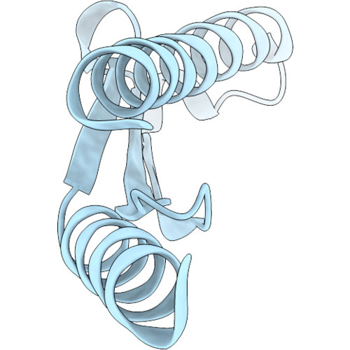 |
Crystal Structure Of The Streptococcus Mutans Ybab Homolog (Smu.394)
Organism: Streptococcus mutans
Method: X-RAY DIFFRACTION Resolution:1.50 Å Release Date: 2026-10-21 Classification: DNA BINDING PROTEIN |
Organism: Streptococcus mutans
Method: X-RAY DIFFRACTION
Release Date: 2026-10-21
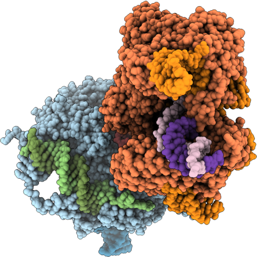 |
Cryo-Em Structure Of Stitchr (R2Tg And Cas9 Nickase) Bound To Guide Rna, Template Rna, And Target Dna In The Target-Primed Reverse Transcription Initiation State
Organism: Taeniopygia guttata, Streptococcus pyogenes, Cloning vector pet-j23100-laco(symr+1)-gfp-lva
Method: ELECTRON MICROSCOPY Resolution:3.30 Å Release Date: 2026-10-14 Classification: DNA BINDING PROTEIN Ligands: ZN, MG, TTP |
Organism: Taeniopygia guttata, Streptococcus pyogenes, Cloning vector pet-j23100-laco(symr+1)-gfp-lva
Method: ELECTRON MICROSCOPY
Release Date: 2026-10-14
Ligands: ZN, MG, TTP
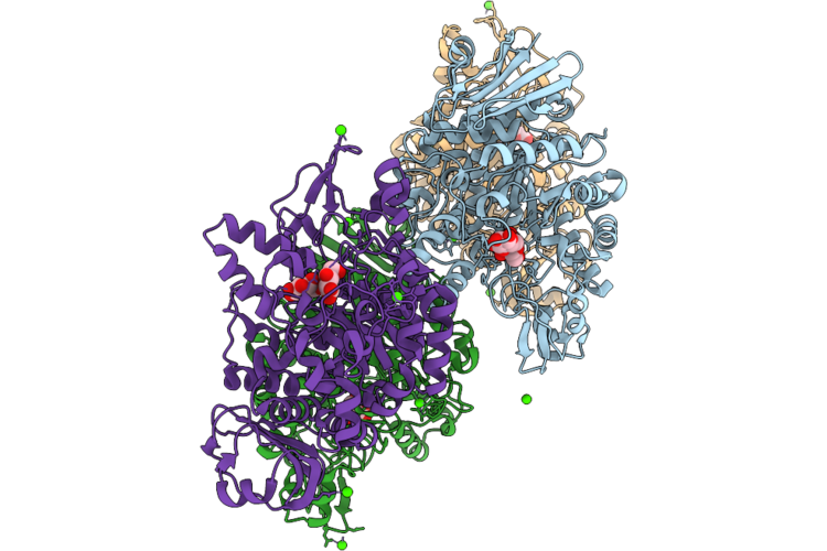 |
Dextran Glucosidase Smdg E236Q In Complex With Isomaltose
Organism: Streptococcus mutans
Method: X-RAY DIFFRACTION Resolution:2.30 Å Release Date: 2026-09-23 Classification: HYDROLASE Ligands: CA |
Organism: Streptococcus mutans
Method: X-RAY DIFFRACTION
Release Date: 2026-09-23
Ligands: CA
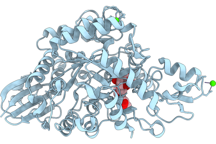 |
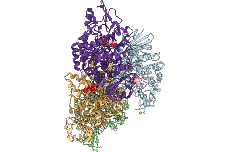
Dextran Glucosidase Smdg E236Q In Complex With Panose
Organism: Streptococcus mutans
Method: X-RAY DIFFRACTION Resolution:2.60 Å Release Date: 2026-09-23 Classification: HYDROLASE Ligands: CA |
Organism: Streptococcus mutans
Method: X-RAY DIFFRACTION
Release Date: 2026-09-23
Ligands: CA
 |
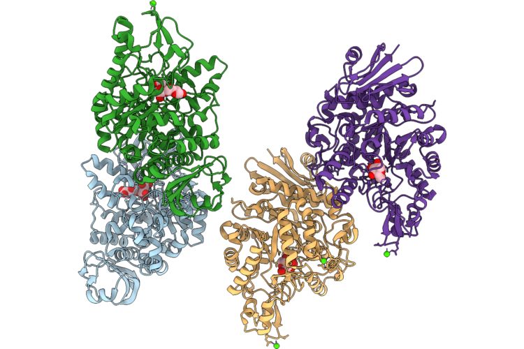
Dextran Glucosidase Smdg E236S In Complex With Isomaltose
Organism: Streptococcus mutans
Method: X-RAY DIFFRACTION Resolution:2.20 Å Release Date: 2026-09-23 Classification: HYDROLASE Ligands: CA |
Organism: Streptococcus mutans
Method: X-RAY DIFFRACTION
Release Date: 2026-09-23
Ligands: CA
 |
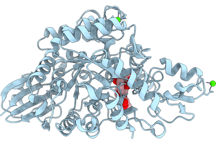
Dextran Glucosidase Smdg E236S In Complex With Isomaltotriose
Organism: Streptococcus mutans
Method: X-RAY DIFFRACTION Resolution:2.40 Å Release Date: 2026-09-23 Classification: HYDROLASE Ligands: CA, GLC |
Organism: Streptococcus mutans
Method: X-RAY DIFFRACTION
Release Date: 2026-09-23
Ligands: CA, GLC
 |
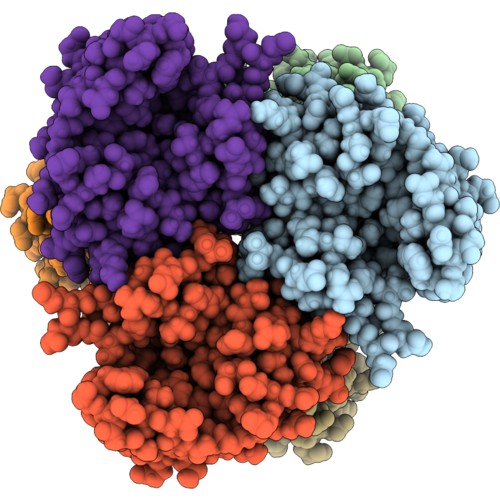
Dextran Glucosidase Smdg E236S In Complex With Panose
Organism: Streptococcus mutans
Method: X-RAY DIFFRACTION Resolution:2.40 Å Release Date: 2026-09-23 Classification: HYDROLASE Ligands: CA |
Organism: Streptococcus mutans
Method: X-RAY DIFFRACTION
Release Date: 2026-09-23
Ligands: CA
 |
Cryoem Structure Of Nucleoside Diphosphate Kinase (Ndk) From Streptococcus Pneumoniae
Organism: Streptococcus pneumoniae r6
Method: ELECTRON MICROSCOPY Resolution:2.47 Å Release Date: 2026-09-16 Classification: TRANSFERASE |
Organism: Streptococcus pneumoniae r6
Method: ELECTRON MICROSCOPY
Release Date: 2026-09-16
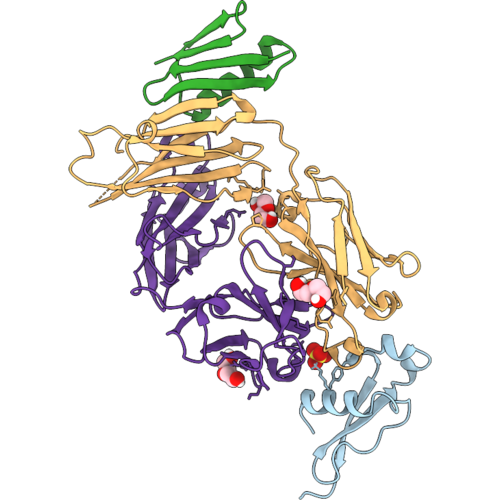 |
Fab1525 In Complex With The C-Terminal Alpha-Tsr Domain Of The P. Falciparum Circumsporozoite Protein
Organism: Homo sapiens, Plasmodium falciparum 3d7, Streptococcus sp. group g
Method: X-RAY DIFFRACTION Resolution:2.20 Å Release Date: 2026-09-09 Classification: IMMUNE SYSTEM Ligands: PEG, SO4 |
Organism: Homo sapiens, Plasmodium falciparum 3d7, Streptococcus sp. group g
Method: X-RAY DIFFRACTION
Release Date: 2026-09-09
Ligands: PEG, SO4
 |
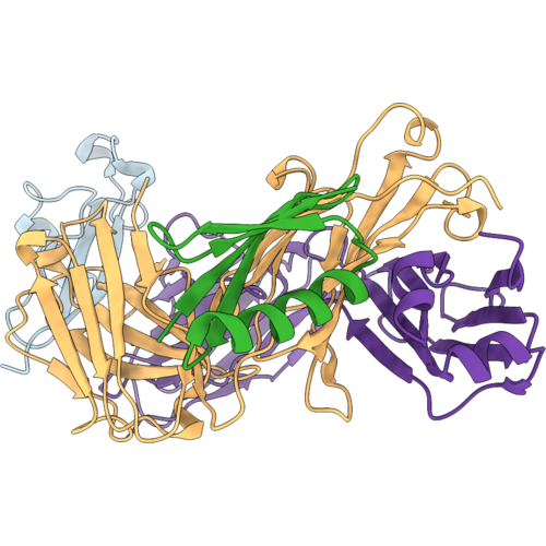
Fab1550 In Complex With The C-Terminal Alpha-Tsr Domain Of The P. Falciparum Circumsporozoite Protein
Organism: Homo sapiens, Plasmodium falciparum 3d7, Streptococcus sp. 'group g'
Method: X-RAY DIFFRACTION Resolution:2.00 Å Release Date: 2026-09-09 Classification: IMMUNE SYSTEM |
Organism: Homo sapiens, Plasmodium falciparum 3d7, Streptococcus sp. 'group g'
Method: X-RAY DIFFRACTION
Release Date: 2026-09-09
 |
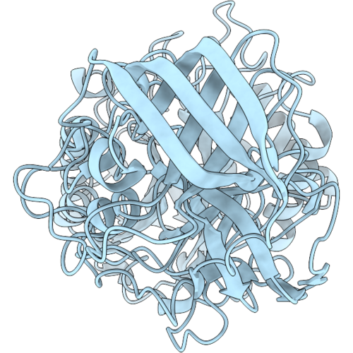
Fab1393 In Complex With The C-Terminal Alpha-Tsr Domain Of The P. Falciparum Circumsporozoite Protein
Organism: Homo sapiens, Plasmodium falciparum 3d7, Streptococcus sp. group g
Method: X-RAY DIFFRACTION Resolution:1.80 Å Release Date: 2026-09-09 Classification: IMMUNE SYSTEM |
Organism: Homo sapiens, Plasmodium falciparum 3d7, Streptococcus sp. group g
Method: X-RAY DIFFRACTION
Release Date: 2026-09-09
 |
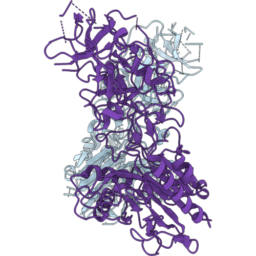
Monomeric Form Of The Streptococcus Pyogenes Nuclease A (Spna)
Organism: Streptococcus pyogenes
Method: ELECTRON MICROSCOPY Resolution:2.60 Å Release Date: 2026-09-02 Classification: DNA BINDING PROTEIN |
Organism: Streptococcus pyogenes
Method: ELECTRON MICROSCOPY
Release Date: 2026-09-02
 |
Dimeric Form Of The Streptococcus Pyogenes Nuclease A (Spna)
Organism: Streptococcus pyogenes
Method: ELECTRON MICROSCOPY Resolution:2.80 Å Release Date: 2026-09-02 Classification: DNA BINDING PROTEIN |
Organism: Streptococcus pyogenes
Method: ELECTRON MICROSCOPY
Release Date: 2026-09-02
 |
Hsa Bound To Neu5Gc Alpha2,3 Gal Beta Ome
Organism: Streptococcus
Method: X-RAY DIFFRACTION Resolution:1.45 Å Release Date: 2026-08-26 Classification: SUGAR BINDING PROTEIN |
Organism: Streptococcus
Method: X-RAY DIFFRACTION
Release Date: 2026-08-26
 |
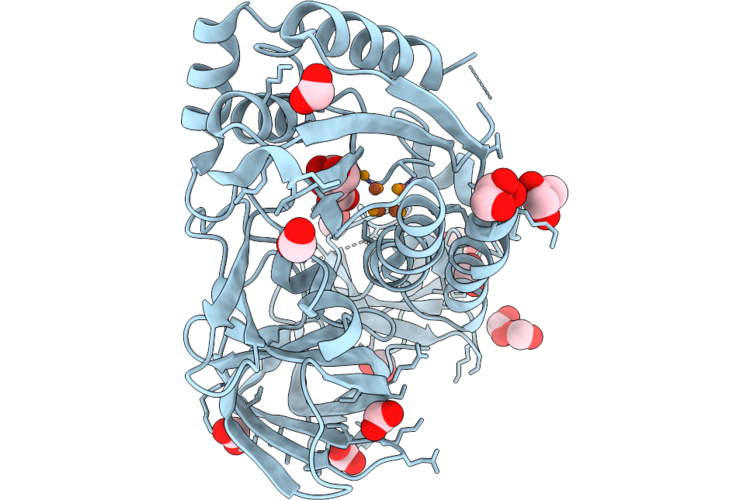
Structure Of Spcas9 With Engineered Disulfide Bonds (R1210C/V1280C And L1119C/P1128C)
Organism: Streptococcus pyogenes serotype m1
Method: ELECTRON MICROSCOPY Resolution:3.72 Å Release Date: 2026-08-26 Classification: HYDROLASE |
Organism: Streptococcus pyogenes serotype m1
Method: ELECTRON MICROSCOPY
Release Date: 2026-08-26
 |
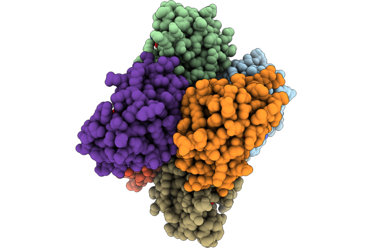
Crystal Structure Of Mnma D100C Mutant From Streptococcus Pneumoniae With [4Fe-5Se] Cluster
Organism: Streptococcus pneumoniae tigr4
Method: X-RAY DIFFRACTION Resolution:2.37 Å Release Date: 2026-08-19 Classification: TRANSFERASE Ligands: SFS, FMT, GOL, SE |
Organism: Streptococcus pneumoniae tigr4
Method: X-RAY DIFFRACTION
Release Date: 2026-08-19
Ligands: SFS, FMT, GOL, SE
 |
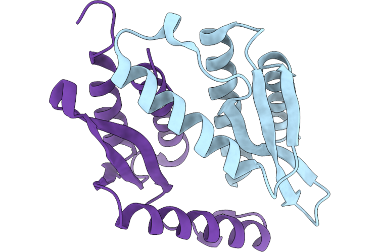
Crystal Structure Of Nucleoside Diphosphate Kinase (Ndk) From Streptococcus Pneumoniae In Complex With Adp And Vanadate
Organism: Streptococcus pneumoniae r6
Method: X-RAY DIFFRACTION Resolution:3.42 Å Release Date: 2026-08-19 Classification: TRANSFERASE Ligands: ADP, VN4, MG, VO4 |
Organism: Streptococcus pneumoniae r6
Method: X-RAY DIFFRACTION
Release Date: 2026-08-19
Ligands: ADP, VN4, MG, VO4
 |
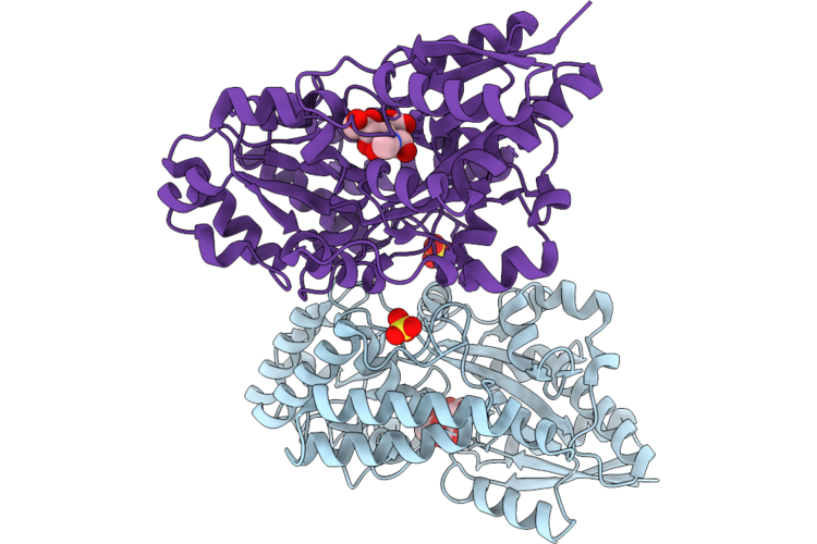
Type Ii-A Anti-Crispr Protein Acriia17 From Streptococcus Gallolyticus
Organism: Streptococcus gallolyticus
Method: X-RAY DIFFRACTION Resolution:2.03 Å Release Date: 2026-08-12 Classification: IMMUNE SYSTEM |
Organism: Streptococcus gallolyticus
Method: X-RAY DIFFRACTION
Release Date: 2026-08-12
 |
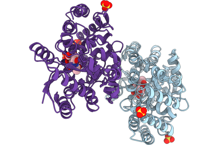
Crystal Structure Of A Sialic Acid Binding Protein From Streptococcus Pneumoniae Bound To Neu5Ac
Organism: Streptococcus pneumoniae tigr4
Method: X-RAY DIFFRACTION Resolution:1.80 Å Release Date: 2026-07-29 Classification: SUGAR BINDING PROTEIN Ligands: SIA, SO4 |
Organism: Streptococcus pneumoniae tigr4
Method: X-RAY DIFFRACTION
Release Date: 2026-07-29
Ligands: SIA, SO4
 |
Crystal Structure Of A Sialic Acid Binding Protein, R113A Mutant, From Streptococcus Pneumoniae Bound To Neu5Ac
Organism: Streptococcus pneumoniae tigr4
Method: X-RAY DIFFRACTION Resolution:1.70 Å Release Date: 2026-07-29 Classification: SUGAR BINDING PROTEIN Ligands: SIA, SO4, CL |
Organism: Streptococcus pneumoniae tigr4
Method: X-RAY DIFFRACTION
Release Date: 2026-07-29
Ligands: SIA, SO4, CL