

Search Count: 996
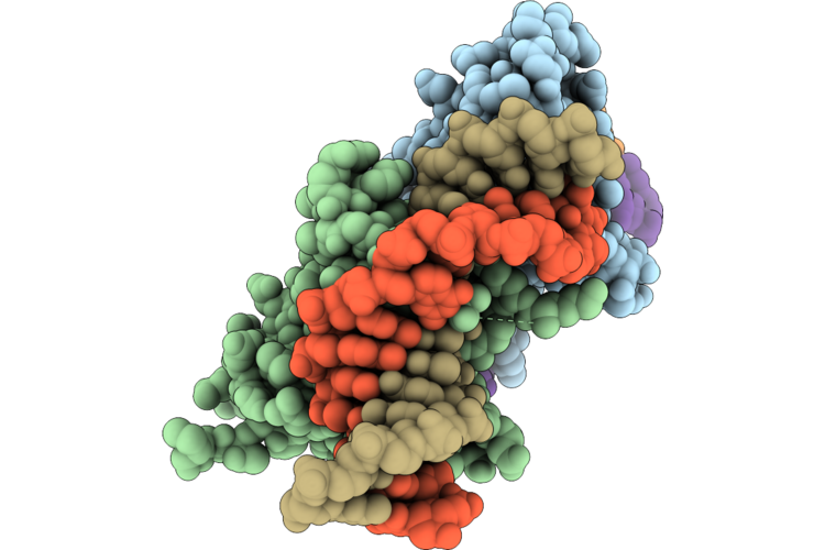 |
Crystal Structure Of Taylorella Equigenitalis Badtf3 Deaminase Toxin Dddb Bound To Double-Stranded Dna
Organism: Taylorella equigenitalis
Method: X-RAY DIFFRACTION Resolution:2.39 Å Release Date: 2026-08-05 Classification: GENE REGULATION/DNA Ligands: ZN, CL |
Organism: Taylorella equigenitalis
Method: X-RAY DIFFRACTION
Release Date: 2026-08-05
Ligands: ZN, CL
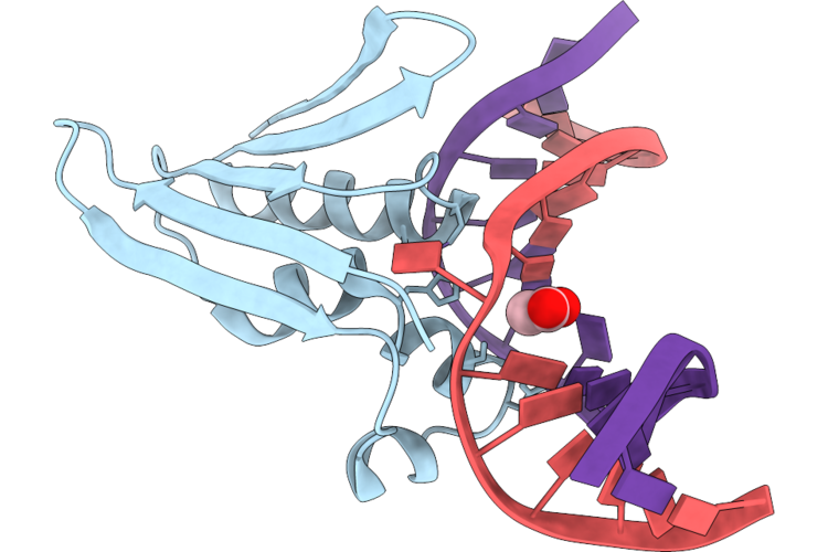 |
Crystal Structure Of Taylorella Equigenitalis Badtf3 Deaminase Toxin Dddb (N-Terminally Truncated) Bound To Double-Stranded Dna
Organism: Taylorella equigenitalis
Method: X-RAY DIFFRACTION Resolution:2.91 Å Release Date: 2026-08-05 Classification: GENE REGULATION/DNA Ligands: ZN, ACT |
Organism: Taylorella equigenitalis
Method: X-RAY DIFFRACTION
Release Date: 2026-08-05
Ligands: ZN, ACT
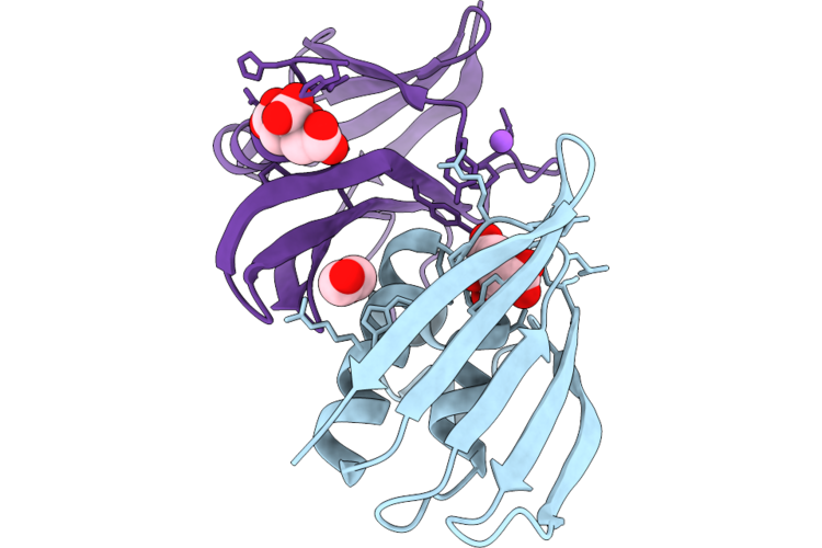 |
Taylorella Equigenitalis Badtf3 Deaminase Toxin Dddb Bound To The Immunity Protein
Organism: Taylorella equigenitalis
Method: X-RAY DIFFRACTION Resolution:1.61 Å Release Date: 2026-08-05 Classification: GENE REGULATION Ligands: CIT, EDO, NA |
Organism: Taylorella equigenitalis
Method: X-RAY DIFFRACTION
Release Date: 2026-08-05
Ligands: CIT, EDO, NA
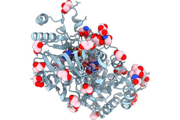 |
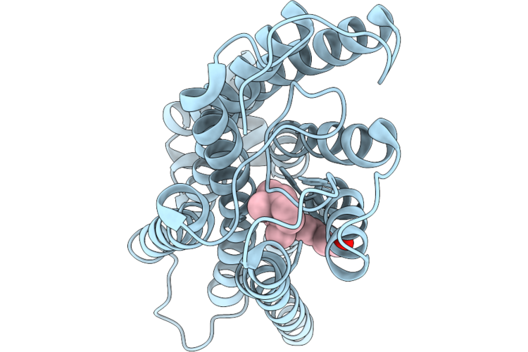
Glucooligosaccharide Oxidase From Hapsidospora Chrysogenum, Recombinant Wild-Type Expressed In Komagataella Phaffii
Organism: Hapsidospora chrysogenum atcc 11550
Method: X-RAY DIFFRACTION Resolution:1.65 Å Release Date: 2026-07-01 Classification: FLAVOPROTEIN Ligands: FAD, NAG, GOL, TRS |
Organism: Hapsidospora chrysogenum atcc 11550
Method: X-RAY DIFFRACTION
Release Date: 2026-07-01
Ligands: FAD, NAG, GOL, TRS
 |
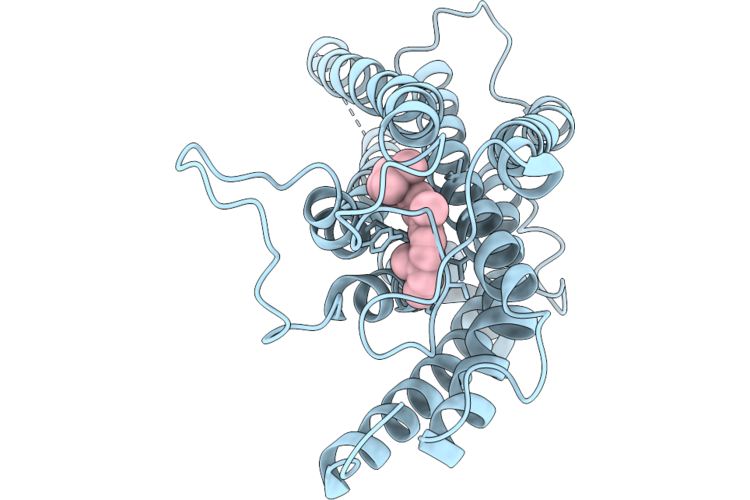
Cryo-Em Structure Of Macaque Red Cone Pigment With Q114N Mutation
Organism: Macaca fascicularis, Escherichia coli
Method: ELECTRON MICROSCOPY Resolution:3.21 Å Release Date: 2026-06-24 Classification: MEMBRANE PROTEIN Ligands: RET |
Organism: Macaca fascicularis, Escherichia coli
Method: ELECTRON MICROSCOPY
Release Date: 2026-06-24
Ligands: RET
 |
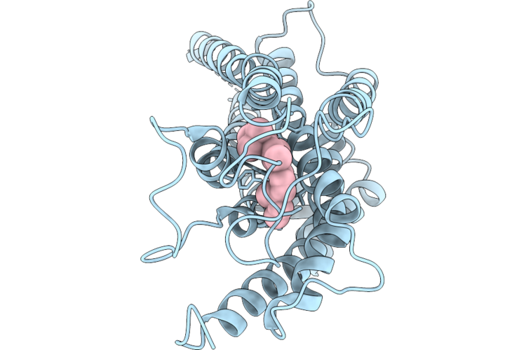
Cryo-Em Structure Of Macaque Green Cone Pigment With Q114N Mutation
Organism: Macaca fascicularis, Escherichia coli
Method: ELECTRON MICROSCOPY Resolution:3.37 Å Release Date: 2026-06-24 Classification: MEMBRANE PROTEIN Ligands: RET |
Organism: Macaca fascicularis, Escherichia coli
Method: ELECTRON MICROSCOPY
Release Date: 2026-06-24
Ligands: RET
 |
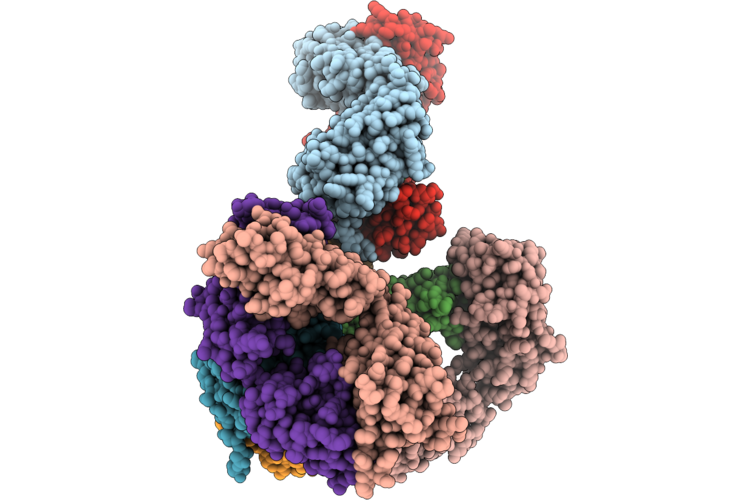
Cryo-Em Structure Of Macaque Green Cone Pigment Wild Type
Organism: Macaca fascicularis, Escherichia coli
Method: ELECTRON MICROSCOPY Resolution:3.71 Å Release Date: 2026-06-24 Classification: MEMBRANE PROTEIN Ligands: RET |
Organism: Macaca fascicularis, Escherichia coli
Method: ELECTRON MICROSCOPY
Release Date: 2026-06-24
Ligands: RET
 |
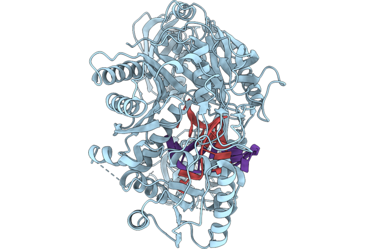
X-Ray Crystal Structure Of A High-Affinity Monoclonal Antibody Sequesters Xylazine
Organism: Homo sapiens
Method: X-RAY DIFFRACTION Resolution:1.99 Å Release Date: 2026-06-10 Classification: IMMUNE SYSTEM Ligands: A1C4R, CL |
Organism: Homo sapiens
Method: X-RAY DIFFRACTION
Release Date: 2026-06-10
Ligands: A1C4R, CL
 |
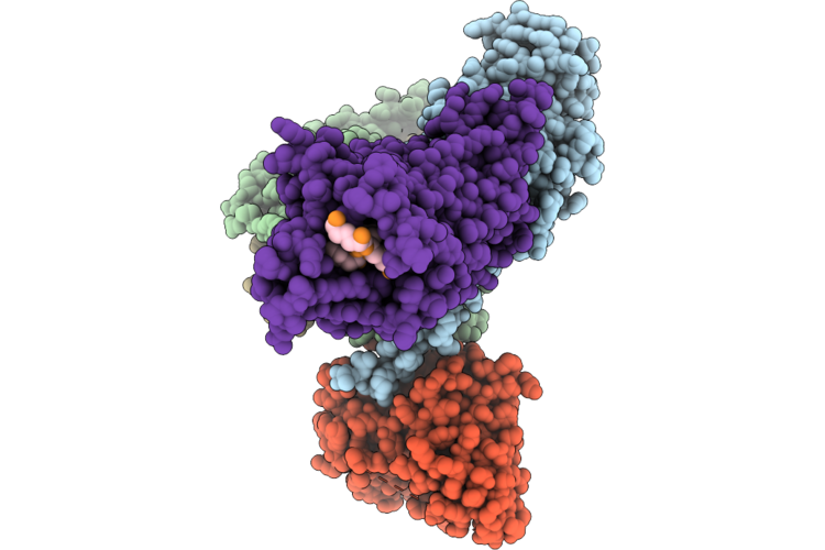
Human Ago2 Bound To A Mir-20A Guide And A Position 10-11 Mismatched Target
Organism: Homo sapiens
Method: ELECTRON MICROSCOPY Resolution:3.02 Å Release Date: 2026-06-03 Classification: RNA BINDING PROTEIN |
Organism: Homo sapiens
Method: ELECTRON MICROSCOPY
Release Date: 2026-06-03
 |
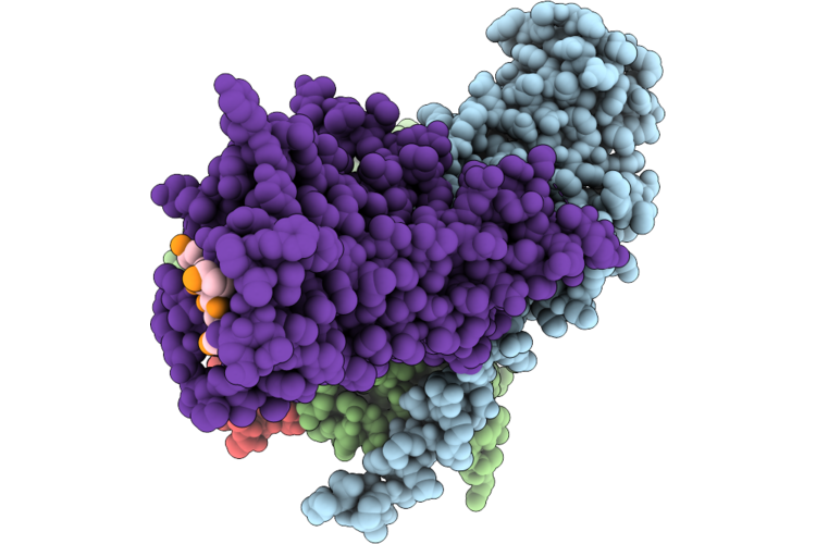
Cryo-Em Structure Of Human Neurotensin Receptor 1 (Hntsr1)-Gi1 (Delipidated) Complex In Nucleotide-Free C State
Organism: Homo sapiens, Mus musculus, Synthetic construct
Method: ELECTRON MICROSCOPY Resolution:2.60 Å Release Date: 2026-06-03 Classification: MEMBRANE PROTEIN |
Organism: Homo sapiens, Mus musculus, Synthetic construct
Method: ELECTRON MICROSCOPY
Release Date: 2026-06-03
 |
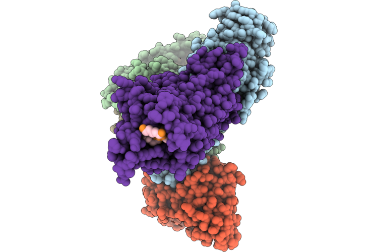
Cryo-Em Structure Of Human Neurotensin Receptor 1 (Hntsr1)-Gi1 (Delipidated) Complex In Nucleotide-Free Nc State
Organism: Homo sapiens, Synthetic construct
Method: ELECTRON MICROSCOPY Resolution:2.80 Å Release Date: 2026-06-03 Classification: MEMBRANE PROTEIN |
Organism: Homo sapiens, Synthetic construct
Method: ELECTRON MICROSCOPY
Release Date: 2026-06-03
 |
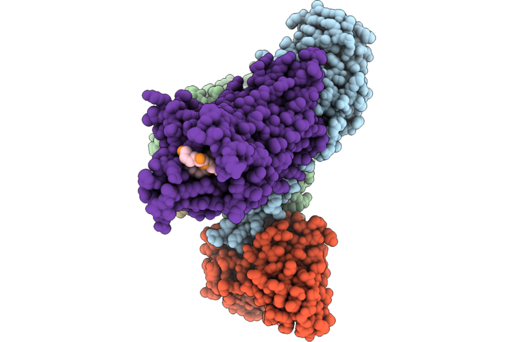
Cryo-Em Structure Of The Human Neurotensin Receptor 1 (Hntsr1)-Gi1 Complex In The Gtp-Bound, Ahd-Open C State 1, Plunge-Frozen 0-5 Seconds After Gtp Addition
Organism: Homo sapiens, Mus musculus, Synthetic construct
Method: ELECTRON MICROSCOPY Resolution:2.40 Å Release Date: 2026-06-03 Classification: MEMBRANE PROTEIN |
Organism: Homo sapiens, Mus musculus, Synthetic construct
Method: ELECTRON MICROSCOPY
Release Date: 2026-06-03
 |
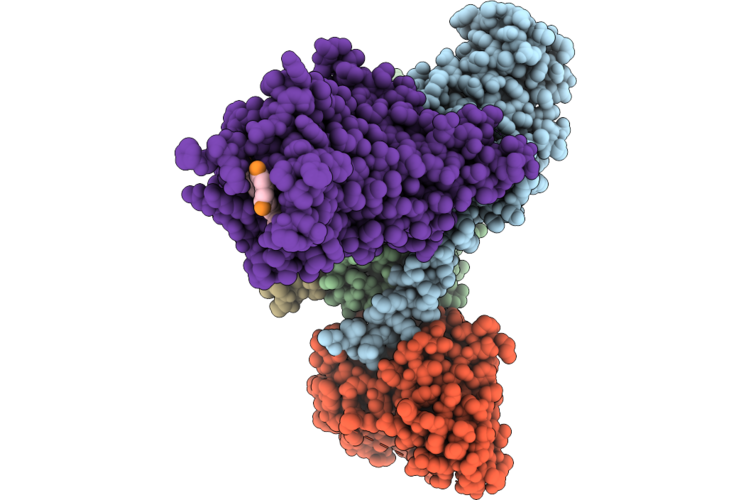
Cryo-Em Structure Of The Human Neurotensin Receptor 1 (Hntsr1)-Gi1 Complex In The Gtp-Bound, Ahd-Open C State 2, Plunge-Frozen 0-5 Seconds After Gtp Addition
Organism: Homo sapiens, Mus musculus, Synthetic construct
Method: ELECTRON MICROSCOPY Resolution:2.40 Å Release Date: 2026-06-03 Classification: MEMBRANE PROTEIN |
Organism: Homo sapiens, Mus musculus, Synthetic construct
Method: ELECTRON MICROSCOPY
Release Date: 2026-06-03
 |
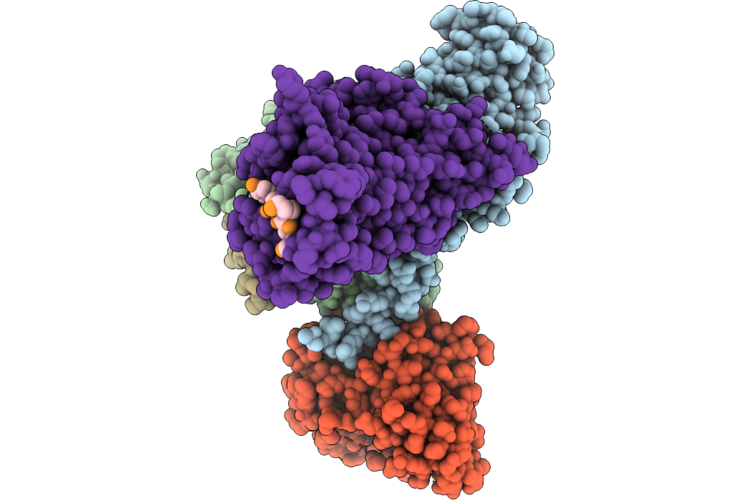
Cryo-Em Structure Of The Human Neurotensin Receptor 1 (Hntsr1)-Gi1 Complex In The Gtp-Bound, Ahd-Open Nc State 1, Plunge-Frozen 0-5 Seconds After Gtp Addition
Organism: Homo sapiens, Mus musculus, Synthetic construct
Method: ELECTRON MICROSCOPY Resolution:2.70 Å Release Date: 2026-06-03 Classification: MEMBRANE PROTEIN |
Organism: Homo sapiens, Mus musculus, Synthetic construct
Method: ELECTRON MICROSCOPY
Release Date: 2026-06-03
 |
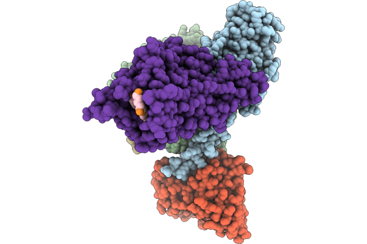
Cryo-Em Structure Of The Human Neurotensin Receptor 1 (Hntsr1)-Gi1 Complex In The Gtp-Bound, Ahd-Open Nc State 2, Plunge-Frozen 0-5 Seconds After Gtp Addition
Organism: Homo sapiens, Mus musculus, Synthetic construct
Method: ELECTRON MICROSCOPY Resolution:2.90 Å Release Date: 2026-06-03 Classification: MEMBRANE PROTEIN |
Organism: Homo sapiens, Mus musculus, Synthetic construct
Method: ELECTRON MICROSCOPY
Release Date: 2026-06-03
 |
Cryo-Em Structure Of The Human Neurotensin Receptor 1 (Hntsr1)-Gi1 Complex In The Gtp-Bound, Ahd-Open Nc State 3, Plunge-Frozen 0-5 Seconds After Gtp Addition
Organism: Homo sapiens, Mus musculus, Synthetic construct
Method: ELECTRON MICROSCOPY Resolution:2.70 Å Release Date: 2026-06-03 Classification: MEMBRANE PROTEIN |
Organism: Homo sapiens, Mus musculus, Synthetic construct
Method: ELECTRON MICROSCOPY
Release Date: 2026-06-03
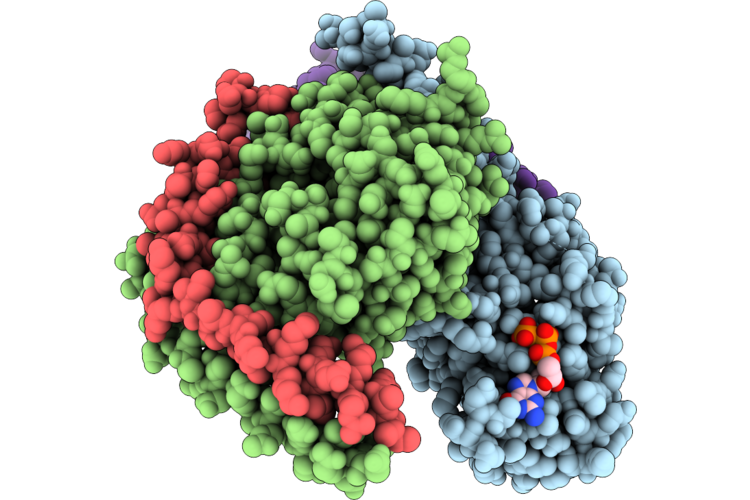 |
Cryo-Em Structure Of The Human Neurotensin Receptor 1 (Hntsr1)-Gi1 Complex In The Gtp-Bound, Ahd-Open Nc State 4, Plunge-Frozen 0-5 Seconds After Gtp Addition
Organism: Homo sapiens, Synthetic construct
Method: ELECTRON MICROSCOPY Resolution:3.10 Å Release Date: 2026-06-03 Classification: MEMBRANE PROTEIN Ligands: GTP |
Organism: Homo sapiens, Synthetic construct
Method: ELECTRON MICROSCOPY
Release Date: 2026-06-03
Ligands: GTP
 |
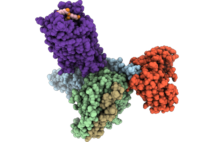
Cryo-Em Structure Of Human Neurotensin Receptor 1 (Hntsr1)-Go Complex In Nucleotide-Free C State
Organism: Homo sapiens, Mus musculus, Synthetic construct
Method: ELECTRON MICROSCOPY Resolution:3.37 Å Release Date: 2026-06-03 Classification: MEMBRANE PROTEIN |
Organism: Homo sapiens, Mus musculus, Synthetic construct
Method: ELECTRON MICROSCOPY
Release Date: 2026-06-03
 |
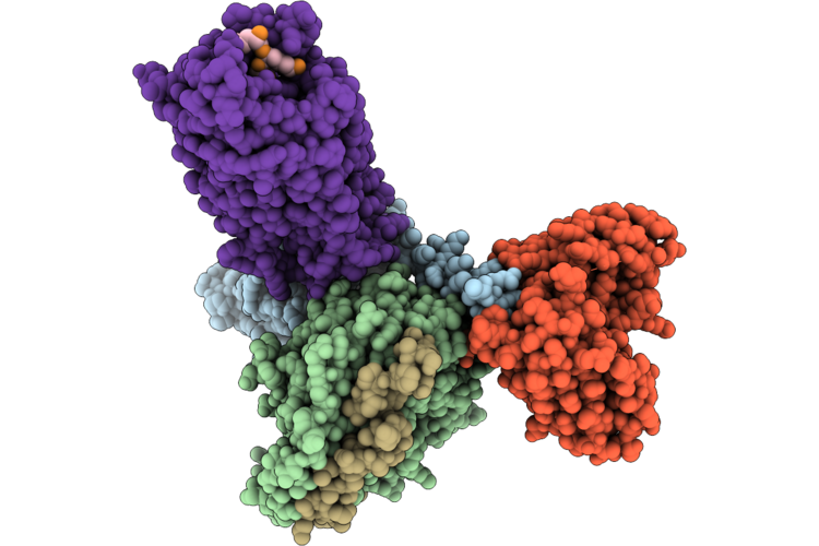
Cryo-Em Structure Of Human Neurotensin Receptor 1 (Hntsr1)-Go Complex In Nucleotide-Free Nc State 1
Organism: Homo sapiens, Mus musculus, Synthetic construct
Method: ELECTRON MICROSCOPY Resolution:2.95 Å Release Date: 2026-06-03 Classification: MEMBRANE PROTEIN |
Organism: Homo sapiens, Mus musculus, Synthetic construct
Method: ELECTRON MICROSCOPY
Release Date: 2026-06-03
 |
Cryo-Em Structure Of Human Neurotensin Receptor 1 (Hntsr1)-Go Complex In Nucleotide-Free Nc State 2
Organism: Homo sapiens, Mus musculus, Synthetic construct
Method: ELECTRON MICROSCOPY Resolution:2.99 Å Release Date: 2026-06-03 Classification: MEMBRANE PROTEIN |
Organism: Homo sapiens, Mus musculus, Synthetic construct
Method: ELECTRON MICROSCOPY
Release Date: 2026-06-03