

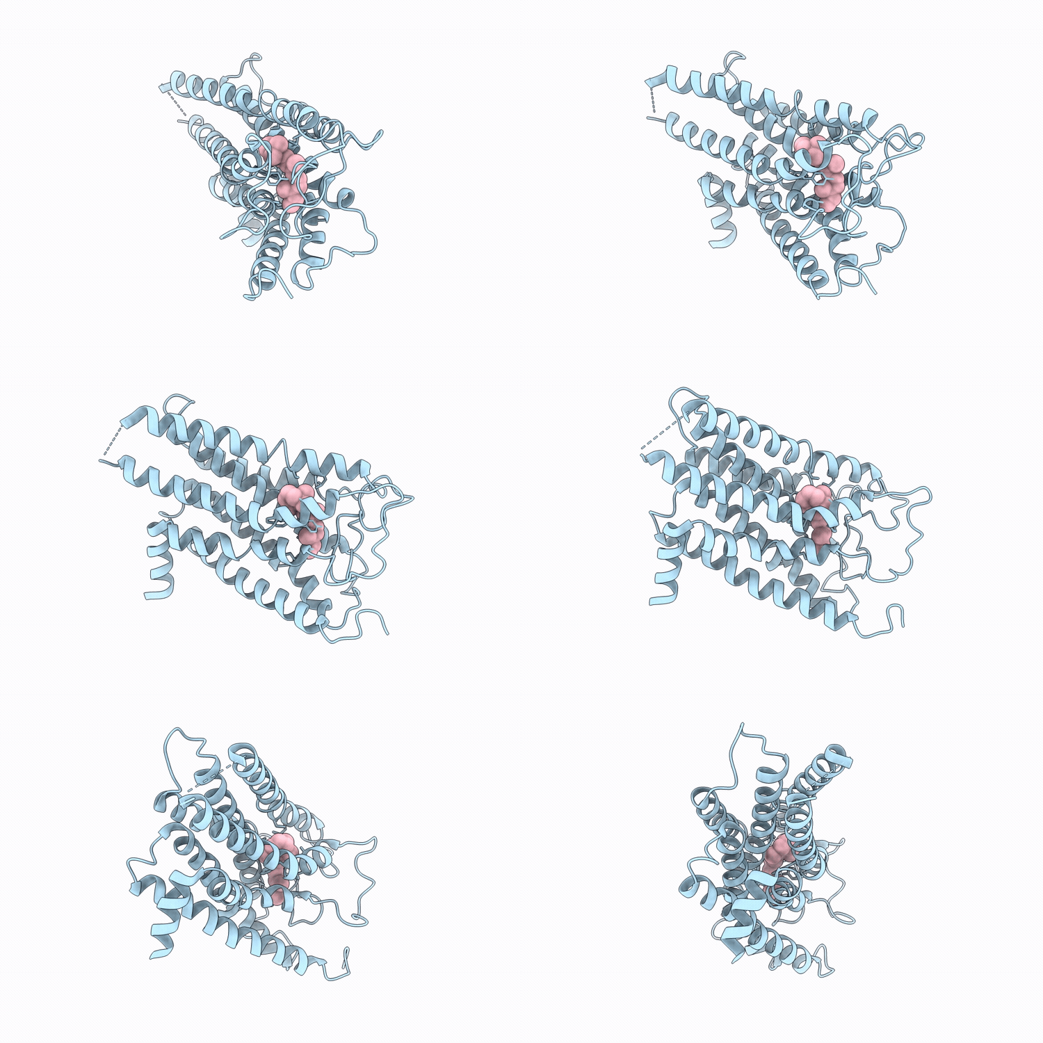
9UPO image
Deposition Date 2025-04-28
Release Date 2026-06-24
Last Version Date 2026-07-08
Entry Detail
PDB ID:
9UPO
Title:
Cryo-EM structure of macaque green cone pigment wild type
Biological Source:
Source Organism(s):
Expression System(s):
Method Details:
Experimental Method:
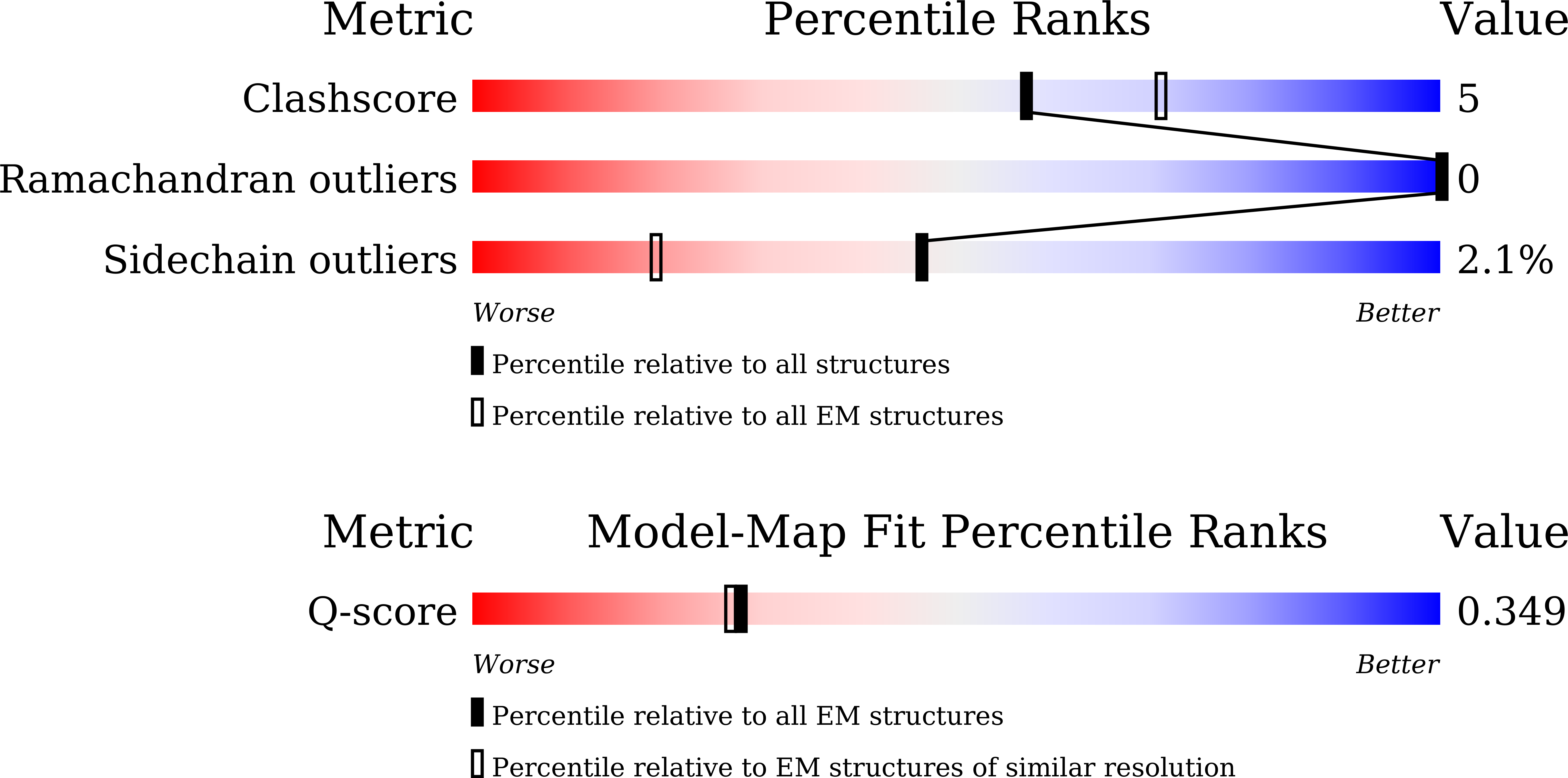
Resolution:
3.71 Å
Aggregation State:
PARTICLE
Reconstruction Method:
SINGLE PARTICLE
Macromolecular Entities
Structural Superimposition Protein Blast
Polymer Type:polypeptide(L)
Molecule:MW opsin,Soluble cytochrome b
Gene (Uniprot):cybC
Mutagens:M257W,R331I,H335I
Chain IDs:A
Chain Length:434
Number of Molecules:1
Biological Source:Macaca fascicularis, Escherichia coli
Ligand Molecules
Primary Citation
Structural insights into spectral tuning and retinal exchange in cone visual pigments.
Science 392 eadz3996 eadz3996 (2026)
PMID: 42348681 DOI: 10.1126/science.adz3996

Abstact

Color vision in catarrhine primates relies on red-, green-, and blue-sensitive cone pigments that share an 11-cis-retinal chromophore but differ in absorption maxima. Red and green pigments arose by recent gene duplication and differ at only a few residues. Here, we report cryo-electron microscopy structures of red and green cone pigments from the cynomolgus macaque (Macaca fascicularis) integrated with low-temperature vibrational spectroscopy and quantum mechanical and molecular mechanical modeling. The red-green spectral shift is dominated by threonine 285, the hydroxyl dipole of which modulates chromophore electrostatics, whereas steric effects appear modest. We also identified membrane-facing lateral openings in cone pigments but not in inactive rhodopsin. Comparisons with active-state structures suggest activation-dependent gating, and mutational and spectroscopic analyses support a role for this opening in retinal uptake and rapid pigment regeneration.

Legend

Protein

Chemical

Disease

Primary Citation of related structures
Feedback Form
Name
Email
Institute
Feedback