

Search Count: 780
 |
Lx-2D6 (Fab) - Polio 1 (Vlp) Complex
Organism: Human poliovirus 1 mahoney, Homo sapiens
Method: ELECTRON MICROSCOPY Resolution:1.70 Å Release Date: 2026-09-30 Classification: VIRUS LIKE PARTICLE Ligands: PLM, MYR |
Organism: Human poliovirus 1 mahoney, Homo sapiens
Method: ELECTRON MICROSCOPY
Release Date: 2026-09-30
Ligands: PLM, MYR
 |
1B8 (Fab) - Polio 2 (Vlp) Complex
Organism: Poliovirus 2, Homo sapiens
Method: ELECTRON MICROSCOPY Resolution:1.60 Å Release Date: 2026-09-30 Classification: VIRUS LIKE PARTICLE Ligands: PLM |
Organism: Poliovirus 2, Homo sapiens
Method: ELECTRON MICROSCOPY
Release Date: 2026-09-30
Ligands: PLM
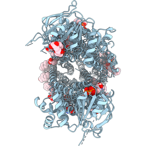 |
V-Shaped (Channel-Formed), Atp-Bound, Vx809-Bound Conformation Of Wild-Type Human Cftr (Composite Map From Phenix Based On Consensus And Local Refinement Maps From Cryosparc)
Organism: Homo sapiens
Method: ELECTRON MICROSCOPY Resolution:3.14 Å Release Date: 2026-09-16 Classification: MEMBRANE PROTEIN Ligands: AJP, CLR, P5S, PTY, OLA, MYS, PLM, D12, CPL, OCT, HEX, ATP, MG, VX8 |
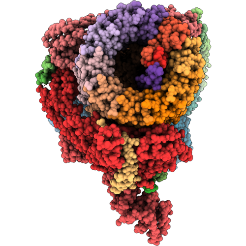 |
Arabidopsis Thaliana V-Type Atpase, State 1
Organism: Arabidopsis thaliana
Method: ELECTRON MICROSCOPY Resolution:3.00 Å Release Date: 2026-09-09 Classification: HYDROLASE Ligands: PLM, ADP, MG |
Organism: Arabidopsis thaliana
Method: ELECTRON MICROSCOPY
Release Date: 2026-09-09
Ligands: PLM, ADP, MG
 |
Alternative Nbd1-Binding Geometry In Channel-Formed, Atp-Bound, Vx809-Bound, T2A-Nanobody-Bound Wild-Type Human Cftr (Composite Map From Phenix)
Organism: Homo sapiens, Lama glama
Method: ELECTRON MICROSCOPY Resolution:3.86 Å Release Date: 2026-09-09 Classification: MEMBRANE PROTEIN Ligands: AJP, CLR, P5S, PC1, 3PH, PTY, PLM, MYR, D12, D10, HEX, OCT, ATP, MG, VX8 |
Organism: Homo sapiens, Lama glama
Method: ELECTRON MICROSCOPY
Release Date: 2026-09-09
Ligands: AJP, CLR, P5S, PC1, 3PH, PTY, PLM, MYR, D12, D10, HEX, OCT, ATP, MG, VX8
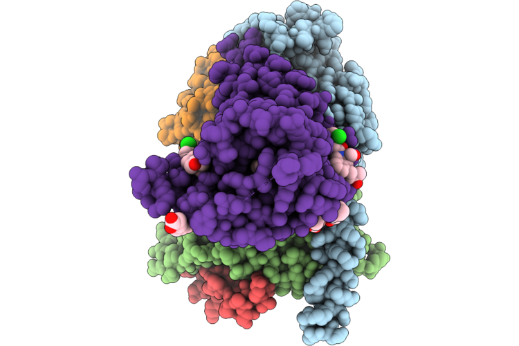 |
D1R-Gs In Complexed With Skf81297/Ly3154207/Bmsa1
Organism: Homo sapiens, Rattus norvegicus, Bos taurus, Vicugna pacos, Oplophorus gracilirostris (luminous shrimp)
Method: ELECTRON MICROSCOPY Release Date: 2026-08-26 Classification: STRUCTURAL PROTEIN Ligands: CLR, A1EV3, SK0, G4C, PLM |
Organism: Homo sapiens, Rattus norvegicus, Bos taurus, Vicugna pacos, Oplophorus gracilirostris (luminous shrimp)
Method: ELECTRON MICROSCOPY
Release Date: 2026-08-26
Ligands: CLR, A1EV3, SK0, G4C, PLM
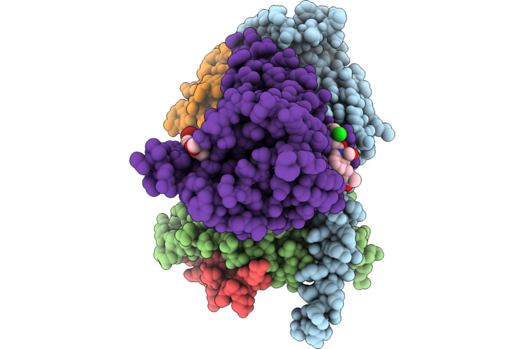 |
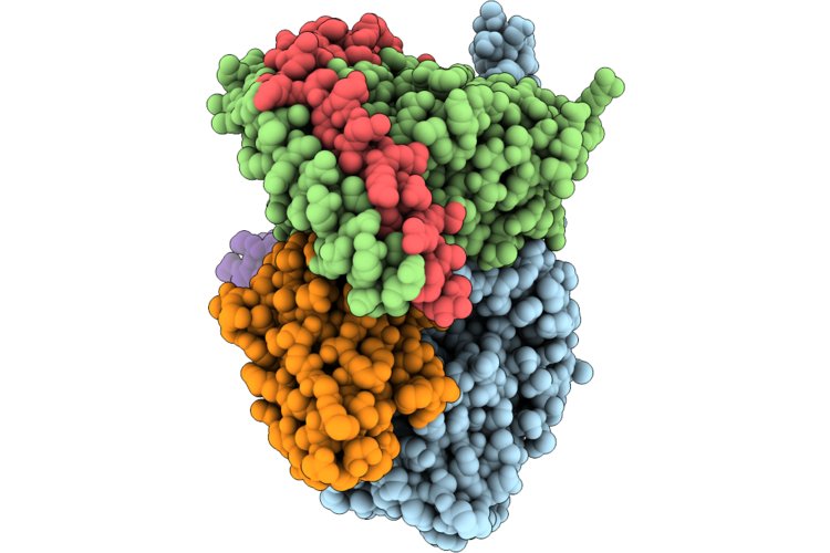
D1R-Gs In Complexed With Skf81297/Ly3154207/Bmsa1/Unc10062
Organism: Homo sapiens, Rattus norvegicus, Bos taurus, Vicugna pacos, Oplophorus gracilirostris
Method: ELECTRON MICROSCOPY Release Date: 2026-08-26 Classification: STRUCTURAL PROTEIN Ligands: A1EWL, A1EV3, SK0, G4C, PLM |
Organism: Homo sapiens, Rattus norvegicus, Bos taurus, Vicugna pacos, Oplophorus gracilirostris
Method: ELECTRON MICROSCOPY
Release Date: 2026-08-26
Ligands: A1EWL, A1EV3, SK0, G4C, PLM
 |
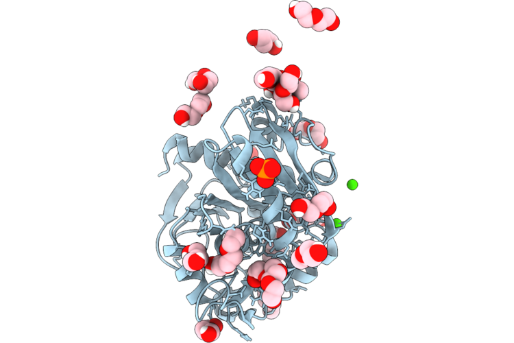
D1R-Gs In Complexed With Dopamine/Ly3154207/Bmsa1/Unc9815
Organism: Homo sapiens, Rattus norvegicus, Bos taurus, Vicugna pacos, Oplophorus gracilirostris
Method: ELECTRON MICROSCOPY Release Date: 2026-08-26 Classification: STRUCTURAL PROTEIN Ligands: LDP, A1EWF, A1EV3, G4C, PLM |
Organism: Homo sapiens, Rattus norvegicus, Bos taurus, Vicugna pacos, Oplophorus gracilirostris
Method: ELECTRON MICROSCOPY
Release Date: 2026-08-26
Ligands: LDP, A1EWF, A1EV3, G4C, PLM
 |
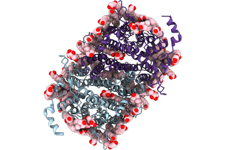
Crystal Structure Of Thermomyces Lanuginosa Lipase With Acyl Intermediate: Space Group I222
Organism: Thermomyces lanuginosus
Method: X-RAY DIFFRACTION Resolution:1.45 Å Release Date: 2026-08-05 Classification: LIPID BINDING PROTEIN Ligands: CA, PLM, NAG, PO4, PG4, PEG, CL |
Organism: Thermomyces lanuginosus
Method: X-RAY DIFFRACTION
Release Date: 2026-08-05
Ligands: CA, PLM, NAG, PO4, PG4, PEG, CL
 |
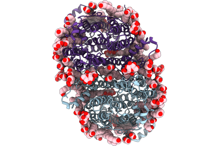
Uapa (Wt) In Ddm, Apo State
Organism: Aspergillus nidulans fgsc a4
Method: ELECTRON MICROSCOPY Resolution:2.06 Å Release Date: 2026-07-01 Classification: MEMBRANE PROTEIN Ligands: LMT, MYR, PLM, ERG, DAO, DGA |
Organism: Aspergillus nidulans fgsc a4
Method: ELECTRON MICROSCOPY
Release Date: 2026-07-01
Ligands: LMT, MYR, PLM, ERG, DAO, DGA
 |
Uapa (Q408E) In Ddm, Apo State
Organism: Aspergillus nidulans fgsc a4
Method: ELECTRON MICROSCOPY Resolution:2.59 Å Release Date: 2026-07-01 Classification: MEMBRANE PROTEIN Ligands: LMT, MYR, PLM, ERG, DAO, FAW |
Organism: Aspergillus nidulans fgsc a4
Method: ELECTRON MICROSCOPY
Release Date: 2026-07-01
Ligands: LMT, MYR, PLM, ERG, DAO, FAW
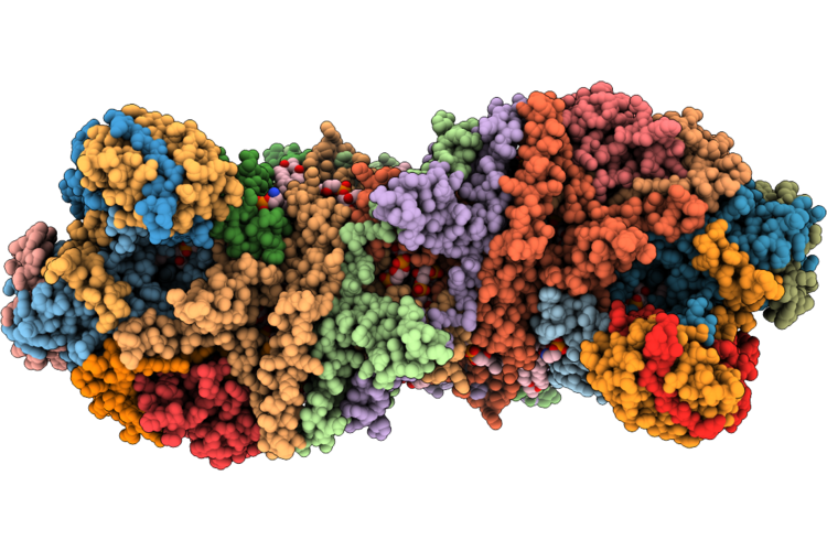 |
Cryo-Em Structure Of The Mycobacterium Abscessus Cytochrome Bcc:Aa3 Supercomplex
Organism: Mycobacteroides abscessus
Method: ELECTRON MICROSCOPY Resolution:2.66 Å Release Date: 2026-07-01 Classification: OXIDOREDUCTASE Ligands: 9XX, CDL, 9Y0, HEA, CU, PLM, OXY, HEM, FES, 9YF, MQ9, HEC |
Organism: Mycobacteroides abscessus
Method: ELECTRON MICROSCOPY
Release Date: 2026-07-01
Ligands: 9XX, CDL, 9Y0, HEA, CU, PLM, OXY, HEM, FES, 9YF, MQ9, HEC
 |
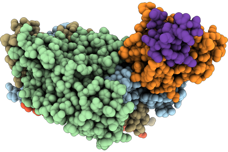
Cryo-Em Structure Of The Mycobacterium Abscessus Cytochrome Bcc:Aa3 Supercomplex In The Presence Of Nd-011458
Organism: Mycobacteroides abscessus
Method: ELECTRON MICROSCOPY Resolution:2.26 Å Release Date: 2026-07-01 Classification: OXIDOREDUCTASE Ligands: CDL, 9Y0, HEA, CU, OXY, PLM, 9YF, HEC, MQ9, FES, A1MBF, HEM, 9XX |
 |
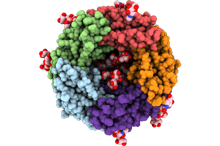
Structure Of Echovirus 18 In Complex With Neonatal Fc Receptor
Organism: Homo sapiens, Echovirus e18
Method: ELECTRON MICROSCOPY Release Date: 2026-06-24 Classification: VIRUS Ligands: PLM |
Organism: Homo sapiens, Echovirus e18
Method: ELECTRON MICROSCOPY
Release Date: 2026-06-24
Ligands: PLM
 |
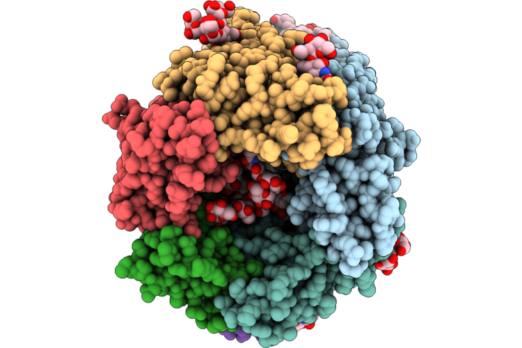
Cryoem Structure Of Human Alpha1Beta3Gamma2L Gaba(A)R In Cl47A
Organism: Homo sapiens
Method: ELECTRON MICROSCOPY Release Date: 2026-06-17 Classification: MEMBRANE PROTEIN Ligands: D10, PT5, R16, EPE, PX2, OLC, PLM, HEX, NAG, PGW, CL |
Organism: Homo sapiens
Method: ELECTRON MICROSCOPY
Release Date: 2026-06-17
Ligands: D10, PT5, R16, EPE, PX2, OLC, PLM, HEX, NAG, PGW, CL
 |
Cryoem Structure Of Human Alpha1Beta3Gamma2 Gaba(A)R In Complex With Garlh4 And The Neuroligin2 Transmembrane Helix, In Cl47A
Organism: Homo sapiens
Method: ELECTRON MICROSCOPY Release Date: 2026-06-17 Classification: MEMBRANE PROTEIN Ligands: PIO, PLM, HEX, PGW, R16, NAG, P1L, D10, CLR |
Organism: Homo sapiens
Method: ELECTRON MICROSCOPY
Release Date: 2026-06-17
Ligands: PIO, PLM, HEX, PGW, R16, NAG, P1L, D10, CLR
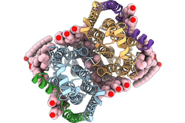 |
Cryo-Em Structure Of Human Organic Solute Transporter In Apo State
Organism: Homo sapiens
Method: ELECTRON MICROSCOPY Resolution:2.81 Å Release Date: 2026-06-10 Classification: TRANSPORT PROTEIN Ligands: CLR, PLM, POV |
Organism: Homo sapiens
Method: ELECTRON MICROSCOPY
Release Date: 2026-06-10
Ligands: CLR, PLM, POV
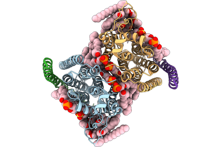 |
Cryo-Em Structure Of Human Organic Solute Transporter In Complex With Dheas
Organism: Homo sapiens
Method: ELECTRON MICROSCOPY Resolution:2.99 Å Release Date: 2026-06-10 Classification: TRANSPORT PROTEIN Ligands: CLR, PLM, POV, ZWY |
Organism: Homo sapiens
Method: ELECTRON MICROSCOPY
Release Date: 2026-06-10
Ligands: CLR, PLM, POV, ZWY
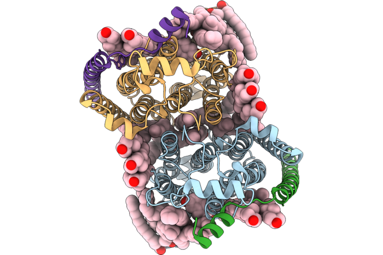 |
Cryo-Em Structure Of Human Organic Solute Transporter In Complex With Tlca
Organism: Homo sapiens
Method: ELECTRON MICROSCOPY Resolution:3.11 Å Release Date: 2026-06-10 Classification: TRANSPORT PROTEIN Ligands: CLR, PLM, POV, A1EPX |
Organism: Homo sapiens
Method: ELECTRON MICROSCOPY
Release Date: 2026-06-10
Ligands: CLR, PLM, POV, A1EPX
 |
Cryo-Em Structure Of Human Organic Solute Transporter In Complex With Ethinylestradiol
Organism: Homo sapiens
Method: ELECTRON MICROSCOPY Resolution:2.68 Å Release Date: 2026-06-10 Classification: TRANSPORT PROTEIN Ligands: CLR, PLM, POV, 3WF |
Organism: Homo sapiens
Method: ELECTRON MICROSCOPY
Release Date: 2026-06-10
Ligands: CLR, PLM, POV, 3WF