
Search Count: 782
All
Selected
 |
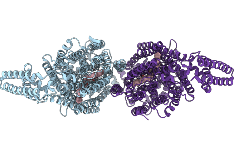
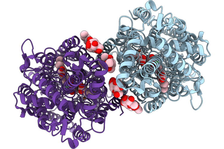
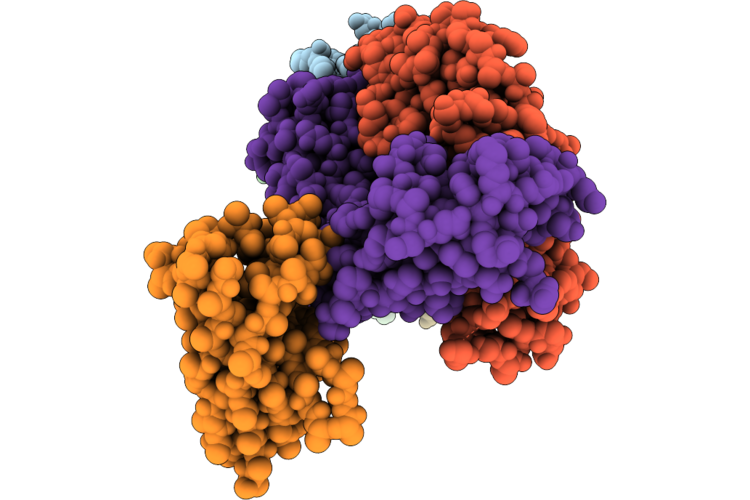
Cryoem Structure Of Native Quinol Dependent Nitric Oxide Reductase At Ph 8.0.
Organism: Achromobacter xylosoxidans
Method: ELECTRON MICROSCOPY Release Date: 2026-05-20 Classification: MEMBRANE PROTEIN Ligands: HEM, FE, CA, LMT, CU, UQ5 |
 |
Organism: Achromobacter xylosoxidans
Method: ELECTRON MICROSCOPY Release Date: 2026-05-20 Classification: MEMBRANE PROTEIN Ligands: HEM, FE, CA |
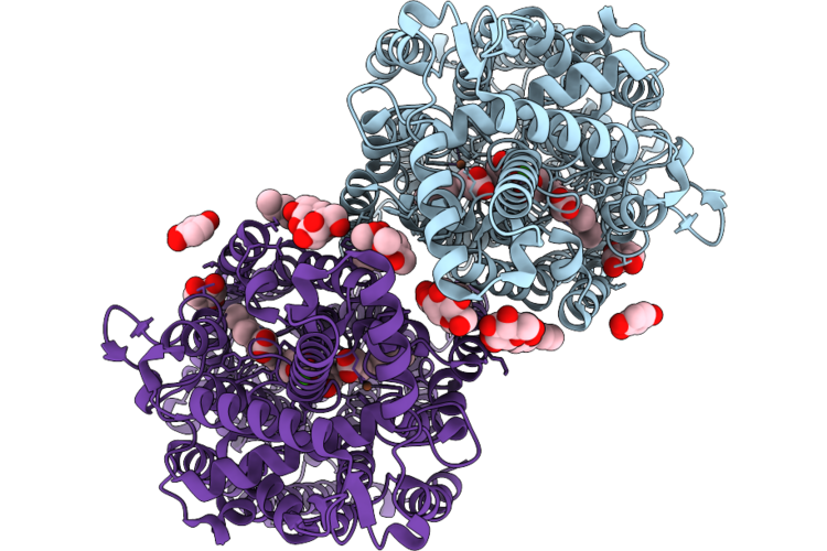 |
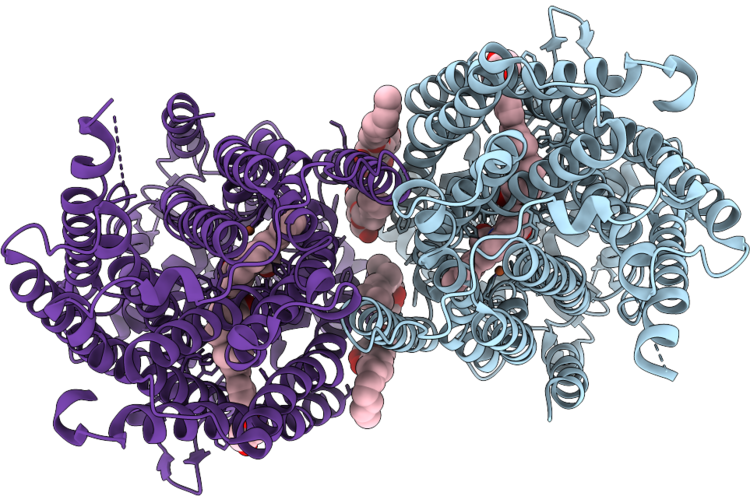
Cryoem Structure Of Native Quinol Dependent Nitric Oxide Reductase With Hqn At Ph 6.5
Organism: Achromobacter xylosoxidans
Method: ELECTRON MICROSCOPY Resolution:2.30 Å Release Date: 2026-05-20 Classification: MEMBRANE PROTEIN Ligands: HEM, CA, FE, LMT, HQN, UQ5 |
 |
Cryoem Structure Of Native Quinol Dependent Nitric Oxide Reductase At Ph 8.0 On Gold Grid.
Organism: Achromobacter xylosoxidans
Method: ELECTRON MICROSCOPY Resolution:2.70 Å Release Date: 2026-05-06 Classification: MEMBRANE PROTEIN Ligands: HEM, CA, FE |
 |
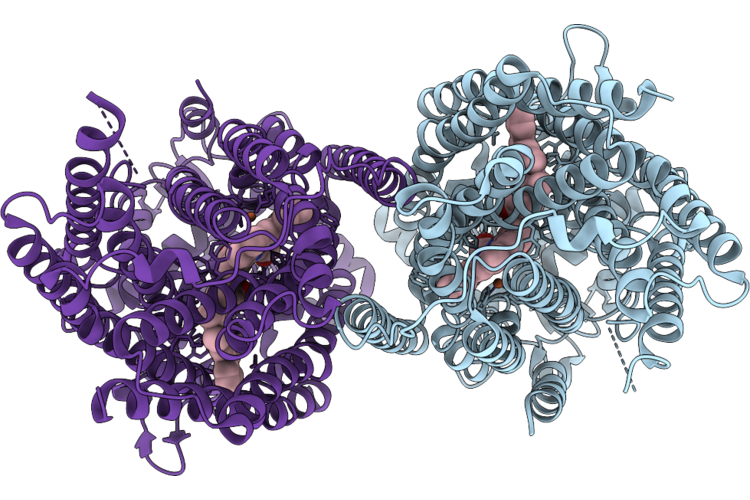
Cryoem Structure Of Native Quinol Dependent Nitric Oxide Reductase Arg720Ala Variant At Ph 6.5 On Gold Grid.
Organism: Achromobacter xylosoxidans
Method: ELECTRON MICROSCOPY Resolution:2.90 Å Release Date: 2026-05-06 Classification: MEMBRANE PROTEIN Ligands: HEM, CA, FE |
 |
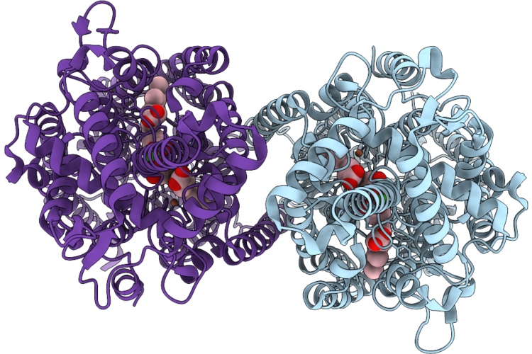
Cryoem Structure Of Native Quinol Dependent Nitric Oxide Reductase Trp718Ala Variant At Ph 6.5 On Gold Grid.
Organism: Achromobacter xylosoxidans
Method: ELECTRON MICROSCOPY Resolution:2.40 Å Release Date: 2026-05-06 Classification: MEMBRANE PROTEIN Ligands: HEM, CA, FE, LMT, UQ5 |
 |
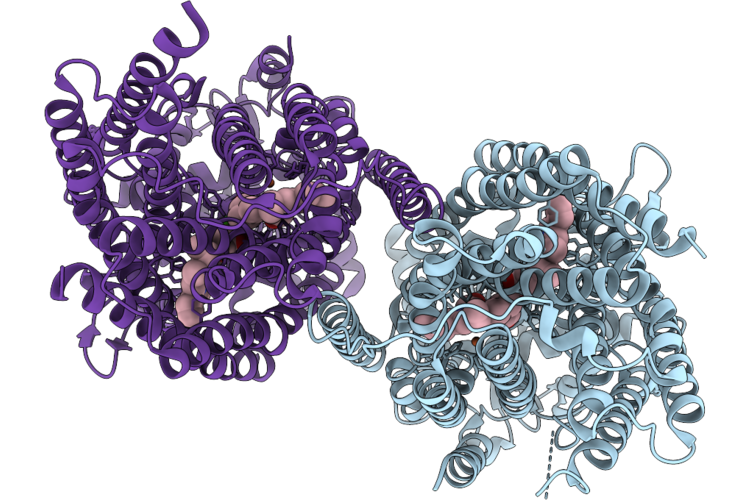
Cryoem Structure Of Native Quinol Dependent Nitric Oxide Reductase Trp718Ala Variant With Quino At Ph 6.5 On Gold Grid.
Organism: Achromobacter xylosoxidans
Method: ELECTRON MICROSCOPY Resolution:2.40 Å Release Date: 2026-05-06 Classification: MEMBRANE PROTEIN Ligands: HEM, CA, FE, UQ5, HQE, LMT |
 |
Cryoem Structure Of Native Quinol Dependent Nitric Oxide Reductase At Ph 6.5
Organism: Achromobacter xylosoxidans
Method: ELECTRON MICROSCOPY Release Date: 2026-05-06 Classification: MEMBRANE PROTEIN Ligands: HEM, CA, FE |
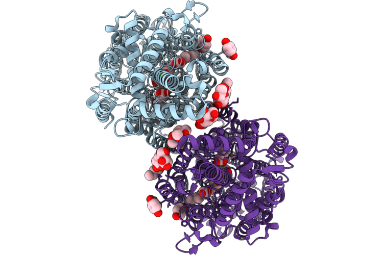 |
Cryoem Structure Of Native Quinol Dependent Nitric Oxide Reductase With Hqe At Ph 6.5
Organism: Achromobacter xylosoxidans
Method: ELECTRON MICROSCOPY Release Date: 2026-05-06 Classification: MEMBRANE PROTEIN Ligands: HEM, CA, FE, LMT, HQE, UQ5 |
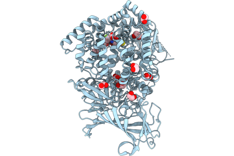 |
Erap1 In Complex With 1-[2-(5-Bromo-7-Fluoro-2-Oxo-2,3-Dihydro-1,3-Benzothiazol-3-Yl)Acetamido]-4,4-Difluorocyclohexane-1-Carboxylic Acid
Organism: Homo sapiens
Method: X-RAY DIFFRACTION Resolution:1.55 Å Release Date: 2026-05-06 Classification: PEPTIDE BINDING PROTEIN Ligands: ZN, SIN, PO4, EDO, A1JVE |
 |
Organism: Homo sapiens, Mus musculus
Method: ELECTRON MICROSCOPY Release Date: 2026-04-29 Classification: MEMBRANE PROTEIN |
 |
Organism: Homo sapiens, Mus musculus
Method: ELECTRON MICROSCOPY Release Date: 2026-04-29 Classification: MEMBRANE PROTEIN |
 |
Organism: Homo sapiens, Mus musculus
Method: ELECTRON MICROSCOPY Release Date: 2026-04-29 Classification: MEMBRANE PROTEIN |
 |
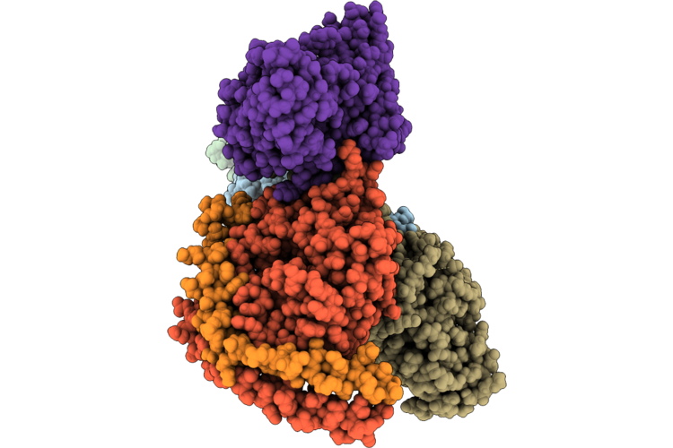
Organism: Human immunodeficiency virus 1, Macaca mulatta, Homo sapiens
Method: ELECTRON MICROSCOPY Resolution:2.70 Å Release Date: 2026-04-29 Classification: VIRAL PROTEIN Ligands: NAG |
 |
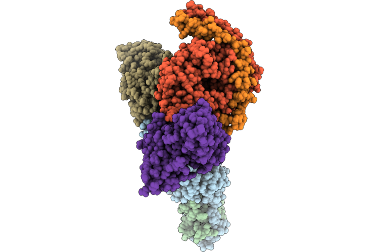
Organism: Human immunodeficiency virus 1, Macaca mulatta, Homo sapiens
Method: ELECTRON MICROSCOPY Resolution:2.70 Å Release Date: 2026-04-29 Classification: VIRAL PROTEIN Ligands: NAG |
 |
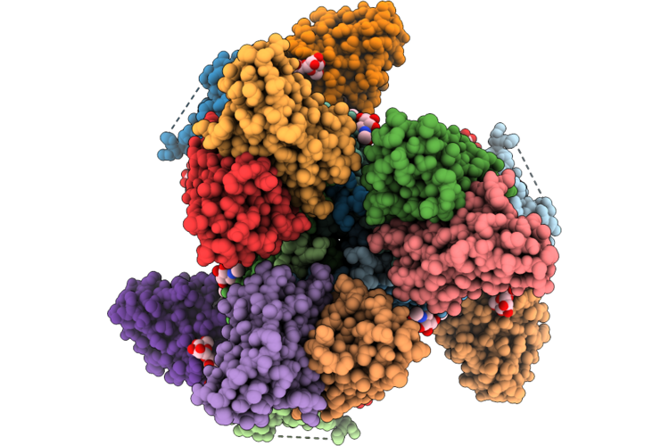
Organism: Homo sapiens, Lama glama
Method: ELECTRON MICROSCOPY Release Date: 2026-04-22 Classification: IMMUNE SYSTEM/Hydrolase |
 |
Erap1 In Complex With 1-[2-(6-Bromo-2,2-Difluoro-3-Oxo-3,4-Dihydro-2H-1,4-Benzoxazin-4-Yl)Acetamido]Cyclohexane-1-Carboxylic Acid
Organism: Homo sapiens
Method: X-RAY DIFFRACTION Resolution:1.57 Å Release Date: 2026-04-22 Classification: PEPTIDE BINDING PROTEIN Ligands: ZN, PO4, EDO, A1JVD |
 |
Erap1 In Complex With 1-[2-(2-Oxo-5-Phenyl-2,3-Dihydro-1,3-Benzoxazol-3-Yl)Acetamido]Cyclohexane-1-Carboxylic Acid
Organism: Homo sapiens
Method: X-RAY DIFFRACTION Resolution:1.73 Å Release Date: 2026-04-22 Classification: PEPTIDE BINDING PROTEIN Ligands: ZN, MLT, EDO, A1JVG |
 |
Erap1 In Complex With 1-[2-(5-Chloro-2-Oxo-2,3-Dihydro-1,3-Benzothiazol-3-Yl)Acetamido]Cyclohexane-1-Carboxylic Acid
Organism: Homo sapiens
Method: X-RAY DIFFRACTION Resolution:1.45 Å Release Date: 2026-04-22 Classification: PEPTIDE BINDING PROTEIN Ligands: ZN, PO4, EDO, A1JVH |
 |
Erap1 In Complex With 1-[2-(3-Oxo-6-Phenyl-3,4-Dihydro-2H-1,4-Benzoxazin-4-Yl)Acetamido]Cyclohexane-1-Carboxylic Acid
Organism: Homo sapiens
Method: X-RAY DIFFRACTION Resolution:1.37 Å Release Date: 2026-04-22 Classification: PEPTIDE BINDING PROTEIN Ligands: ZN, PO4, EDO, A1JVF |