

Search Count: 1,528
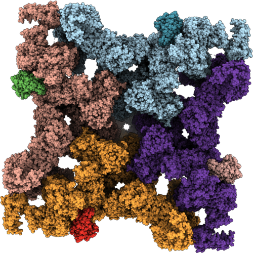 |
Structure Of Ryr1-Toxin Complex
Organism: Homo sapiens, Oryctolagus cuniculus
Method: ELECTRON MICROSCOPY Release Date: 2026-10-07 Classification: TRANSPORT PROTEIN/ISOMERASE/TOXIN Ligands: ATP, CFF, CA, ZN, A1CXI |
Organism: Homo sapiens, Oryctolagus cuniculus
Method: ELECTRON MICROSCOPY
Release Date: 2026-10-07
Ligands: ATP, CFF, CA, ZN, A1CXI
 |
Structure Of Ryr1 In Presence Of Rosuvastatin (Including Auxiliary Transmembrane Helices Tmx1 And Tmx2; Ca2+/Cff/Atp Dataset; Closed Pore)
Organism: Oryctolagus cuniculus
Method: ELECTRON MICROSCOPY Resolution:2.70 Å Release Date: 2026-09-30 Classification: TRANSPORT PROTEIN Ligands: ATP, CA, ZN, CFF, PCW |
Organism: Oryctolagus cuniculus
Method: ELECTRON MICROSCOPY
Release Date: 2026-09-30
Ligands: ATP, CA, ZN, CFF, PCW
 |
Structure Of Ryr1 In Complex With Rosuvastatin (Ca2+/Cff/Atp Dataset; Open Pore)
Organism: Oryctolagus cuniculus
Method: ELECTRON MICROSCOPY Resolution:2.90 Å Release Date: 2026-09-30 Classification: TRANSPORT PROTEIN Ligands: ATP, CA, ZN, CFF, PCW, A1C3Z |
Organism: Oryctolagus cuniculus
Method: ELECTRON MICROSCOPY
Release Date: 2026-09-30
Ligands: ATP, CA, ZN, CFF, PCW, A1C3Z
 |
Structure Of Ry12 Domain Of Rabbit Ryr1 Complexed With Adp And Mg2+ (Mimicking Physiological Conditions; Bound And Closed Ry12)
Organism: Oryctolagus cuniculus
Method: ELECTRON MICROSCOPY Release Date: 2026-09-16 Classification: MEMBRANE PROTEIN Ligands: ADP, MG |
Organism: Oryctolagus cuniculus
Method: ELECTRON MICROSCOPY
Release Date: 2026-09-16
Ligands: ADP, MG
 |
Structure Of Ry12 Domain Of Rabbit Ryr1 In The Presence Of Dantrolene And Mg2+ (Open Ry12 Conformation)
Organism: Oryctolagus cuniculus
Method: ELECTRON MICROSCOPY Release Date: 2026-09-16 Classification: MEMBRANE PROTEIN |
Organism: Oryctolagus cuniculus
Method: ELECTRON MICROSCOPY
Release Date: 2026-09-16
 |
Structure Of Ry12 Domain Of Rabbit Ryr1 In The Presence Of Atp, Mg2+, And Ca2+ (Open Ry12 Conformation)
Organism: Oryctolagus cuniculus
Method: ELECTRON MICROSCOPY Release Date: 2026-09-16 Classification: MEMBRANE PROTEIN |
Organism: Oryctolagus cuniculus
Method: ELECTRON MICROSCOPY
Release Date: 2026-09-16
 |
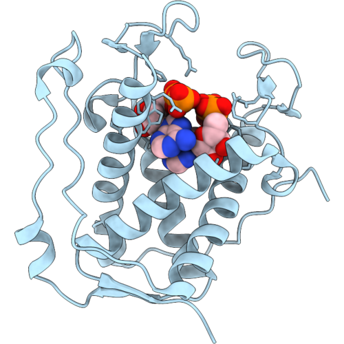
Structure Of Ry12 Domain Of Rabbit Ryr1 In The Presence Of Atp, Adp, And Mg2+ (Mimicking Physiological Conditions; Unbound And Open Ry12)
Organism: Oryctolagus cuniculus
Method: ELECTRON MICROSCOPY Release Date: 2026-09-16 Classification: MEMBRANE PROTEIN |
Organism: Oryctolagus cuniculus
Method: ELECTRON MICROSCOPY
Release Date: 2026-09-16
 |
Structure Of Ry12 Domain Of Rabbit Ryr1 Complexed With Adp And Mg2+ (Mimicking Muscle Fatigue)
Organism: Oryctolagus cuniculus
Method: ELECTRON MICROSCOPY Release Date: 2026-09-16 Classification: MEMBRANE PROTEIN Ligands: ADP, MG |
Organism: Oryctolagus cuniculus
Method: ELECTRON MICROSCOPY
Release Date: 2026-09-16
Ligands: ADP, MG
 |
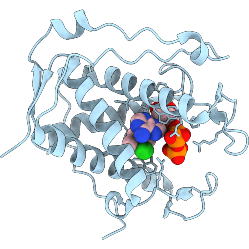
Structure Of Ry12 Domain Of Rabbit Ryr1 Complexed With Arm210 And Atp
Organism: Oryctolagus cuniculus
Method: ELECTRON MICROSCOPY Release Date: 2026-09-16 Classification: MEMBRANE PROTEIN Ligands: ATP, KVR |
Organism: Oryctolagus cuniculus
Method: ELECTRON MICROSCOPY
Release Date: 2026-09-16
Ligands: ATP, KVR
 |
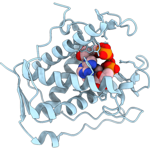
Structure Of Ry12 Domain Of Rabbit Ryr1 Complexed With Atp And 4-Chloro-M-Cresol
Organism: Oryctolagus cuniculus
Method: ELECTRON MICROSCOPY Resolution:2.77 Å Release Date: 2026-09-16 Classification: MEMBRANE PROTEIN Ligands: 43M, ATP |
Organism: Oryctolagus cuniculus
Method: ELECTRON MICROSCOPY
Release Date: 2026-09-16
Ligands: 43M, ATP
 |
Structure Of Rabbit Ryr1 Ry12 Domain Complexed With Adp And Mg2+
Organism: Oryctolagus cuniculus
Method: ELECTRON MICROSCOPY Resolution:2.52 Å Release Date: 2026-09-16 Classification: MEMBRANE PROTEIN Ligands: ADP, MG |
Organism: Oryctolagus cuniculus
Method: ELECTRON MICROSCOPY
Release Date: 2026-09-16
Ligands: ADP, MG
 |
Structure Of Ry12 Domain Of Rabbit Ryr1 Complexed With Dantrolene And Atp
Organism: Oryctolagus cuniculus
Method: ELECTRON MICROSCOPY Release Date: 2026-09-16 Classification: MEMBRANE PROTEIN Ligands: U1C, ATP |
Organism: Oryctolagus cuniculus
Method: ELECTRON MICROSCOPY
Release Date: 2026-09-16
Ligands: U1C, ATP
 |
Structure Of Ry12 Domain Of Rabbit Ryr1 Complexed With Dantrolene And Adp
Organism: Oryctolagus cuniculus
Method: ELECTRON MICROSCOPY Release Date: 2026-09-16 Classification: MEMBRANE PROTEIN Ligands: ADP, U1C |
Organism: Oryctolagus cuniculus
Method: ELECTRON MICROSCOPY
Release Date: 2026-09-16
Ligands: ADP, U1C
 |
Structure Of Rabbit Ryr1 Complexed With Fkbp12.6 And Calmodulin In The Presence Of Dantrolene, Adp, 4-Chloro-M-Cresol, Caffeine, And Mg2+
Organism: Homo sapiens, Oryctolagus cuniculus
Method: ELECTRON MICROSCOPY Release Date: 2026-09-16 Classification: MEMBRANE PROTEIN Ligands: ZN, ADP, U1C, 43M, CA, CFF |
Organism: Homo sapiens, Oryctolagus cuniculus
Method: ELECTRON MICROSCOPY
Release Date: 2026-09-16
Ligands: ZN, ADP, U1C, 43M, CA, CFF
 |
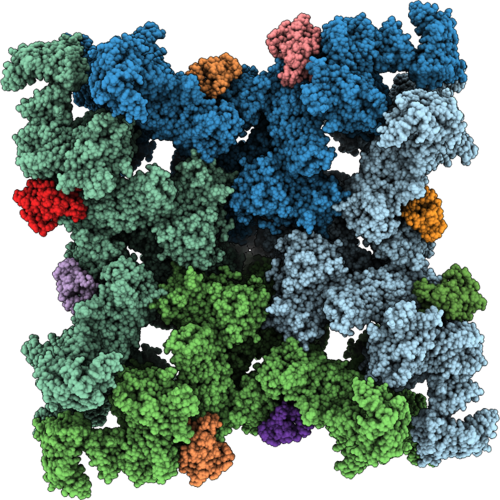
Structure Of Rabbit Ryr1 Complexed With Fkbp12.6 And Calmodulin In The Presence Of Atp, Adp, And Mg2+ (Mimicking Muscle Fatigue)
Organism: Homo sapiens, Oryctolagus cuniculus
Method: ELECTRON MICROSCOPY Release Date: 2026-09-16 Classification: MEMBRANE PROTEIN Ligands: ZN, ADP, ATP, MG |
Organism: Homo sapiens, Oryctolagus cuniculus
Method: ELECTRON MICROSCOPY
Release Date: 2026-09-16
Ligands: ZN, ADP, ATP, MG
 |
Structure Of Rabbit Ryr1 Complexed With Fkbp12.6 And Calmodulin In The Presence Of Atp, Adp, And Mg2+ (Mimicking Physiological Conditions)
Organism: Homo sapiens, Oryctolagus cuniculus
Method: ELECTRON MICROSCOPY Release Date: 2026-09-16 Classification: MEMBRANE PROTEIN Ligands: ZN, ATP |
Organism: Homo sapiens, Oryctolagus cuniculus
Method: ELECTRON MICROSCOPY
Release Date: 2026-09-16
Ligands: ZN, ATP
 |
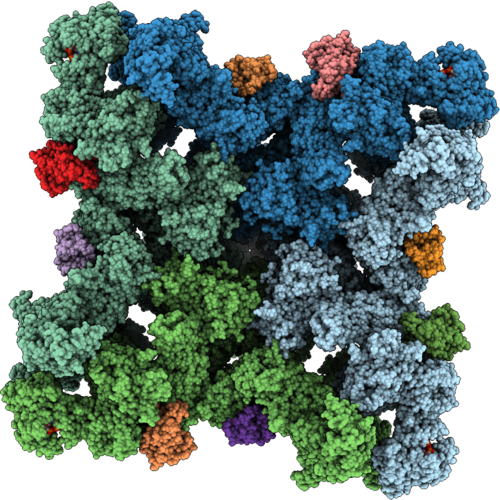
Structure Of Rabbit Ryr1 Complexed With Fkbp12.6, Calmodulin, Atp, 4-Chloro-M-Cresol, And Caffeine
Organism: Homo sapiens, Oryctolagus cuniculus
Method: ELECTRON MICROSCOPY Release Date: 2026-09-16 Classification: MEMBRANE PROTEIN Ligands: 43M, CFF, CA, ATP, ZN |
Organism: Homo sapiens, Oryctolagus cuniculus
Method: ELECTRON MICROSCOPY
Release Date: 2026-09-16
Ligands: 43M, CFF, CA, ATP, ZN
 |
Structure Of Rabbit Ryr1 Complexed With Fkbp12.6, Calmodulin, Adp, 4-Chloro-M-Cresol, Caffeine, And Mg2+
Organism: Homo sapiens, Oryctolagus cuniculus
Method: ELECTRON MICROSCOPY Release Date: 2026-09-16 Classification: MEMBRANE PROTEIN Ligands: 43M, ADP, ZN, PLC, CFF, MG |
Organism: Homo sapiens, Oryctolagus cuniculus
Method: ELECTRON MICROSCOPY
Release Date: 2026-09-16
Ligands: 43M, ADP, ZN, PLC, CFF, MG
 |
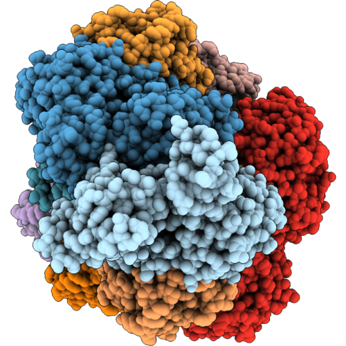
Structure Of Rabbit Ryr1 Complexed With Fkbp12.6 And Calmodulin In The Presence Of Dantrolene, Atp, 4-Chloro-M-Cresol, Caffeine, And Mg2+
Organism: Homo sapiens, Oryctolagus cuniculus
Method: ELECTRON MICROSCOPY Resolution:3.09 Å Release Date: 2026-09-16 Classification: MEMBRANE PROTEIN Ligands: CA, ZN, U1C, 43M, ATP, CFF, CPS |
Organism: Homo sapiens, Oryctolagus cuniculus
Method: ELECTRON MICROSCOPY
Release Date: 2026-09-16
Ligands: CA, ZN, U1C, 43M, ATP, CFF, CPS
 |
Cofilactin Filament With One Aip1 Bound To Middle Of The Filament
Organism: Caenorhabditis elegans, Oryctolagus cuniculus
Method: ELECTRON MICROSCOPY Resolution:2.29 Å Release Date: 2026-09-09 Classification: STRUCTURAL PROTEIN Ligands: ADP, MG |
Organism: Caenorhabditis elegans, Oryctolagus cuniculus
Method: ELECTRON MICROSCOPY
Release Date: 2026-09-09
Ligands: ADP, MG