
Search Count: 1,395
All
Selected
 |
Organism: Lotus japonicus
Method: X-RAY DIFFRACTION Resolution:1.60 Å Release Date: 2026-05-13 Classification: OXYGEN BINDING Ligands: HEM, CYN, PO4 |
 |
Cm1-Activated Gturc In Complex With Nascent Alpha-E254D Mutant Microtubules
Organism: Saccharomyces cerevisiae s288c, Homo sapiens
Method: ELECTRON MICROSCOPY Release Date: 2026-05-13 Classification: STRUCTURAL PROTEIN Ligands: GTP |
 |
Organism: Homo sapiens
Method: X-RAY DIFFRACTION Resolution:1.80 Å Release Date: 2026-04-22 Classification: OXIDOREDUCTASE Ligands: FAD, GOL |
 |
Organism: Homo sapiens, Human immunodeficiency virus 1
Method: ELECTRON MICROSCOPY Release Date: 2026-04-22 Classification: VIRAL PROTEIN/IMMUNE SYSTEM Ligands: NAG |
 |
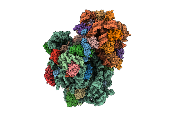
Structure Of The Wild-Type Staphylococcus Aureus 70S Ribosome Complexed With Clincelin
Organism: Staphylococcus aureus
Method: ELECTRON MICROSCOPY Release Date: 2026-04-15 Classification: RIBOSOME Ligands: ZN, MG, FME, K, A1I09 |
 |
Structure Of The A2058-Dimethylated Staphylococcus Aureus 70S Ribosome Complexed With Clincelin
Organism: Staphylococcus aureus
Method: ELECTRON MICROSCOPY Release Date: 2026-04-15 Classification: RIBOSOME Ligands: ZN, MG, FME, K, A1I09 |
 |
Organism: Homo sapiens
Method: X-RAY DIFFRACTION Resolution:1.65 Å Release Date: 2026-04-08 Classification: METAL BINDING PROTEIN Ligands: CA, 0LI, NA, ACY |
 |
Organism: Homo sapiens
Method: X-RAY DIFFRACTION Resolution:2.28 Å Release Date: 2026-04-08 Classification: METAL BINDING PROTEIN Ligands: CA, NA, ACY, A1IO4, P6G, 1PE |
 |
Crystal Structure Of Feruloyl Esterase From Fusarium Oxysporum G122S Variant In Complex With Benzoic Acid
Organism: Fusarium oxysporum
Method: X-RAY DIFFRACTION Resolution:1.71 Å Release Date: 2026-04-01 Classification: HYDROLASE Ligands: NAG, PEG, EDO, BEZ, CA |
 |
Crystal Structure Of Feruloyl Esterase From Fusarium Oxysporum G122S Variant
Organism: Fusarium oxysporum
Method: X-RAY DIFFRACTION Resolution:1.90 Å Release Date: 2026-04-01 Classification: HYDROLASE Ligands: NAG, PEG, EDO, PGE, CA |
 |
Organism: Homo sapiens
Method: X-RAY DIFFRACTION Resolution:2.50 Å Release Date: 2026-03-18 Classification: IMMUNE SYSTEM Ligands: SO4 |
 |
Organism: Human hepatitis a virus
Method: ELECTRON MICROSCOPY Release Date: 2026-03-18 Classification: VIRUS |
 |
Organism: Human hepatitis a virus
Method: ELECTRON MICROSCOPY Resolution:1.70 Å Release Date: 2026-03-18 Classification: VIRUS |
 |
Organism: Pedobacter sp. kp-2
Method: X-RAY DIFFRACTION Resolution:2.00 Å Release Date: 2026-03-11 Classification: HYDROLASE |
 |
Organism: Homo sapiens
Method: X-RAY DIFFRACTION Resolution:1.91 Å Release Date: 2026-03-11 Classification: METAL BINDING PROTEIN Ligands: CA, PEG, P6G, H9F, EOH, NA |
 |
The Structure Of A Bacterial Cyanide Dihydratase From Bacillus Safensis Per-Urp-08
Organism: Bacillus safensis
Method: ELECTRON MICROSCOPY Release Date: 2026-02-25 Classification: HYDROLASE |
 |
Organism: Microdochium sorghi
Method: ELECTRON MICROSCOPY Release Date: 2026-02-25 Classification: HYDROLASE |
 |
Organism: Homo sapiens, Coxsackievirus b3 (strain nancy)
Method: X-RAY DIFFRACTION Resolution:3.00 Å Release Date: 2026-02-18 Classification: RNA/IMMUNE SYSTEM |
 |
Organism: Homo sapiens
Method: X-RAY DIFFRACTION Resolution:2.51 Å Release Date: 2026-02-11 Classification: OXIDOREDUCTASE Ligands: NAD, FAD, ACT |
 |
Organism: Macaca mulatta, Human immunodeficiency virus 1
Method: ELECTRON MICROSCOPY Release Date: 2026-02-11 Classification: VIRAL PROTEIN Ligands: NAG |